UNIVERSITÀ DEGLI STUDI DI MESSINA
Dipartimento CHIBIOFARAM
TESI DI DOTTORATO DI RICERCA IN SCIENZE CHIMICHE
Curriculum: progettazione, sintesi, analisi e proprietà di sistemi
molecolari funzionali
XXXII CICLO
2016-2019
Synthetic Strategies, Photophysical and First Biological Applications
of New Glycoamino OPEs
Aurora Mancuso
Supervisor
Prof.ssa Anna Barattucci
Index
Abstract
1
Chapter 1 - Introduction
51.1 Carbohydrates in drug delivery 5
1.2 Luminescent dyes for biomedical applications 12
1.2A Bioimaging 12
1.2B Photodynamic Therapy 15
1.2C DNA-targeted drugs and sensors 24
1.2D pH fluorescent indicators 28
1.3 Oligo(phenilene etynylene)s 33
Chapter 2 – Results and Discussion
452.1 OPE_Glucose 1 and 2 synthesis 48
2.2 Chain elongation 54
2.3 Sugar change 62
2.4 Dimethylamino group quaternarization 67
2.5 Chain desymmetrization 74
2.5A Introduction of strong electronwithdrawing groups 74
2.5B Anchoring to nanoparticles 82
Chapter 3 – Photophysical studies and preliminary biological
applications
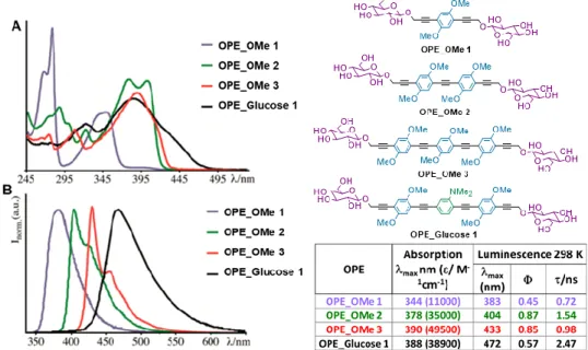
943.1 Photophysical studies 94
3.1A Photophysical properties of 38 and 39 94
3.1C Effect of chain desymmetrization and introduction of strong electron withdrawing groups 101 3.2 Preliminary biological studies on selected compounds 105 3.2A Studies of DNA binding of 38 and 39 105 3.2B Effect of sugar change: preliminary internalization and PDT
studies on OPE_Maltose 111
Conclusions
116Chapter 4 – Experimental methods
1184.1 General synthetic methods 118
4.2 Synthetic methodologies and characterizations 121
Pubblications 174
Comunications 174
1
Abstract
Oligo(phenyleneethynylenes) (OPEs) and the corresponding polymers, Poly(phenyleneethynylenes) (PPEs), are organic molecules with π‐conjugated backbone, which could be designed to display high electrical conductivity, outstanding photophysical properties, and excellent biocompatibility. While they have been extensively explored for a wide range of applications, from electronics and optoelectronic devices, to energy harvesting and nanotechnology, they attracted attention in the biomedical field only in the past decades. Specifically, they have found innovative applications in a variety of biotechnologies, as biosensors, probes for cell imaging, biocides and many others.
In the last years, my research group has widely studied the photophysical and biological properties of a new class of end-only glucose functionalized oligo(phenyleneethynylenes): thanks to the modulation of substituents and length of the central conjugated chain, some of the obtained OPEs have demonstrated to be promising dyes in bioimaging and superficial Photodynamic Therapy (PDT) (Figure A1).
On the basis of these assumptions, my PhD work consisted in the synthesis of differently modified glycosilated OPEs with the aim to better modulate their photophysical features and biological behaviour, studying new possible applications in the biomedical field.
2
In particular, Chapter 1 consists in a literature survey and gives an overview on general principles and previous works upon which this research draws. In paragraph 1.1, carbohydrates conjugation, as an efficient strategy for delivering and targeting drugs, is discussed. Paragraph 1.2 deals with the main applications of luminescent dyes in the biomedical field: at first, in 1.2.A, a general view on the principles of bioimaging and the design of suitable fluorescent probes is reported; in 1.2.B, the mechanisms that are the basis of photodynamic therapy (PDT) and a brief literature overview on different classes of dyes and nanoparticles, which have found application as photosensitizers in this type of treatments, are described; 1.2.C deals with drugs interactions with nucleic acids, with particular attention to the principal binding modes and to the possible uses of luminescent dyes for DNA labelling; finally, in 1.2.D, applications of specific kinds of fluorescent dyes, whose luminescence is pH dependent, as intracellular pH indicators are reported. To conclude, paragraph 1.3 provides an overall picture of Oligo(phenyleneethynylenes) (OPEs) features and biomedical applications, mainly focusing on the previous works of my research group.
Chapter 2 deals with the discussion of the synthetic pathways used for the
obtainment of the new glycosylated OPE systems and the structural modifications introduced during the PhD work. In fact, in paragraph 2.1 the synthetic approach and the optimization of yields and methods for the obtainment of the already reported OPE_Glucose 1 and OPE_Glucose 2 (Figure A1), which had shown the best results in terms of cell internalization and applications as photosensitizers in PDT, are described. In paragraph 2.2, the synthetic routes which led to the elongation of the OPE chain are reported: the extension of the π-conjugated system is meant to further study the influence of chain length on the photophysical behaviour. The synthesis of three new amino-OPE systems, bearing different sugar terminations (monosaccharide galactose and mannose, and disaccharide maltose) are described in paragraph 2.3: the presence of different carbohydrate residues could, in fact, influence the biological behaviour of the new compounds,
3
with respect to the glucosylated systems. Furthermore, the synthesis of several tetralkylammonium OPEs is the subject of paragraph 2.4: the disappearance of the lone pair on the amino nitrogen and the formation of a positive charge on the OPE chain could, in fact, strongly influence the photophysical OPE features but also the water solubility, opening the way to other biological applications. Finally, the last modifications reported in paragraph 2.5 are focused on the desymmetrization of the OPE chain. In part 2.5A the introduction of electron withdrawing fluorinated aromatic groups as chain termination is discussed: the new moiety, which is able to interact with the methoxy and amino electron donating groups already present as substituents on the OPE conjugated chain, could influence the photophysical features, leading to the obtainment of new push-pull systems. In part 2.5B the synthesis of OPEs, bearing suitable termination for the functionalization of upconverting nanoparticles (UCNPs), is described. These nanoparticles, after excitation with NIR light (980 nm), emit in a region of UV that matches the absorption of our OPEs (~380 nm): an energy transfer process from the NPs to our conjugated system could extend their applications in NIR-PDT.
Chapter 3 deals with the preliminary photophysical and biological studies
performed on some selected compounds. In particular, in paragraph 3.1 the effects of the different modification on our OPE systems (chain elongation, nitrogen quaternarization and desymmetrization) on the spectroscopic features are discussed, in comparison with the already reported OPE_Glucose 1 and
OPE_Glucose 2. In fact, it has been demonstrated that each structural change in
the OPE chain causes different variations in the photophysical properties; moreover, preliminary studies on the fluorinated compounds show that the emission of one of these derivatives is pH-sensitive. On the other hand, the first biological tests, carried out on some of the synthesized compounds, are described in paragraph 3.2. It has been found that the two positive charged tetralkylammonium OPEs, analogues of OPE_Glucose 1 and OPE_Glucose 2,
4
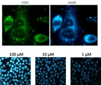
strongly bind the double helix of DNA: this interaction is also responsible for their absorption and emission switching-off and reduction of cancer cells proliferation. Moreover, the first internalization and PDT tests on healthy and cancer cells performed with the synthesized compound bearing two maltosidic residues are also reported.
Finally, in Chapter 4 experimental synthetic methods, spectral and analytical characterizations of the obtained compounds, equipment, materials and methods for the photophysical and biological experiments are reported.
5
CHAPTER 1
Introduction
1.1 CARBOHYDRATES IN DRUG DELIVERY
Carbohydrates represent one of the most abundant and pervasive class of biomolecules. Besides being important for survival of living organisms as an important dietary component, their role in many biological processes is fundamental for life. Their role is polyvalent: a) they can serve as immediate source of energy in the form of table sugar, starch, and fibres; b) they are part of the structural building blocks of genetic materials (DNA and RNA) and c) play a key role in cell recognition events, acting as extracellular and intracellular components in molecular recognition. Thanks to advancements in carbohydrate research and to the fact that carbohydrates are now recognized as important players in numerous processes such as cell recognition (facilitated by cell surface saccharides), cell migration, and immune defence, the use of carbohydrates as direct drug delivery systems has been a fundamental subject in the last decades.1
The process of their absorption is cell-selective and sometimes limited to few saccharides. For example, among the large number of theoretically existing simple carbohydrates, glucose remains the principal food or 'fuel' for most cells; in fact, other carbohydrates are routinely transformed to glucose before their storage as glycogen in the liver.
1 Mishra, S.; Upadhay, K.; Mishra, K. B.; Shukla, A. K.; Tripathi, R. P.; Tiwari, V. K. Studies in
6
Due to its polar nature and size, glucose molecules cannot traverse the lipid membrane of the cell by simple diffusion; the regulation of glucose transport in humans is complex and involves two classes of membrane proteins:
1) Na+-dependent glucose transporters, which are involved in the active transport of glucose from the lumen of the small intestine and from the proximal kidney tubule in the process of reabsorption;
2) the facilitative glucose transporters, which are still not fully characterized proteins but present on all the cells.
These transporters possess distinct physiological and biochemical properties, which endow them with specific functions in the tissues in which they are expressed. For these reasons, glucose-selective proteins and other proteins, which exhibit selectivity for a variety of simple carbohydrates are the preferred choice for an emerging new form of cell-specific drug delivery system.2
Carbohydrates with structural and functional diversity have enabled chemists to synthesize novel carbohydrate-based therapeutic agents or to make cardinal modifications to improve the activity and selectivity of existing drugs. In fact, many natural and semisynthetic glycoconjugates are clinically used as antimicrobials and anticancer agents. Carbohydrates can guarantee water solubility, minimized toxicity and can enhance pharmacokinetics to conjugated drugs with respect to their aglycones.3 In addition to this, carbohydrates also establish stereochemically defined scaffolds to project pharmacophores, that can give rise to a number of carbohydrate-based compound libraries.
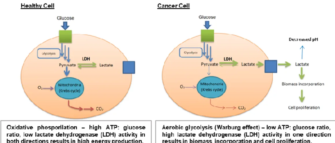
Carbohydrates are also extremely important for processes involved in cell transformation and tumour genesis. Almost a century ago, the scientist Otto Warburg discovered that cancerous tissues consume larger amounts of glucose compared to healthy tissues. Glucose transporters (GLUTs), which are located on
2 Palomino, E. Advanced Drug Delivery Reviews, 1994, 13, 311.
3 Mishra, S.; Upadhay, K.; Mishra, K. B.; Shukla, A. K.; Tripathi, R. P.; Tiwari, V. K. Studies in
7
membranes of endothelial cells and facilitate the entry and the diffusion of glucose and other carbohydrates into cells, are widely overexpressed in human cancers.4
Figure 1.1. Metabolism in healthy and cancer cells.
In order to sustain the uncontrolled growth rate, cancer cells have adopted a different unconventional mechanism to produce energy. Usually, healthy cells do not stop glucose metabolism to lactate when oxygen is available, but they undergo oxidative phosphorylation, producing high ATP level: they exploit anaerobic glycolysis (metabolism of glucose to lactic acid) only when the oxygen is absent or limited. In contrast, cancer cells metabolize glucose to lactate even in the presence of oxygen (aerobic glycolysis), producing low levels of ATP; because of the great quantity of produced lactate, it is ejected outside the cell membrane, also causing a decreasing of the pH in the external microenviroment.5
(Figure 1.1) Thus, altered cancer cell metabolism can modulate the tumor microenvironment, which plays important roles in cancer cell somatic evolution, metastasis, and therapeutic response.6 Today this phenomenon is known as the “Warburg effect” and is recognized as one of the hallmarks of cancer and gives
4 Warburg, O.; Wind, F.; Negelein, E. J. Gen. Physiol., 1927, 8, 519–530; Warburg, O. Science,
1956, 123, 309.
5 Koppenol, W. H.; Bounds, P. L.; Dang, C. V. Nat. Rev. Cancer 2011, 11, 325. 6 Justus, C.; Sanderlin, E. J.; Yang, L. V. Int. J. Mol. Sci. 2015, 16, 11055.
8
us the information that cancer cells consume huge amounts of glucose for their proliferation. Increased understanding of this dysfunctional metabolism has led to an interest in targeting it for cancer therapy. For this reason, one promising strategy for drug design is glycoconjugation: the linking of a drug to glucose or other mono- and disaccharides. Monosaccharide-conjugated analogues of diverse agents, designed to improve the water solubility, serum stability, and targeting of their aglycones, have been reported in the chemical literature since the early 1990s.
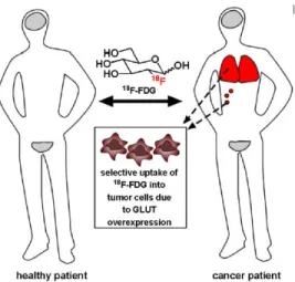
One of the first clinically used carbohydrates-based drug is 2-deoxy-2-(18F)fluoro-D-glucose (18F-FDG; Figure 1.2), a radiolabelled glucose analogue.
18F-FDG is used to visualize tumours and their metastases due to the tendency of
these cancerous tissues to uptake glucose at a higher rate than most normal tissues.
Moreover, positron emission tomography (PET) with 18F-FDG has been used to assess alterations in glucose metabolism in brain, cancer, cardiovascular diseases, Alzheimer’s disease and other central nervous system disorders, infectious, autoimmune and inflammatory diseases.
Figure 1.2. Localization of 18F-FDG in healthy (grey) and cancer (red) patient.
In healthy patients, 18F-FDG is taken up only by tissues that constitutively consume glucose, such as the brain and bladder (grey in figure). In a sick patient, tumour cells preferentially uptake 18F-FDG, allowing clinicians to identify sites
9
of tumours and metastases (red in figure), as well as stage cancer and monitor response to treatment. Currently, lung, breast, colorectal, and endometrial carcinomas, as well as bone and soft tissue sarcomas and Hodgkin’s and non-Hodgkin’s lymphomas, are commonly staged based on their ability to preferentially uptake this radiolabelled glucose analogue compared to noncancerous tissues.
Since then, this field has grown exponentially and in the last decades several cytotoxic agents, e.g., glufosfamide,7 chlorambucil,8 busulfan,9 paclitaxel,10 have been glycoconjugated and found to be cancer selective and less toxic to normal cells than the parent aglycons.11 Furthermore, other sugars such as galactose, mannose, fucose, etc., have been used as powerful scaffolds, installed on drug molecules, for targeting specific tissues.12 In fact, it is known that other non-glucosidic monosaccharides can be substrates for GLUT transporters, and thus can be considered candidates for a GLUT-targeting approach. Specifically, using light diffraction measurement in human erythrocytes that express GLUT-1 as their sole glucose transporter, 2-deoxy-glucose, galactose, mannose, D-xylose, 2-deoxy-D-galactose, L-arabinose, D-ribose, D-fucose, and D-lyxose, in order of decreasing affinity, were all found to be transported into cells in a transporter mediated mechanism. In contrast, D-arabinose, fucose, and L-rhamnose were poor substrates, and the enantiomers of several GLUT substrates, L-glucose and L-xylose, were not GLUT substrates.13 Thus, it seems that many sugars can be conjugated to anticancer agents to take advantage of GLUT-mediated cellular uptake; the choice of which sugar to utilize may depend on the desired cleavage mechanism of the compound. For instance, D-galactose, the C4
7 Pohl, J.; Bertram, B.; Hilgard, P.; Nowrousian, M. R.; Stuben, J.; Wiessler, M. Cancer
Chemother Pharmacol. 1995, 35,364.
8 Halmos, T.; Santarromana, M.; Antonakis, K.; Scherman, D. Eur J Pharmacol. 1996, 18, 477. 9 Halmos, T.; Santarromana, M.; Antonakis, K.; Scherman, D. Carbohydr. Res. 1997, 299, 15. 10 Lin, Y. S.; Tungpradit, R.; Sinchaikul, S.; An, F. M.; Liu, D. Z.; Phutrakul, S.; Chen, S. T. J.
Med. Chem. 2008, 51, 7428.
11 Calvaresi, E. C.; Hergenrother, P. J. Chem. Sci. 2013, 4, 2319.
12 Yuan, S. S.; Li, M. L.; Chen, J. S.; Zhou, L.; Zhou, W. ChemMedChem., 2018, 13, 764. 13 LeFevre, P.G. Pharmacol Rev. 1961, 13, 39.
10
epimer of glucose, has been reported to possess an equivalent affinity and uptake rate by GLUT-1 compared to glucose.14 Galactose-conjugated drugs may be used to selectively target certain types of cancers known to highly express galactosidase enzymes, such as breast and colon cancers.15 In the absence of substantial tumour galactosidase expression, galactose-conjugated prodrugs can be used in conjunction with tumour-selective monoclonal antibodies linked to galactosidases, which ideally cleave the inactive conjugate to its active form in tumour tissue. Thus, glycocojugation generally offers selective targeting to cancer cells, if the glycoside of choice is a GLUT substrate. For instance, in 2001, Mikuni, Mandai, and coworkers16 reported the synthesis and cancer cell potency of several glycoconjugates of docetaxel, including conjugates with glucose, galactose, mannose, and xylose. These compounds were shown to have a 3- to 18-fold improvement in activity compared to the aglycone against B16 murine melanoma cells and it was particularly highlighted the in vivo potency of galactose-conjugated docetaxel, compared to the aglycone.
Moreover, with the great development of carbohydrate synthesis technology, carbohydrate chain analysis methods and nanotechnology, research on the biological effects associated with carbohydrate has become a hot topic in recent years. Nanomaterials as the carriers of carbohydrates have been gradually developed since the first synthesis of carbohydrate functionalized gold nanoparticles in 200117 and related reports have gradually increased. They have shown great potential for applications in biomedical imaging, diagnosis, and treatment.18 Compared with the use of other molecules as carriers, nanoparticles can regulate the density of ligands on the surface by adjusting their size and shape.
14 Melisi, D.; Curcio, A.; Luongo, E.; Morelli, E.; Rimoli, M. G. Curr. Top. Med. Chem. 2011, 11, 2288.
15 Devalapally, H.; Rajan, K. S.; Akkinepally, R. R.; Devarakonda. R. K. Drug Dev Ind Pharm.
2008, 34, 789.
16 Mandai, T.; Okumoto, H.; Oshitari, T.; Nakanishi, K.; Mikuni, K.; Hara, K.; Iwatani, W.; Amano, T.; Nakamura, K.; Tsuchiya, Y. Heterocycles 2001; 54, 561.
17 de la Fuente, J.M.; Barrientos, A. G.; Rojas, T. C.; Rojo, J.; Cañada, J.; Fernández, A.; Penadés S. Angew. Chem. Int. Ed. 2001, 40, 2257.
11
In addition, nanoparticles have unique optical, electrical, magnetic, mechanical, and chemical activities. These properties make glyconanoparticles not only useful for studying sugar-related biological effects but also for cell imaging and tumour cell-targeted drug delivery.19
Among the large number of nanomaterials, gold, an extremely inert and biocompatible material, is one of the main exploited scaffolds for producing glyconanoparticles.20 At nanosize, gold nanoparticles (AuNPs) are characterized by unique optical features, which result extremely useful in many diagnosis and affinity studies or protocols. Moreover, gold can be easily and covalently decorated on its surface by exploiting the strong soft–soft interaction between an Au atom and sulphur.21 Therefore, glyco-gold nanoparticles (GAuNPs) represent a smart, multimodal and versatile nanoplatform to develop carbohydrate-based nanotechnology and nanomedicine systems.22
Also glycoside lanthanide ion-doped upconversion nanoparticles (UCNPs) (paragraph 1.2.B), which can convert low-energy infrared photons into high-energy visible and ultraviolet photons, have recently found several applications in advanced biomedical and biophotonics fields.23For instance, in 2010, Bogdan
et al.24 reported the synthesis of mannose-coated lanthanide-doped upconverting
nanoparticles (Figure 1.3) for the selective detection of proteins based on selective carbohydrate-protein complementary recognition, succeeding in interacting and detecting bacteria or virus infections.
19 Jafari Malek, S.; Khoshchehreh, R.; Goodarzi, N.; Khoshayand, M. R.; Amini, M.; Atyabi, F.; Esfandyari-Manesh, M.; Tehrani, S.; Mohammad Jafari, R.; Maghazei, M. S.; Alvandifar, F.; Ebrahimi, M.; Dinarvand, R. Colloids Surf. B: Biointerfaces 2014, 122, 350 .
20 Compostella, F.; Pitirollo, O.; Silvestri, A.; Polito, L. Beilstein J. Org. Chem. 2017, 13, 1008. 21 Saha, K.; Agasti, S. S.; Kim, C.; Li, X.; Rotello, V. M. Chem. Rev. 2012, 112, 2739.
22 Chaudhary, P. M.; Sangabathuni, S.; Murthy, R. V.; Paul, A.; Thulasiram, H. V.; Kikkeri, R.
Chem. Commun. 2015, 51, 15669; Sangabathuni, S.; Vasudeva Murthy, R.; Chaudhary, P. M.;
Surve, M.; Banerjee, A.; Kikkeri, R. Nanoscale 2016, 8, 12729. 23 Gee, A.; Xu, X. Surfaces 2018, 1, 96.
12 Figure 1.3. Mannose-coated UCNPs
1.2 LUMINESCENT DYES FOR BIOMEDICAL
APPLICATIONS
1.2.A Bioimaging. Methods to “see into the body” or “see into cells” are
essential for the diagnosis and treatment of disease, as well as for research into the basic processes of life. It is desirable that the used methods should not be invasive, i.e., should not involve cutting into the body or isolating cellular constituents. Therefore, techniques to visualize physiological or pathophysiological changes in the body and cells have become increasingly important in biomedical sciences.25 Compared to other technologies such as radioisotope labelling, magnetic resonance imaging (MRI), electron spin resonance (ESR) spectroscopy, and electrochemical detection, fluorescence bioimaging is the most versatile and widely used visualization method to study the structure and function of biological systems and the molecular process in living organisms without perturbing them. This technique offers many advantages for this purpose, because it is characterized by high sensitivity, high resolution,
13
less-invasive and safe detection using readily available instruments. Today, fluorescent probes based on small organic molecules have become indispensable tools in modern biology because they provide dynamic information concerning the localization and quantity of the molecules of interest, without the need for genetic engineering of the sample.
The design of new fluorophores for biological applications is essential, but often suffers from intrinsic problems: keeping high structural homology with natural molecules is generally useful to provide biocompatibility but often prevents the achievement of remarkable results in term of fluorescence properties (e.g. fluorescence quantum yield, bathochromic emission); on the other hand, more elaborate modifications of the molecular skeleton could induce perturbation of normal cellular activity or, in the worst case, be lethal for living systems.
Figure 1.4
For this reason, only few fluorophores are currently approved by the U.S. Food and Drug Administration (FDA) for medical use such as indocyanine green
14
(ICG),26 methylene blue27 and fluorescein (Figure 1.4).28 Another fluororophore,
rhodamine B, approved for use in 1966, was subsequently banned in 1987 after being linked to cancer in mice and rats.29
A successful optical molecular probe for medical imaging has to possess specific features, including wavelength, brightness, bio- and photostability, and pharmacokinetics including nonspecific tissue accumulation.30 First of all, fluorophores require excitation light to give emission: excitation in the UV range can cause direct tissue damage, whereas excitation in the IR can lead to tissue heating; blue and/or green excitation light is compatible with surface imaging applications because it has poor tissue penetration. On the other hand, yellow or red light (at around 600 nm) leads to excessive autofluorescence, because of naturally occurring endogenous fluorophores (mostly haemoglobin and related), also excited in this range. The optimal excitation wavelength of a fluorophore is in the deep red or near-infrared range because of the combined virtues of good tissue penetration and low autofluorescence.31 However, for surface applications, such as detecting tumours on a mucosal or epithelial surface, lower wavelength (e.g., blue, green, yellow) emitters with high quantum efficiency may produce as good or better results compared to NIR probes.
The sensitivity and resolution for bioimaging depend also by the difference between excitation and emission wavelengths of the probe, known as the Stokes shift: in fact a small Stokes shift may result in self-quenching and back scattering from biological samples.32 Other important considerations in choosing a
26 Yoshida, M.; Kubota, K.; Kuroda, J.; Ohta, K.; Nakamura, T.; Saito, J.; Kobayashi, M.; Sato, T.; Beck, Y.; Kitagawa, Y.; Kitajima, M. J. Gastroenterol. Hepatol. 2012, 3, 29.
27 Verbeek, F. P.; van der Vorst, J. R.; Schaafsma, B. E.; Swijnenburg, R. J.; Gaarenstroom, K. N.; Elzevier, H. W.; van de Velde, C. J.; Frangioni, J. V.; Vahrmeijer. A. L. J. Urol. 2013, 190, 574.
28 McGinty, J. J. Jr.; Hogle, N.; Fowler, D. L. D. Surg Endosc. 2003, 17, 1140.
29 European Food Safety Authority. Review of the toxicology of a number of dyes illegally present in food in the EU. EFSA J. 2005, 263, 1.
30 Kobayashi, H.; Ogawa, M.; Alford, R.; Choyke, P. L.; Urano, Y. Chem. Rev. 2010, 110, 2620. 31 Ntziachristos, V.; Bremer, C.; Weissleder, R. Eur. Radiol. 2003, 13, 195.
15
fluorophore are the emission quantum yield, which needs to be high enough to guarantee low excitation light to obtain fluorescence, and the stability in vivo, which can be extremely different from the one in vitro, leading for instance to a loss of fluorescence or degradation after internalization.33
An array of low molecular weight synthetic fluorophores with various core structures including fluorescein, BODIPY, rhodamine, and cyanine derivatives, ranging from 300 to 2000 Da, are available from commercial providers and span the emission spectrum from blue to NIR. Furthermore, low molecular weight fluorophores can be designed to be sensitive to enzymatic catalysis so that they activate in specific environments.34
Other conditions such as pH range variation and production of singlet oxygen or other reactive oxygen species can influence the performance of small molecule fluorophores.35 Furthermore, due to the developing field in material science and nanotechnology, in the last years the synthesis of nanocrystals, such as quantum dots 36 and upconverting nanoparticles, 37 gained particular attention too.
1.2.B Photodynamic Therapy.
General principles. Photodynamic therapy (PDT) is a relatively new, non-invasive therapeutic method used for the destruction of various cells and tissues consisting in the administration of a photosensitizing drug followed by irradiation of light and generation of reactive oxygen species (ROS) which lead to cell
33 Longmire, M. R.; Ogawa, M.; Hama, Y.; Kosaka, N.; Regino, C. A.; Choyke, P. L.; Kobayashi, H. Bioconjugate Chem. 2008, 19, 1735.
34 Pham, W.; Weissleder, R.; Tung, C.-H. Angew. Chem., Int. Ed. 2002, 41, 3659.
35 Koide, Y.; Urano, Y.; Kenmoku, S.; Kojima, H.; Nagano, T. J. Am. Chem. Soc. 2007, 129, 10324.
36 Burda, C.; Chen, X.; Narayanan, R.; El-Sayed, M. A. Chem. Rev. 2005, 105, 1025; Giepmans, B. N. G.; Deerinck, T. J.; Smarr, B. L.; Jones, Y. Z.; Ellisman, M. H. Nat. Methods 2005, 2, 743; Giepmans, B. N. G.; Deerinck, T. J.; Smarr, B. L.; Jones, Y. Z.; Ellisman, M. H. Nat. Methods
2005, 2, 743.
37 Wang, X.; Yu, W. W.; Zhang, J.; Aldana, J.; Peng, X.; Xiao, M. Phys. Rev. B 2003, 68, 125318; Ortgies, D. H.; Tan, M.; Ximendes, E. C.; del Rosal, B.; Hu, J.; Xu, L.; Wang, X.; Rodríguez, E. M.; Jacinto, C.; Fernandez, N.; Chen, G. Jaque, D. ACS Nano 2018, 12, 4362.
16
death.38 It has been employed in several medical fields including dermatology,
urology, ophthalmology, pneumology, cardiology, dentistry and immunology. Moreover, antimicrobial and antiviral PDT have been found useful for the treatment of various infectious diseases, water sterilization and inactivation of pathogens in blood products.39
PDT requires the simultaneous presence of three components: a photosensitizer (PS), a light source and the presence of oxygen.40 The PS preferentially accumulates in tumour cells and in macrophages; when it is exposed to light of specific wavelength it becomes activated to the short-live (nanoseconds) excited singlet state (S1). This state can decay to the ground state
or undergo intersystem crossing to the long-live (microseconds) triplet state (T1).
Figure 1.5
The PS in the triplet state interacts with the surrounding molecules through twotypes of reactions (Figure 1.5). For type I pathway, electron transfer and/or hydrogen abstraction occur between PSs and substrates, generating free radicals.
38 Tampa, M.; Sarbu, M.-I.; Matei, C.; Mitran, C.-I.; Mitran, M.-I.; Caruntu, C.; Constantin, C.; Neagu, M.; Georgescu, S.-R. Oncol. Lett. 2019, 17, 4085.
39 Benov, L. Med. Princ. Pract. 2015, 24, 14.
40 Matei, C.; Tampa, M.; Poteca, T.; Panea-Paunica, G.; Georgescu, S.-R.; Ion, R. M.; Popescu, S. M.; Giurcaneanu, C. J. Med. Life. 2013, 6, 50.
17
These radicals have very short lifetime (about 1 ns) and instantaneously react with molecules such as water and oxygen, producing hydrogen peroxide (H2O2),
superoxide anion (O2•–) and hydroxyl radical (•OH);41 ideal PSs for type I process
possess good electron donating ability, i.e. low oxidation potential. As for type II process, energy transfer occurs from PSs at T1 to triplet oxygen (3O2), producing
cytotoxic singlet oxygen (1O2). Type I and type II processes generally occur in
parallel in PDT. Their ratio depends on PSs concentrations, properties of PSs and substrates, and is closely related to environment.
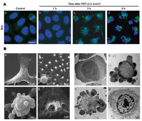
Figure 1.6. (A) Apoptosis induction after 1 h treatment with Ps followed by irradiation in HeLa cells. (B) Cell morphological changes of HeLa cells incubated 1 h with PS
and subjected to light irradiation, visualized by scanning electron microscopy (SEM).
In general, appropriate concentration of ROS supports normal cellular proliferation and homeostasis.42 They could modify protein structures and protect
41 Wang, Y.-Y.; Liu, Y.-C.; Sun, H.; Guo, D.-S. Coord. Chem. Rev. 2019, 395, 46. 42 Zhou, Z.; Song, J.; Nie, L.; Chen, X. Chem. Soc. Rev. 2016, 45, 6597.
18
cells from invading organisms. In order to regulate the concentration of ROS at a moderate level, an antioxidant system is involved in cells, including enzymes such as superoxide dismutase and non enzymatic chemicals such as glutathione and vitamin C. However, once the balance between ROS generation and detoxification is disturbed, cellular constituents would be damaged by oxidation, which is the core idea of PDT. PDT-mediated oxidative cytotoxicity induces tumour tissue destruction via cell apoptosis or necrosis, vascular damage, and inflammation-mediated immune response (Figure 1.6).43
Photosensitizers. A PS should ideally be a single pure compound with low manufacturing costs and good stability in storage. It should have a strong absorption peak in the red to near-infrared spectral region (between 650 and 800 nm) and should possess a substantial triplet quantum yield leading to good production of ROS upon irradiation. It should have no dark toxicity and relatively rapid clearance from normal tissues, thereby minimizing the side effects of phototoxicity.44
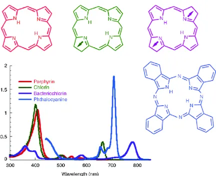
In the past few years, many molecules have been reported for PDT, especially porphyrins, phtalocyanines, etc (Figure 1.7). Tetrapyrrole backbones occur naturally in several important biomolecules such as haemoglobine, chlorophyll and bacteriochlorophyll. As the double-bonds are successively reduced when moving from backbones that are porphyrins to chlorins to bacteriochlorins the emission band is substantially red-shifted and the height of the band also increases. Phthalocyanines also have a large band in the 670 nm region.45 Several compounds belonging to this first generation of PSs, such as the hematoporphyrin derivative HpD (Photofrin), are already in clinical use or in clinical trials to treat cancer patients and are approved by the Food and Drug
43 Dolmans, D. E.; Fukumura, D.; Jain, R. K. Nat. Rev. Cancer 2003, 3, 380; Acedo, P.; Stockert, J. C.; Cañete, M.; Villanueva, A. Cell Death Dis. 2014, 5, 1122.
44 Allison, R. R.; Sibata, C. H. Photodiagn. Photodyn. 2010, 7, 61. 45 Abrahamse, H.; Hamblin, M. R. Biochem J. 2016, 473, 347.
19
Administration for the palliative treatment of obstructive lung and esophageal cancers.46
Figure 1.7. Absorption spectra of porphyrins, chlorins, bacteriochlorins, and phthalocyanines.
Moreover, porphyrins possess the characteristic of producing singlet molecular oxygen with high quantum yields after photoexcitation. Nowadays, numerous porphyrin-derivatives have been prepared for PDT, based on substitution pattern of the chromophore and inner heavy atom. Furthermore, several other natural products, such as, riboflavin47 (vitamin B2, λ
abs=360 nm and 440 nm) and
hypericin48 (a long-known medicinal plant, λ
abs=600 nm) have been extensively
explored as PSs. Moreover, in recent years, PDT has been improved by the synthesis of many other synthetic dyes (Figure 1.8), including methylene blue
46 Agostinis, P.; Berg, K.; Cengel, K. A.; Foster, T. H.; Girotti, A. W.; Gollnick, S. O.; Hahn, S. M.; Hamblin, M. R.; Juzeniene, A.; Kessel, D.; Korbelik, M.; Moan, J.; Mroz, P.; Nowis, D.; Piette, J.; Wilson, B. C.; Golab, J. Cancer. J. Clin. 2011, 61, 250.
47 Makdoumi, K.; Backman, A.; Mortensen, J.; Crafoord, S. Graefes Arch. Clin. Exp. Ophthalmol.
2010, 248, 207; Ettinger, A.; Miklauz, M. M.; Bihm, D. J.; Maldonado-Codina, G.; Goodrich, R.
P. Transfus Apher Sci. 2012, 46, 153.
48 Theodossiou, T. A.; Hothersall, J. S.; De Witte, P. A.; Pantos, A.; Agostinis, P. Mol Pharm.
20
(phenothiazinium salt, Figure 1.4), rose Bengal and eosine Y (water soluble xanthene dyes), neutral red and acridine orange (phenazine dyes), crystal violet (triarylmethane), BODIPY dyes and transitions metal coordination complexes.49
Figure 1.8
Recently, there also have been a large number of targeting studies in which PSs are covalently attached to various molecules that have some affinity for neoplasia or to receptors expressed on specific tumours.50 The intention is to rely on the ability of the targeting vehicle to control localization so that the PS can be chosen based on its photochemical properties rather than on its tumour targeting properties, which are often unimpressive. These targeting vehicles include monoclonal antibodies,51 peptides and proteins,52 various carbohydrates,53 folic acid54 and many others.
49 Ghorbani, J.; Rahban,D.; Aghamiri, S.; Teymouri, A.; Bahador, A. Laser Ther. 2018, 27, 293. 50 Sibani, S. A.; McCarron, P. A.; Woolfson, A. D.; Donnelly, R. F. Expert Opin. Drug. Deliv.
2008, 5, 1241.
51 Sato, K.; Nagaya, T.; Choyke, P. L.; Kobayashi, H. Theranostics. 2015, 5, 698.
52 You, H.; Yoon, H. E.; Jeong, P. H.; Ko, H.; Yoon, J. H.; Kim, Y. C. Bioorg Med Chem. 2015,
23, 1453.
53 Park, S. Y.; Baik, H. J.; Oh, Y. T.; Oh, K. T.; Youn, Y. S.; Lee, E. S. Angew. Chem. Int. Ed.
Engl. 2011, 50, 1644.
21
The first applications of glyco-OPEs in PDT are described in paragraph
1.3.55
Up-converting nanoparticles and PDT. Many nanostructures such as plasmonic gold nanoparticles, mesoporous silica nanoparticles, carbon nanotubes, graphene, upconversion nanoparticles, fullerenes and quantum dots have found uses in PDT.56
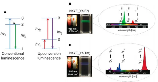
In particular, upconverting nanoparticles (UCNPs) are a class of luminescent nanomaterials that can emit light with shorter wavelength than the excitation light.57 This photophysical phenomenon is based on an anti-Stokes process and referred to as photon upconversion; that is, sequential absorption of two or more low-energy photons to populate real, intermediate excited electronic states followed by emission of a single high-energy photon (Figure 1.9).
Figure 1.9. (A) Schematic representation of the conventional luminescence and upconversion luminescence processes. (B and C) Emission spectra of typical UCNPs upon Near-Infrared
(NIR) excitation.
55 Deni, E.; Zamarrόn, A.; Bonaccorsi, P.; Carreño, M. C.; Juarranz, A.; Puntoriero, F.; Sciortino, M. T.; Ribagorda, M.; Barattucci A. Eur. J. Med. Chem. 2016, 111, 58.
56 Huang, Y. Y.; Sharma, S. K.; Dai, T.; Chung, H.; Yaroslavsky, A; Garcia-Diaz, M.; Chang, J.; Chiang, L. Y.; Hamblin, M. R. Nanotechnol. Rev. 2012, 1, 111.
22
In conventional luminescence (Figure 1.9.A), the absorption of a high-energy photon (hν1) by a system in the ground state can lead to promotion of the system
to the excited state. The system can then undergo non-radiative decay to a lower-excited state, followed by relaxation to the ground state accompanied by the emission of a lower-energy photon (hν2). In the upconversion process, the system
in the ground state is initially promoted into the first excited state by an excitation photon and then is further excited into a higher-energy level by receiving energy from another excitation photon. Radiative transition of the excited state into the ground state leads to emission of a photon with higher energy than the individual excitation photon.58
In recent years, lanthanide-doped upconversion nanoparticles (Ln-UCNPs) have been developed as a new class of luminescent optical labels that have become promising for applications in biological assays and medical imaging. Generally, Ln-UCNPs are composed of a crystalline inorganic host and lanthanide dopants added to the host lattice in low concentrations [NaMF4:Ln3+, Yb3+ (M =
Y or Gd) is the general structure], leading to the preparation of nanomaterials with well defined optical properties. These systems offer the possibility to irradiate with Near-Infrared (NIR) light (which deeper penetrates tissues), low autofluorescence background, large anti-Stokes shifts, sharp emission bands and high resistance to photobleaching. These optical properties make Ln-UCNPs attractive candidates for a variety of broad applications, including sensing, imaging, diagnosis, and therapy, as well as photovoltaic, security, and display technology.59
However, in comparison to conventional PSs, quantum yields of lanthanide-doped UCNPs are generally lower. Moreover, knowing that most effective PSs tend to be insoluble and hydrophobic, with a high propensity to aggregate, their conjugation and encapsulation in nano-drug carriers, could be useful to overcome
58 Liu, Q.; Feng, W.; Yang, T.; Yi, T.; Li, F. Nat. Protoc. 2013, 8, 2033. 59 Wilhelm, S. ACS Nano 2017, 11, 10644.
23
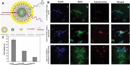
their complementary limitations. For this reason, UCNP-based PS delivery systems have recently been developed.60 UCNPs are easy to synthesize, can support high loading volumes of therapeutic drugs (due to there are-to-volume ratios), have a small size (so easily accumulate in cells) and their surface chemistry allows further functionalization (e.g. with biomolecules for selective targeting of cancer tissues). In a UCNP drug delivery-based approach, a PS is either encapsulated or immobilised on the NPs surface using covalent or non-covalent interactions to form a nano-photosensitizer (NPPS).61 Then, the surface functionalization of PS nano-drug with biomolecules, polymers, lipids, protein, polysaccharides or targeting receptors moieties, overexpressed in tumour cells, guarantees high selectivity, allowing active uptake only in cancer cells and minimizing damage and toxicity to normal tissues.
Figure 1.10. (A) Schematic picture of the lipid micelles encapsulating both the photosensitizer ZnPc and UCNs for simultaneous PDT and fluorescence imaging. (B)ROS production in cells PDT treated with various lipid micelle nanoparticles. Cells were labelled with
carboxy-H2DCFDA that fluoresces when oxidized in the presence of reduced oxygen species (green). Nuclei were counterstained with DAPI (blue), and the nanoparticles are shown by their
red fluorescence colour. (C) Efficacy of PDT treatment assessed by measuring cell viability.
60 Kruger, C. A.; Abrahamse, H. Molecules 2018, 23, 2628.
61Calixto, G. M.; Bernegossi, J.; de Freitas, L. M.; Fontana, C. R.; Chorilli, M. Molecules 2016,
24
For instance, in 2014 Wang et al.62 reported the encapsulation of UCNPs,
functionalized with the photosensitizer zinc phthalocyanine (ZnPc) into lipid micelles (Figure 1.10). It was demonstrated that by increasing the amount of ZnPc loaded in the nanoparticles, their red fluorescence is significantly quenched because of the energy transfer from the UCNs to the Ps ZnPc, while the lipid shell is essential to help the system to cross the cell membrane.
1.2.C DNA-targeted drugs and sensors. Deoxyribonucleic acid (DNA) is a natural product of an enormous importance for understanding the mechanism of genetic processes such as growth, differentiation and aging of the cell. Binding of small organic and inorganic molecules to DNA can influence numerous biological processes in which DNA participate, like transcription and replication. These processes begin when DNA receives the signal from regulatory protein, which binds to its particular part. If, instead of the regulatory protein, some other small molecule binds to DNA, its function is artificially changed – inhibited or activated.63 Such interference can retard or even prevent the cell growth, or, on
the other hand, it can lead to excessive production of some protein and uncontrolled cell growth. Extensive chemical and biochemical studies have characterised a variety of DNA interacting molecules which are classified as antibiotic, antitumor, antiviral or antiprotozoal agents.
Drugs interact with DNA helix both covalently and non-covalently. Covalent binding in DNA is irreversible and undoubtedly leads to complete inhibition of DNA functions and subsequent cell death. A major advantage of covalent binders is the high binding strength. Moreover, covalent bulky adducts can cause DNA backbone distortion, which in turn can affect both transcription and replication, by disrupting protein complex recruitment.64 The most famous
62 Wang, H. J.; Shrestha, R.; Zhang, Y. Part. Part. Syst. Charact. 2014, 31, 228. 63 Aleksić, M. M.; Kapetanović, V. Acta Chim. Slov. 2014, 61, 555.
25
covalent binder is cisplatin, 65 which is used as an anticancer drug since it is able
to induce apoptosis and necrosis of the cancer cell. On the other hand, non-covalent binding of drugs to DNA is reversible, and considering drug metabolism and potential toxic effects, it is more desirable comparing to covalent. However, non-covalent DNA interacting agents can change DNA conformation, DNA torsional tension, interrupt protein–DNA interaction, and potentially lead to DNA strand breaks. One of the main principles of DNA chemistry is molecular recognition, the process when molecules (small or large) selectively recognize each other. This is manifested through a couple of interaction modes: electrostatic, hydrogen bonds, and van der Waals (dipole–dipole) interaction. The stability of the formed complex DNA–drug depends on the intensity of the mentioned interactions.
Drugs that react non-covalently with DNA are classified in the following categories (Figure 1.11):66
1. Intercalating agents 2. Minor groove binders 3. Major groove binders 4. Electrostatic binders
Figure 1.11
65 Jakupec, M. A.; Galanski, M.; Keppler, B. K. Rev. Physiol. Biochem. Pharmacol. 2003, 146, 1. 66 Ni, Y.; Lin, D.; Kokot, S. Anal. Biochem. 2006, 352, 231.
26
Intercalators are molecules that stack perpendicular to the DNA backbone without forming covalent bonds and without breaking up the hydrogen bonds between the DNA bases. The only known forces that sustain the stability of the DNA–intercalator complex, even more than DNA alone, are van der Waals, hydrogen bonding, hydrophobic, and/or charge transfer forces. Intercalation stabilizes, lengthens, stiffens, and unwinds the DNA double helix.67 Intercalators contain planar heterocyclic groups, which stack between adjacent DNA base pairs, forming complex which is stabilized by π–π stacking interactions between the drug and DNA. The phenomenon of intercalation involves the aromatic part of a drug molecule positioning itself between base pairs. These agents introduce strong structural perturbations in DNA molecule by increasing the distance between the adjacent base pairs. In order for an intercalator to fit between base pairs, the DNA must dynamically open a space between its base pairs. Stacking interactions between the bases and the intercalating molecule are the major stabilising factors for the complex formed.
The most famous groove binding drugs usually have shape which is complementary to the shape of the groove,68 and facilitates binding by promoting
van der Waals interactions. These drugs typically have several aromatic rings, such as pyrrole, furan or benzene connected by bonds possessing torsional freedom. Additionally, these drugs can form hydrogen bonds to bases, typically to adenine/thymine rich sequences. In all complexes, the drug fits closely into the minor groove and displaces the hydration layer. The loss of water-mediated hydrogen bonding, which acts as a major complex stabilising force in DNA, leads to serious perturbations of the DNA helix from the normal geometry and induces changes in the conformation of the groove.
Finally, some ligands are capable of forming non-specific, external electrostatic interactions with the DNA phosphate backbone. This mode usually
67 Bauer, W.; Vinograd, J. J. Mol. Biol. 1970, 54, 281.
27
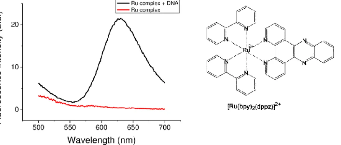
occurs when the ligand self-associates to form higher-order aggregates, which may stack on the anionic DNA backbone in order to reduce charge–charge repulsion between ligand molecules. Some metal complexes interact with DNA through external binding. This association mode was proposed for [Ru(bpy)3]2+
as the luminescence enhancement of this complex upon binding to DNA is strongly dependent on the ionic strength.69
DNA is highly polymorphic and can adopt different motifs such as the A-form, left-handed Z-A-form, triplexes, G-quadruplexes, i-motif and unpaired DNA structures.70 These unique DNA conformations make DNA a critical binding target for small molecules and allow for regulation of DNA function. As a consequence, enormous interest exists in developing small molecules that can bind and react with DNA. Over the past few decades, small molecules that bind to DNA have shown significant promise as diagnostic probes, reactive agents and therapeutics.71
Figure 1.12. Fluorescence emission spectra of 10 μM Ru(bpy)2(dppz)2+ in the absence (red line) and presence (black line) of 10 μM DNA.
In particular, in 1990, Barton et al. first reported that [Ru(bpy)2dppz]2+ (bpy
= 2,2′-bipyridine, dppz = dipyrido[3,2-a:2′,3′-c]phenazine) could serve as a
non-69 Kelly, J. M.; Tossi, A. B.; McConnell, D. J.; OhUigin, C. Nucl. Acids Res. 1985, 13, 6017. 70 Gueron M.; Leroy, J. L. Curr. Opin. Struct. Biol. 2000, 10, 326.
28
radioactive luminescent DNA probe.72 This complex shows no
photoluminescence in aqueous solution at ambient temperature but displays intense photoluminescence in the presence of double-helix DNA, with an enhancement factor of >104 (Figure 1.12). This phenomenon, which is known as the DNA “light-switch” effect, has attracted much attention and has been used extensively to study the interaction of small molecules and metal complexes with DNA.
1.2.D pH fluorescent indicators. Intracellular pH (pHi) plays many critical
roles in cell, enzyme, and tissue activities, including proliferation and apoptosis, ion transport, endocytosis, and muscle contraction.73 Monitoring pH changes inside living cells is also important for studying cellular internalization pathways, such as phagocytosis, endocytosis, and receptor ligand internalization. Cells and their components usually have different pH values, such as cytoplasm (pH 6.8−7.6), nucleus (around pH 7.6), lysosomes and endosomes (pH 4.5−6.0), mitochondria (pH 7.8–7.9), and Golgi (pH 6.4–6.8).74 Intracellular pH (pH
i)
regulates a number of cell metabolism processes: for instance, low intracompartmental pH values can serve to denature proteins or to activate enzyme and protein functions that would be too slow around pH 7.0. Cellular dysfunction is often associated with abnormal pH values in cells and organelles: for example, changes of pHi effect the nervous system, by influencing synaptic
transmission, neuronal excitability and signal cascades. Moreover, abnormal pHi
values are associated with inappropriate cell function, growth, and division and are observed in some common disease types such as cancer and Alzheimer’s. In fact, acidic extracellular pH is a major feature of tumour tissue; extracellular acidification is primarily considered to be due to lactate secretion from anaerobic
72 Friedman, A. E.; Chambron, J. C.; Sauvage, J.-P.; Turro, N. J.; Barton, J. K. J. Am. Chem. Soc.
1990, 112, 4960.
73 Han, J.; Burgess, K. Chem. Rev. 2010, 110, 2709.
74 Cody, S. H.; Dubbin, P. N.; Beischer, A. D.; Duncan, N. D.; Hill, J. S.; Kaye, A. H.; Williams, D. A. Micron 1993, 24, 573.
29
glycolysis (paragraph 1.1) and it is thought to increases the expression of some genes involved with pro-metastatic factors.75 Thus, intimate connections between the cell functions with intracellular pH means that precise measurement of intracellular pH can provide critical information for studying physiological and pathological processes down to cells and organelles.
As seen in paragraph 1.2.A, fluorescence techniques have high sensitivities, tend to be operationally simple, and are, in most cases, non-destructive to cells. Compared to other methods such as microelectrodes, NMR, and absorption spectroscopy for sensing pHi, fluorescence spectroscopy has several advantages:
(1) high spatial-temporal resolution, which can monitor the dynamic change of pHi; (2) non-invasive feature in most cases, which would not disturb the normal
pHi; (3) simplicity.76 These advantages make fluorescence spectroscopy a widely
used method for sensing pHi, and thus excellent fluorescent pH probes attract the
extensive and frequent attention. Qualitative measurements of pHi can be
achieved using fluorescent indicators that switch (ON/OFF or OFF/ON) or shift their emission at sharply defined pH values. To achieve high sensitivity and accuracy, the designed pH probe should have a pKa value that matches the pH of the specific cell organelle. Moreover, in addition to sensitivity and selectivity, some special properties of fluorescent probes are highly desired for sensing pHi,
such as wavelength, brightness, permeability, and so on.77 The excitation and emission wavelengths are the key parameters of fluorophores: light with short wavelength often causes significant photobleaching, severe biological autofluorescence and damage; on the contrary, fluorophores with long wavelength can effectively avoid these problems. In addition, large difference between excitation and emission wavelength (Stokes shift) is favourable for decreasing autofluorescence as well. Therefore, various fluorescent pH probes
75 Kato, Y.; Ozawa, S.; Miyamoto, C.; Maehata, Y.; Suzuki, A.; Maeda, T.; Baba, Y. Cancer Cell
Int. 2013, 13, 89.
76 Wencel, D.; Abel, T.; McDonagh, C. Anal. Chem. 2014, 86, 15. 77 Shi, W.; Li, X.; Ma, H. Methods Appl. Fluoresc. 2014, 2, 042001.
30
with near infrared (NIR) and/or large Stokes shift features have aroused great interest in this area. As regards brightness (Bs) (Bs = ε × Φ; ε=molar absorptivity and Φ=quantum yield), brighter fluorophores show higher signal-to-noise ratios and require less dosage and weaker excitation light, diminishing cell damages.
Typically, three types of response mechanisms for fluorescent probes can be used: fluorescence quenching (ON/OFF), fluorescence enhancement (OFF/ON) and ratiometry, which exploits dyes that can absorb or emit light at two different wavelengths.78 The fluorescence ON/OFF probes show less practicability because of their high background and poor selectivity. Fluorescence OFF/ON probes, as compared to ON/OFF or ratiometric probes, have the advantage of the ease detection of low-concentration contrast relative to a near dark background, which is very desired for qualitative analysis due to the high sensitivity.
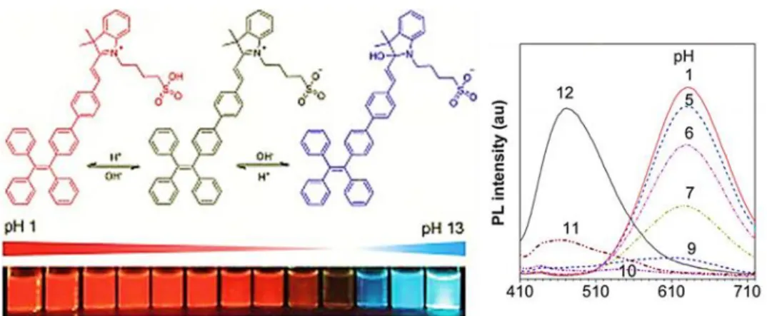
Figure 1.13. Emission spectra of a cyanine-based dye in aqueous solutions at different pH values.
However, for imaging studies in cells, the intensity-based fluorescence probes suffer easily from the influence of many variants, such as probe concentration, instrumental efficiency, and environmental conditions. To address these issues, ratiometric fluorescent probes are preferred. They exhibit different
31
responses to an analyte (e.g. pH) at two different excitation/emission wavelengths. The simultaneous recording the fluorescence intensities at the two wavelengths and the calculation of their ratio can provide more accurate measurement than that at only one wavelength (Figure 1.13).79
Moreover, to obtain the intact information about pHi, fluorescent probes would
better non-destructively enter cells and have less leakage from the cells. A typical strategy to achieve this is to mask the charged groups (e.g. carboxyl) with neutral acetoxymethyl esters. The resulting probes can diffuse into cells and then are hydrolysed by intracellular esterases to afford the free, charged probes, on which the charges facilitate the probe stay in cells.80 Another strategy for delivering a probe is to take advantage of the ligand-receptor interactions, especially for nanoprobes. The receptors in cellular membrane can bind with specific ligands and then transport the ligands into cytoplasm. In this way, if the nanoprobes are modified with these kinds of ligands, they can be easily internalized into cells through the ligand-receptor interactions.
Finally, the pKa values of probes are strongly dependent on the
microenvironment of media, such as ionic strength, viscosity, and coexisting substances.81 For instance, many pH probes can bind with proteins, causing a
change in their pKa values and thus a large error in pH measurements. To decrease
the microenvironmental influence, the interference from coexisting substances (e.g. metal ions, proteins, etc) should be studied carefully, and the pH calibration curve in cells (cytosol) instead of in plain buffer solution should be established for the calculation of pHi values.
79 Li, X. H.; Gao, X. H.; Shi, W. Ma, H. M. Chem. Rev. 2014, 114, 590; Chen, S.; Liu, J.; Liu, Y.; Su, H.; Hong, Y.; Jim, C. K. W.; Kwok, R. T. K.; Zhao, N.; Qin, W.; Lam, J. W. Y.; Sing Wong, K.; Zhong Tang, B. Chem. Sci. 2012, 3, 1804.
80 Tsien, R. Y. Nature 1981, 290, 527.
81 Miao, F.; Song, G. F.; Sun, Y. M.; Liu, Y.; Guo, F. Q.; Zhang, W. J.; Tian, M. G.; Yu, X.
32
Fluorescein82, cyanines83 and their derivatives are the most widely used pH
probes. However, many other organic molecular pH probes have been recently, developed, such as naphthalimide derivatives,84 lanthanide complexes,85 BODIPY dyes, carbazoles,86 quinolines87 and other fluorophores. Most of them exhibit ratiometric response to pH and have been used to sensing the change of pHi under different conditions. However, it should be pointed out that pH probes
with a wide linear response range are still rare. To increase the response range of pH, a useful strategy is to design a probe with multiple H+ binding sites by assembling several electronegative atoms (e.g. N and/or O) in the different positions of a conjugated molecule.88
Moreover, Horiuchi et al.89 recently applied the properties of different pH-sensitive aniline moieties to the synthesis of porphyrin derivatives that could be used as photosensitizers in PDT. In fact, in this kind of therapy, the tumour specificity is very important to minimize photo-induced side effects. One of the major strategies to improve the tumour specificity is to develop activatable photosensitizers.90 The fluorescence and 1O
2 sensitization efficiencies of
activatable photosensitizers are inefficient in normal tissues. However, a certain trigger in the tumour tissue can modify the photosensitizer’s chemical properties, increasing the fluorescence and 1O
2 sensitization efficiencies. As triggers for the
tumour selectivity, activation in low-pH cancer cells condition has been proposed.
82 Venn, A. A.; Tambutté, E.; Lotto, S.; Zoccola, D.; Allemand, D.; Tambutté, S. Proc. Natl Acad.
Sci. USA 2009, 106, 16574.
83 Li, P.; Xiao, H. B.; Cheng, Y. F.; Zhang, W.; Huang, F.; Zhang, W.; Wang, H.; Tang, B. Chem.
Commun. 2014, 50, 7184.
84 Hu, J. L.; Wu, F.; Feng, S.; Xu, J. H.; Xu, Z. H.; Chen, Y. Q.; Tang, T.; Weng, X. C.; Zhou, X.
Sensor. Actuat. B-Chem. 2014, 196, 194.
85 Fan, L.; Liu, Q. L.; Lu, D. T.; Shi, H. P.; Yang, Y. F.; Li, Y. F.; Dong, C.; Shuang, S. M. J.
Mater. Chem. B 2013, 1, 4281.
86 Miao, F.; Song, G. F.; Sun, Y. M.; Liu, Y.; Guo, F. Q.; Zhang, W. J.; Tian, M. G.; Yu, X. Q.
Biosens. Bioelectron. 2013, 50, 42.
87 Huang, W. M.; Lin, W. Y.; Guan, X. Y. Tetrahedron Lett. 2014, 55, 116.
88 Su, M. H.; Liu, Y.; Ma, H. M.; Ma, Q. L.; Wang, Z. H.; Yang, J. L.; Wang, M. X. Chem.
Commun. 2001, 11, 960.
89 Horiuchi, H.; Hirabara, A.; Okutsu, T. J. Photoch. Photobio. A. 2018, 365, 60. 90 Lovell, J. F.; Liu, T. W. B.; Chen, J.; Zheng, G. Chem. Rev. 2010, 110, 2839.
33
These activatable photosensitizers can minimize erroneous diagnoses and photo-induced side effects.
1.3 OLIGO(PHENILENE ETYNYLENE)s
Oligo(phenylene-ethynylenes) (OPEs) and the corresponding polymers, Poly(phenyleneethynylenes) (PPEs), represent a peculiar class of luminescent dyes with stable π-conjugated rigid rod-like skeletons.91 In particular, their
structure is a succession of aromatic rings linked by carbon-carbon triple bonds (Figure 1.14). This feature guarantees a high electronic conjugation and, consequently, their photochemical properties evidence high quantum yields and luminescence features. The synthetic approaches leading to the construction of these oligomers allow easy functional group manipulation and make modulable their functional features. Their tunable properties depend not only on their molecular structure, but also on the supramolecular interactions and on the aggregation of the chains both in solution and in the solid state.92 OPEs and PPEs have attracted for the above mentioned reasons significant attention in the scientific community and, in the last decades, have been widely applied in organic electroluminescent light emitting devices, molecular electronic switches, nonlinear optical materials, biological sensors.
Figure 1.14
The photophysical properties of OPEs are directly connected to their extensive conjugation. Modulation of these properties depends on their structure
91 Bunz, U. H. F. Chem. Rev. 2000, 100, 1605. 92 Waters, M. L. Curr. Opin. Chem. Biol. 2002, 6, 736.
34
and on substitution on the aromatic conjugated skeleton. In 2011, Dehaen et al.93
reported that, on increasing the number of phenyl acetylene units, varying from 1 to 7, the absorption and emission maxima of OPEs are increasingly redshifted; this effect becomes smaller with each addition of a repeating unit. Moreover, it was demonstrated that the introduction on the chain of moieties bearing a lone pair could influence the photophysical properties of phenylenethynylene-based systems.
Figure 1.15
In fact, the synthesis of 2,5-dialkoxy-substituted derivatives of OPEs94 leads to lower-wavelength absorbing and emitting systems, with respect to unsubstituted compounds (Figure 1.15), and to the increase of the molar extinction coefficient and emission quantum yields; for this reason, these molecular systems are now widely used as building blocks for various functional molecular materials.
Furthermore, in 2005 Yamaguchi et al.95 reported a study about spectroscopic features of OPEs (absorption and emission wavelengths, quantum yield Φf, lifetime τ) and their behaviour when the π-conjugated molecular rods
93 Yin, S.; Leen, V.; Jackers, C.; Beljonne , D.; Van Averbeke, B.; Van der Auweraer, M.; Boens, N.; Dehaen, W. Chem. Eur. J. 2011, 17, 13247.
94 James, P. V.; Sudeep, P. K.; Suresh, C. H.; George Thomas, K. J. Phys. Chem. A 2006, 110, 4329.
95 Yamaguchi, Y.; Tanaka, T.; Kobayashi, S.; Wakamiya, T.; Matsubara, Y.; Yoshida, Z.-i. J. Am.
35
consisting of p-(phenylene)ethynylene units were modified by donor (OMe) and/or acceptor (CN) groups situated at the aromatic rings. They reported three different cases: (1) donor modification systems (SD systems), (2) side-acceptor modification systems (SA systems), and (3) systems consisting of donor blocks and acceptor blocks (BL systems, Figure 1.16).
Figure 1.16
The best values in terms of Φf, were obtained for the block modification
systems (BL,Φf>0.95). This result evidenced that the building of a push-pull
system (paragraph 2.5B) by combination of electron donating and electron withdrawing groups in the same skeleton strongly influences the photophysical properties of the OPEs. On the other side, the values of quantum yields obtained for the systems functionalized with –OMe in the side donor moiety (SD) or with –CN in the side acceptor fragment, (SA) were higher (Φf>0.90). Moreover, the
study of the trend of bathochromic shift of emission (λem), compared to the
elongation of aromatic chain and the introduction of the electron withdrawing groups, showed the shift of λem toward higher wavelengths for the largest OPEs
systems and for the ones containing the acceptor moieties.
Another appealing property of OPEs corresponds to their possibility to generate singlet oxygen (1O
36
Andraud,96 reported in 2012, highlighted this characteristic of the OPEs systems
through the synthesis of a series of compounds containing two amino groups as chain terminations.
Figure 1.17
It was demonstrated, by the detection of the characteristic Δ1O
2 → Σ3O2
phosphorescence at 1270 nm, that all the compounds generated singlet oxygen and that the presence of the “heavy-atom” bromine on the aromatic rings has an advantageous effect on the singlet oxygen production yield. Moreover, fluorescence quantum yields and lifetimes evolved opposite to the 1O2 generation
quantum yield. This result gave the possibility to use OPEs as photosensitizers in Photodynamic Therapy (paragraph 1.2.B), and showed that these are molecules possessing an accessible triplet excited state able to generate singlet oxygen by an energy transfer to ground state oxygen, ultimately leading to cell death.
Biological applications of OPEs. While in the last decades OPEs showed a rapid
development in optoelectronic devices, their use for biomedical applications has more recently gained increasing attention.97Specifically, they have found innovative applications in a variety of biotechnologies, ranging from biosensing,98
96Lanoë, P. H.; Gallavardin,T.; Dupin,A.; Maury, O.; Baldeck, P. L.; Lindgren, M.; Monnereau C.; Andraud, C. Org. Biomol. Chem. 2012, 10, 6275.
97 Liu, B. Conjugated Polymers for Biological and Biomedical Applications 2018, Wiley-VCH. 98 Feng, X.; Liu, L.; Wang, S.; Zhu, D. Chem. Soc. Rev. 2010, 39, 2411.