ALMA MATER STUDIORUM - UNIVERSITÀ DI BOLOGNA
CAMPUS DI CESENA
SCUOLA DI INGEGNERIA E ARCHITETTURA
!
!
CORSO DI LAUREA MAGISTRALE IN INGEGNERIA BIOMEDICA
!!
!!
!
MONITORING THE EXTRACELLULAR MATRIX
MINERALIZATION PROCESSES IN BIOLOGICAL
SCAFFOLDS USING BIOREACTOR
X-RAY µCT TECHNOLOGY AND 3D MODELING
METHODS
!
!
TESI IN
!
MECCANICA DEI TESSUTI BIOLOGICI
!
!!
!!
!
Relatore
Presentata da
!
Prof. Luca Cristofolini Joseph Lovecchio!
Correlatore
!
Prof. Paolo Gargiulo
Dott. Ólafur E. Sigurjónsson
!
!
!
III° Sessione!
Anno Accademico 2012/2013!
!
!
!
!
!
!
!
!
!
!
!
!
!
!
Ai miei Genitori…
!
!
!
!
!
!
!
!
!
!
!
!
!
!
!
!
!
!
“
La geniale invenzione nasce sempre dall’uomo isolato,
ma solo l’opera tenace di pazienti ricercatori, con mezzi larghi e adatti,
può efficacemente svilupparla e utilizzarla „
B.M.
Abstract
...
3
1. Introduction
...
6
1.1 Stem Cell
...
9
1.2 Scaffold
...
11
1.2.1 Temporary Matrices for Bone Growth
...
12
1.2.2 Essential Properties
...
13
1.2.3 Mechanical Properties and Biodegradability
...
15
1.2.4 Biomaterials used as Scaffolds
...
15
1.2.5 Processing Techniques
...
17
1.2.6 Scaffold Coating
...
17
1.3 Bioreactor
...
19
1.4 Morphometric Analysis
...
20
1.4.1 Scaffolds 3D Modeling
...
23
2. Materials and Methods
...
24
2.1 Bioreactor Development
...
24
2.1.1 Control Unit
...
28
2.1.2 Interface
...
30
2.2 Computational Fluid Dynamic Modeling
...
32
2.2.1 Viscosity
...
32
2.2.2 Model
...
35
2.2.3 Pressure in the Bioreactor
...
38
2.3 Stem Cell Culture
...
39
2.3.1 Counting Cells
...
39
2.3.2 Scaffold Coating Preparation
...
40
2.3.3 Static and Bioreactor Culture
...
40
2.4 Data Acquisition
...
41
3. Results
...
47
3.1 Bone Tissue Formation
...
47
3.2 Static and Bioreactor Culture Comparison
...
48
3.2.1 Gray Values Gradient Distribution
...
50
3.2.2 Statistical Analysis
...
51
4. Discussion
...
54
4.1 Future Directions
...
56
5. Conclusion
...
58
Appendixes
...
59
References
...
73
Acknowledgments
...
79
Abstract!
"
To improve in vitro cell cultures, bioreactor systems have been widely used, e.g. in bone tissue engineering. Spinner flasks, rotating wall bioreactors, and flow perfusion systems have all been used, and each system has advantages and disadvantages. This thesis describes the development of a simple perfusion bioreactor system and the results from the assessment methodology employed which is based on x-ray µCT analysis and 3D Modeling Techniques. "
A simple bioreactor with flow generator propeller was designed and built with the aim of improving differentiation of human embryonic derived mesenchymal stem cells (hES-MP) seeded on porous titanium scaffolds in order to improve mineralized matrix deposition. The bioreactor allows three types of flow: forward (clockwise), backward (counterclockwise) and pulse mode (back and forth) controlled by a micro-controller and a graphical interface. "
We designed a simple model to calculate the pressure generated by the flow of the bioreactor on the scaffold (3•10-2 Pa). "
We compared 3 scaffolds under static culture condition to 3 scaffolds within the bioreactor. The scaffolds were incubated for 21 days, fixed in paraformaldehyde (4% w/v) and subjected to acquisition by x-ray μCT."
Images obtained were processed using 3D imaging software; a “virtual" sectioning of the scaffolds was performed in order to obtain samples extracted from the surface and from the inside of the scaffolds in order to obtain the gray value gradient distribution. "
This distribution is used to distinguish the various components present in the images, in this case scaffolds from the hypothetical cellular matrix."
The results show that scaffolds maintained in the bioreactor have higher density of gray values gradient distribution on the scaffold surface which suggests improved mineralized matrix deposition.!
The studies coming from this bioreactor will be used to design a new version that will make it possible to analyze more than 20 scaffolds at the same time making it possible to further analyze the quality of the differentiation using molecular and histochemical methodology."
Abstract!
"
Al fine di migliorare le tecniche di coltura cellulare in vitro, sistemi a bioreattore sono sempre maggiormente utilizzati, e.g. ingegnerizzazione del tessuto osseo. Spinner Flasks, bioreattori rotanti e sistemi a perfusione di flusso sono oggi utilizzati e ogni sistema ha vantaggi e svantaggi. "
Questo lavoro descrive lo sviluppo di un semplice bioreattore a perfusione ed i risultati della metodologia di valutazione impiegata, basata su analisi μCT a raggi-X e tecniche di modellizzazione 3D."
Un semplice bioreattore con generatore di flusso ad elica è stato progettato e costruito con l'obiettivo di migliorare la differenziazione di cellule staminali mesenchimali, provenienti da embrioni umani (HES-MP); le cellule sono state seminate su scaffold porosi di titanio che garantiscono una migliore adesione della matrice mineralizzata. Attraverso un microcontrollore e un'interfaccia grafica, il bioreattore genera tre tipi di flusso: in avanti (senso orario), indietro (senso antiorario) e una modalità a impulsi (avanti e indietro)."
Un semplice modello è stato realizzato per stimare la pressione generata dal flusso negli scaffolds (3•10-2 Pa). "
Sono stati comparati tre scaffolds in coltura statica e tre all’interno del bioreattore. Questi sono stati incubati per 21 giorni, fissati in paraformaldehyde (4% w/v) e sono stati soggetti ad acquisizione attraverso μCT a raggi-X. "
Le immagini ottenute sono state poi elaborate mediante un software di imaging 3D; è stato effettuato un sezionamento “virtuale” degli scaffolds, al fine di ottenere la distribuzione del gradiente dei valori di grigio di campioni estratti dalla superficie e dall’interno di essi."
Tale distribuzione serve per distinguere le varie componenti presenti nelle immagini; in questo caso gli scaffolds dall’ipotetica matrice cellulare. "
I risultati mostrano che sia sulla superficie che internamente agli scaffolds, mantenuti nel bioreattore, è presente una maggiore densità dei gradienti dei valori di grigio ciò suggerisce un migliore deposito della matrice mineralizzata.!
Gli insegnamenti provenienti dalla realizzazione di questo bioreattore saranno utilizzati per progettare una nuova versione che renderà possibile l’analisi di più di 20 scaffolds contemporaneamente, permettendo un’ulteriore analisi della qualità della differenziazione usando metodologie molecolari ed istochimiche."
"
"
"
"
"
"
"
"
"
"
"
"
"
"
"
"
"
"
"
"
"
"
"
"
"
"
"
"
"
"
"
"
"
"
"
"
"
"
"
"
"
"
"
"
"
"
1. Introduction!
"
Tissue Engineering (TE) is a discipline integrating biology with engineering to create tissues or cellular products outside the body (ex vivo) or to make use of gained knowledge to better manage the repair of tissues within the body (in vivo). This discipline requires understanding of different biological fields, including cell and molecular biology, physiology and systems integration, stem cell proliferation and differentiation with lineage attributes, extracellular matrix chemistry and compounds, and endocrinology. It also requires knowledge of many engineering fields, including biochemical and mechanical engineering, polymer sciences, bioreactor design and application, mass transfer analysis of gas and liquid metabolites, and biomaterials. "
The combination of these sciences has spawned the field of regenerative medicine which has, at present, two strategic clinical aims [1]: "
"
• Cell therapies for the repair of damaged tissues, involving injection or engraftment of cells or cellular suspensions, sometimes in combination with scaffolding material"
"
• Establishment of tissue ex vivo for use as grafts or extracorporeal organs to assist or supplement ailing in vivo organs"
"
Tissue Engineering has been defined first as “an interdisciplinary field that applies the principles of engineering and the life sciences toward the development of biological substitutes that restore, maintain, or improve tissue function” [2]. Another definition is “understanding the principles of tissue growth, and applying this to produce functional replacement tissue for clinical use” [3]. "
Thus, Tissue Engineering has always been driven by the need to provide functional equivalents of native tissues that can be used for in vivo implantation in order to restore, maintain, or enhance tissue and organ physiology. To engineer living tissues, cultured cells are coaxed to grow on bioactive degradable scaffolds that provide the physical and chemical cues
to guide their differentiation and assembly into two- or three-dimensional tissues [4]. In contrast to classic biomaterial approach, it is based on the understanding of tissue formation and regeneration, and aims to induce new functional tissues, rather than just to implant new spare parts [5]."
"
Tissue Engineering seeks to rebuild portions of tissue or whole organs through the manipulation of cells, using a wide variety of strategies [6]. These mainly involve the use of synthetic scaffolds, fabricated from biocompatible materials, to carry, support and guide cells towards tissue regeneration. The field of scaffold construction is still in its infancy, and many different approaches, both general and specific, are under investigation. Of particular interest is the ability to mimic the natural extracellular matrix surrounding cells in each tissue. Tissue specific functionality is largely maintained through the interaction of cells with matrix biological ligands, mechanical support and structural interaction. Matrix structure is particularly important for determining the flow of nutrients, signaling factors and waste products about cells, influencing their motility, protein expression and long-term differentiation [7]. In many cases it would be desirable to replace both small and large areas of diseased and damaged tissue with new healthy tissue, either fully generated outside the body, or at the site of damage through use of scaffolds engineered to support and encourage new tissue genesis. The scaffold must provide both the mechanical properties required by the regenerating tissue as well as the cues that cells require. This is a complex challenge. The organs of the body are highly sophisticated biological systems. Each maintains their own unique set of biological conditions, needed to sustain the function of tissue specific cells and enable self repair. These environments are difficult to mimic outside the body. One approach is to use the body as its own incubator, [8] drawing directly on the complexity of natural tissues to enable the generation of new tissue. For example an osteoconductive gel injected into the stem cell rich periostium layer in bone was used to create a space for the generation of new bone tissue, suitable for re‐implantation [9]. Another approach is to use scaffolds that provide an appropriately sophisticated representation of the tissue environment, encouraging cells to generate new tissue (Figure 1) as the scaffold degrades"
"
! "
Figure 1 - The typical tissue engineering approach: 1. Remove cells; 2. Expand number in culture; 3. Seed onto an appropriate scaffold with suitable growth factors and cytokines; 4.
Place into culture; 5. Re-implant engineered tissue repair damaged site [7]"
"
There are three principal strategies for treating diseased or lost tissue in patients [10]: "
"
• In situ tissue regeneration: new tissue formation is induced by specific scaffolds or external stimuli that are used to stimulate the body‘s own cells and promote local tissue repair"
"
• Implantation of freshly isolated or cultured cells: individual cells or small cellular aggregates from the patient or a donor are injected directly into the damaged or lost region with a degradable scaffold"
"
• Implantation of bone like tissue assembled in vitro from cells and scaffold: a complete three-dimensional tissue is grown in vitro using autologous or donor cells within a scaffold, which has to be implanted once it has reached “maturity”"
Bone tissue engineering, as in most other Tissue Engineering areas, exploits living cells in a variety of ways to restore, maintain, or enhance tissue functions. The basic elements for tissue engineered bone are signaling molecules, cells and matrices for cell growth and differentiation. The combination of these three elements may need to be modified according to several variables such as patient age, gender, health, systemic conditions, habits, and anatomical shape of the implant. Furthermore that same strategy should also adapt itself to the area where the tissue engineering construct is needed, because different regions of the body will have different functional loads and vascularity. Up to now several strategies, from scaffolds alone to 3D matrices loaded with growth factors, have been proposed [5]. "
"
"
1.1 Stem Cell
"
Bioreactors utilize materials and cells that have already been proven effective for bone tissue engineering including polymer scaffolds that are biodegradable and mesenchymal stem cells (MSCs), a population of cells that exists in the bone marrow capable of differentiating into osteoblasts, chondrocytes, and adipocytes [11, 12]. This population represents only a small percentage of cells found in the bone marrow, thus expanding MSCs to clinically relevant numbers represents a significant hurdle to the implementation of a tissue engineering strategy utilizing these cells."
"
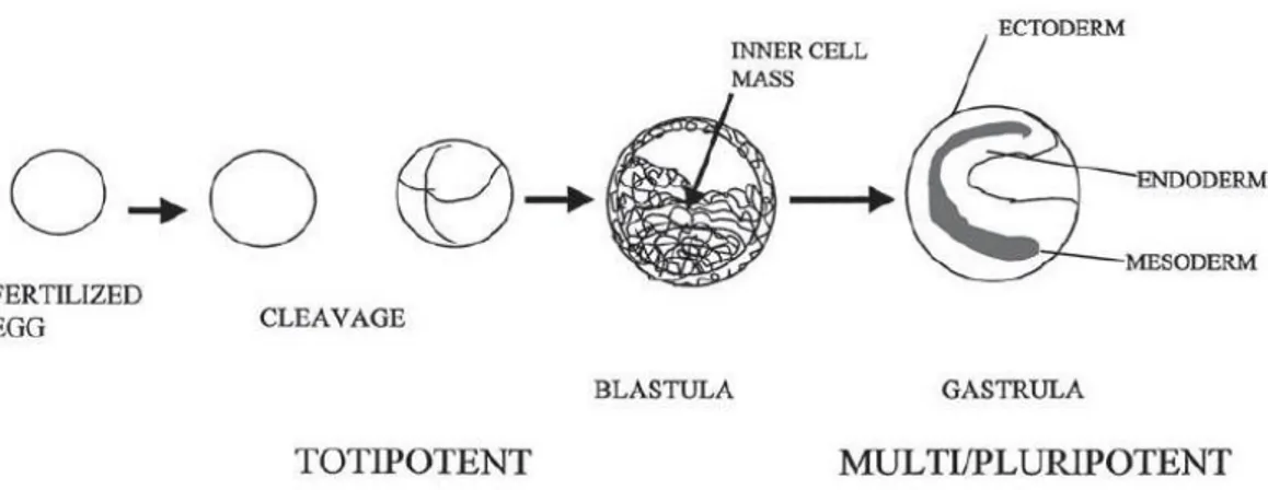
Stem cells are undifferentiated cells with a high proliferation capability, being able of self- renewal, multilineage differentiation and therefore the regeneration of tissues [5]. However, stem cells have varying degrees of differentiation potential. The most primitive derive from the fertilized oocyte (the zygote), more precisely from the very first descendants of the first divisions (two divisions). These cells are totipotent, because they are able to form the embryo and the trophoblast of the placenta. Some days later, these cells start to specialize, forming a hollow ball of cells, the blastocyst, and a cluster of cells called the Inner Cell Mass (ICM) from which the embryo derives. The ICM cells, also known as embryonic stem cells (ES), are
considered to be pluripotent (Figure 2). They can differentiate in almost all cells that arise from the three germ lines, but not the embryo because they are not able to give rise to the placenta and supporting tissues [13 ,14]."
"
! "
"
Figure 2 - Early development of embryo. Division of the fertilized egg results in the formation of a ball-like structure with a cavity on one end (the blastula). Until this stage, the cells divide by symmetric division, and all cells produced are totipotent. Invagination of one pole of the
blastula leads to formation of the gastrula and establishment of the primitive germinal cell layers. During gastrulation and later formation of the fetus, the daughter cells lose potential
as they gain specialized function. The process of the loss of potential and the gain of specialized function is known as determination [15]."
"
Finally, we can find multipotent stem cells, also known as adult stem cells (ASC), in the fully differentiated tissues [13, 14, 16]. Theoretically, and opposing to ES cells, these would only be capable of producing a limited range of differentiated progeny, related to the embryonary origin of the tissue where they are located [13, 14]. However, these cells may have a higher degree of differentiation plasticity (differentiation into other cell lineages that are not related with the embryonary origin of the tissue were the ASCs are found) than expected. The biological mechanisms responsible by the broad developmental potential of stem cells are still not fully understood. Interaction with other cell types and the components of the extracellular matrix are believed to influence the survival and the development of stem cells to the committed lineages [5, 14, 16]."
Tissue engineering strategies that combine scaffolds with cells capable of osteogenesis or bioactive proteins offer a potential alternative to bone grafting for treatment of large, clinically challenging bone defects. Several studies have demonstrated that delivery of osteoprogenitor cells or
osteoblasts significantly improves repair of long bone and cranial defects [17]. Tissue engineering has become a popular field of research for the treatment of many medical conditions. Strategies in tissue engineering for orthopedic disorders including those of bone, cartilage and muscle have been intensively studied. Stem cells are particularly attractive for TE applications due to their ability to self-renew, high proliferation capacity and ability to differentiate into many different cell types [18]."
"
"
1.2 Scaffold
"
Scaffolds for tissue regeneration are defined as: “three dimensional open-cell porous structures synthesized from either natural or synthetic polymers which have the potential to support attachment, migration and multiplication of living cells”. These structural mimics of the extracellular matrix can be made of biodegradable synthetic polymers (e.g. polylactic and polyglycolic acid) or natural polymers (e.g. collagen, collagen– glycosaminoglycan copolymer). It has been demonstrated that scaffold pore size and shape, porosity, specific surface area, biodegradability and stiffness significantly influence cell functions. To control these features, various fabrication techniques have been developed for different biomaterials [19]."
For bone tissue engineering, a wide choice of scaffold materials, including metals, natural and synthetic polymers, and ceramics has been proposed, each of them presenting different mechanical, chemical, and biological characteristics. Particular attention has been given to the manufacturing of porous bioceramics that mimic trabecular bone chemistry and structure. Calcium phosphate (CaP), hydroxyapatite (HA) ceramics and Porous Titanium (Ti) are considered among the most promising bone substitute for their bone-like chemical composition and mechanical properties [20]."
!
Figure 3. Represents 4 types of biocompatible scaffolds which are used in tissue engineering. A) Bioglass is bioactive glass; B) Calcium phosphate scaffold (CaP); C)
Open-cell polylactic acid (OPLA); D) Titanium scaffold [21]"
"
"
1.2.1 Temporary Matrices for Bone Growth!
"
Scaffolds are temporary matrices for bone growth and provide a specific environment and architecture for tissue development [22]. Any tissue consists of a matrix and one, or usually, many cell types. The matrix is, in vivo, a 3D scaffold for cells, and provides them with a tissue specific environment and architecture. Furthermore, it serves as a reservoir of water, nutrients, cytokines, and growth factors. In this sense, and in order to restore function or regenerate tissues one needs a template, a scaffold that will act as a temporary matrix for cell proliferation and extracellular matrix deposition, with consequent bone in-growth until the new bony tissue is totally restored/regenerated. Moreover, would also act as a template for the vascularization of this neo-tissue and they could actively participate in the regenerative process through the release of growth/differentiation factors, present in its structure. It is then logical to say that an appropriate 3D scaffold is an essential component for a tissue engineering strategy [5].
However, it is important to realize that the latter must have a series of properties that make it suitable for TE purposes. Besides the choice of adequate materials the macro and micro-structural properties of the materials are of utmost importance. Such properties effect not only cell
survival, signaling, growth, propagation, and reorganization but also their gene expression and the preservation, or not, of their phenotype [5]. An optimal scaffold for tissue engineering application would mimic the properties of the extracellular matrix (ECM) of those tissues to be regenerated perfectly and completely [23]."
"
1.2.2 Essential Properties!
"
The following properties have been defined has being essential:"
"
• Biocompatibility!
Scaffolds should be well integrated in the host‘s tissue without eliciting an immune response [5]."
"
• Porosity!
Scaffolds must posses an open pore, fully interconnected geometry in a highly porous structure with large surface to area volume ratios that will allow cell in-growth and an accurate cell distribution throughout the porous structure, and will facilitate the neovascularization of the construct from the surrounding tissue. Furthermore, the scaffolds should also exhibit adequate microposity, in order to allow capillary in-growth. Porosity and interconnectivity are also important for an accurate diffusion of nutrients and gases and for the removal of metabolic waste resulting from the activity of the cells that had meanwhile grown into the scaffold. This is of particular importance regarding bone tissue engineering because, due to bone metabolic characteristics, high rates of mass transfer are expected to occur, even under in vitro culture conditions. However, the degree of porosity always influences other properties of the scaffolds such as its mechanical stability, so, its value, should always be balanced with the mechanical needs of the particular tissue that is going to be replaced [5, 24]."
"
"
"
• Pore Size!
Pore size is also a very important issue because, if the pores employed are too small, pore occlusion by the cells will happen. This will prevent cellular penetration, extracellular matrix production, and neovascularization of the inner areas of the scaffold. It is well accepted that for bone tissue engineering purposes, pore size should be within the 200–900 mm range. However, Holly et al. [25] reported a different concept. In the referred case the authors believe that bone reconstruction will only be achieved by having a 3D temporary matrix with a large macro-porous interconnected structure with pore size ranging from 1.2-2.0 mm. This later approach has evident advantages due to its high surface to volume ratios that will facilitate cell, tissue and blood vessels in-growth. However, this affects the mechanical properties avoiding its use in areas which are very demanding from the mechanical point of view [5]."
"
• Surface Properties!
Surface properties, both chemical and topographical, can control and affect cellular adhesion and proliferation. Chemical properties are related with the ability of cells to adhere to the material as well as with the protein interactions with the latter. Topographical properties are of particular interest when the topic is osteoconduction. As defined by Davies et al. [26] osteoconduction is the process by which osteogenic cells migrate to the surface of the scaffold trough a fibrin clot, which is established right after the material implantation. This migration of osteogenic cells trough the clot will cause retraction of the temporary fibrin matrix. Hence, it is of the utmost importance that the fibrin matrix is well secured to the scaffold or, otherwise, when osteogenic cells start to migrate the fibrin will detach from the scaffolds due to wound contraction. It has been previously shown [27, 28] that a more “rough‘‘ surface will be able to imprison the fibrin matrix, better than a smooth surface, and hence facilitate the migration of osteogenic cells to the materials surface."
"
"
"
"
• Osteoinductivity!
Osteoinduction is the process by which stem and osteoprogenitor cells are recruited to a bone healing site, and stimulated to undergo the osteogenic differentiation pathway. However, when the portion of bone to regenerate is large, natural osteoinduction combined with a biodegradable scaffold may be not enough. Because of this the scaffold should be osteoinductive by itself [5, 29]."
"
"
1.2.3 Mechanical Properties and Biodegradability!
"
In vitro, the scaffolds should have sufficient mechanical strength to withstand the hydrostatic pressures and to maintain the spaces required for cell in-growth and matrix production. In vivo, and because bone is always under continuous stress, the mechanical properties of the implanted construct should ideally match those of living bone, so that an early mobilization of the injured site can be made possible. Furthermore, the scaffolds degradation rate must be tuned appropriately with the growth rate of the neotissue, in such a way that by the time the injury site is totally regenerated the scaffold is totally degraded [5, 30]."
"
"
1.2.4 Biomaterials used as Scaffolds!
"
The selection of the most appropriate material to produce a scaffold to be used in bone tissue engineering applications is a very important step towards the construction of a tissue engineered product, since its properties will determine, to a great extent, the properties of the scaffold. Up to now several materials such as metals, ceramics and polymers from both natural or synthetic origins have been proposed [5]."
However, metals and most of the ceramics are not biodegradable, which leaves the researcher‘s choice reduced to a small number of ceramics and to biodegradable polymers. Ceramics have been widely used in the biomedical engineering and bone substitution/regeneration field. They can
be from natural (e.g., coralline hydroxylapatite (HA)) origin or synthetic such as synthetic HA or b-tricalcium phosphate (b-TCP). Due to interesting properties, mainly the fact of being osteoconductive and osteoinductive, have been considered for bone tissue engineering applications [5]. Several works [5, 31–41] have shown that by using ceramics with or without bone marrow cells, good results regarding bone regeneration could be obtained. However, these materials have some major drawbacks."
To begin, ceramics materials are brittle and present a low mechanical stability, which prevents their use in the regeneration of large bone defects. Furthermore, due to factors that happen in vivo, such as osteoclastic activity, their degradation/dissolution rates are difficult to predict. This could present a problem because if degrades too fast will compromise the mechanical stability of the construct, which is low by itself. At the same time, this would dramatically increase the extracellular concentrations of Ca and P, which can cause cellular death, as demonstrated by Adams et al. [42]."
As an alternative to the above referred materials, there are biodegradable polymers, which are believed to be the ideal materials for bone TE. These can be divided in two groups: natural and synthetic.
Natural biodegradable polymers are those obtained from natural sources, either from animal or vegetal source. Within these we can find, among others, collagen, fibrinogen, chitosan, starch, hyaluronic acid, and poly(hydroxybutyrate). The main advantages of these materials are their low immunogenic potential, the potential bioactive behavior and the capability of interacting with the host‘s tissue, chemical versatility, and in some cases their source, as in starch and chitosan, which is almost unlimited [5]."
Synthetic biodegradable polymers are the ones that are more commonly used within the biomedical engineering field. Their chemical versatility and processability varies according to their structure and nature, and hence a direct comparison with the natural polymers can not be established. The most widely used are poly(a-hydroxy acids), poly(e-caprolactone), poly(propylene fumarates), poly(carbonates), poly-(phosphazenes), and poly(anhydrides) [5]."
1.2.5 Processing Techniques!
"
The next step after selecting the adequate materials is to develop or choose an adequate processing technique. In order to do so, and to be sure that all the scaffolds characteristics are fulfilled, the chosen processing technique should obey, in general terms, to the following criteria:"
"
" ." 1) The processing methodology must not adversely affect the materials properties, namely their biocompatibility or chemical properties. "
" ." 2) The technique should be accurate and consistent, regarding porosity, pore size, pore distribution and interconnectivity. "
" ." 3) Different scaffold batches should exhibit minimal variations in their properties when processed from the same set of processing parameters and conditions [5]. "
Through the years a series of processing techniques such as solvent casting, phase inversion, fiber bonding, melt based technologies, high pressure based methods, freeze drying, and rapid prototyping technologies were developed with the aim of producing scaffolds with adequate properties for bone tissue engineering [5, 43-46]. These techniques typically produce stochastically ordered pores and have been used to engineer a variety of tissues. Manual intervention, inconsistent and inflexible processing procedures, use of toxic organic solvents, use of porogens, shape limitations and irreproducibility are the main limitations of these techniques [47]."
"
"
1.2.6 Scaffold Coating!
"
Current interest in regenerative medicine is focused on the three dimensional tissue formation through the interplay of biomaterials and cells. By using scaffolds, cells can be guided to grow into a certain three dimensional shape based on the shape of the scaffolds."
Titanium implants are commonly used in orthopedics today e.g. dental and hip implants. The problem of implant loosening with time and poor integration with the surrounding bone tissue has lead to experiments where cells are cultured on titanium surfaces in attempts to improve implant integration."
Coating porous titanium scaffolds with fibronectin, is an important step for better cell attachment after subsequent cell seeding."
Fibronectin (FN) is a multifunctional, extracellular matrix glycoprotein composed of two nearly identical disulfide-bound polypeptides of molecular weight 220 kDa. Cellular fibronectin is structurally and antigenically similar to cold insoluble globulin from plasma, therefore polyclonal antibodies to either form usually crossreact. "
"
Figure 4 - Fibronectin, functionally and structurally distinct domain"
"
Careful analysis of the fibronectin molecule indicates that it contains several functionally and structurally distinct domains which may bind to cell surfaces, collagen, fibrinogen or fibrin, complement, glycosaminoglycans, proteoglycans and heparin. Numerous studies have shown that fibronectin may enhance cell adhesion and spreading and affect the routes of cell migration both in vivo and in culture [48]. Moreover, it has been shown that upon malignant transformation many cells lose most of their surface bound fibronectin. Fibronectin has been shown to also play a role in cellular morphology, cytoskeletal organization, phagocytosis, hemostasis, embryonic differentiation and wound repair. Fibronectin is produced by a wide variety of epithelial and mesenchymal cells in vitro including: fibroblasts, chondrocytes, 1
Skjalnúmer...:Blbstofn-xxx Útg.d...:27.01.14 Útgáfa...: 1.0 Áb.maður...: SMJB
1. Purpose
To coat porous titanium scaffolds with fibronectin for better cell attachment after subsequent cell seeding
2. Principles and background Fibronectin (FN) is a
multifunctional, extracellular matrix glycoprotein composed of two nearly identical
disulfidebound polypeptides of molecular weight 220 kDa. Cellular fibronectin is
structurally and antigenically similar to cold insoluble globulin from plasma, therefore
polyclonal antibodies to either form usually crossreact. Careful analysis of the fibronectin
molecule indicate that it contains several functionally and structurally distinct domains which may bind to cell surfaces, collagen, fibrinogen or fibrin, complement, glycosaminoglycans, proteoglycans and heparin. Numerous studies have shown that fibronectin may enhance cell adhesion and spreading and affect the routes of cell migration both in vivo and in culture. Moreover, it has been shown that upon malignant transformation many cells lose most of their surface bound fibronectin. Fibronectin has been shown to also play a role in cellular morphology, cytoskeletal organization, phagocytosis, hemostasis, embryonic differentiation and wound repair. Fibronectin is produced by a wide variety of epithelial and mesenchymal cells in vitro including: fibroblasts, chondrocytes, myoblasts, Schwann cells, macrophages, hepatocytes and intestinal epithelial cells. Cellular fibronectin is present in many tissues including spleen, lymph node, tonsil, blood vessel walls, liver, kidney, muscle, skin, brain and peripheral nerves. It is found in basement membranes and in loose connective tissue stroma. It is also present in platelet α-granules and is expressed on the platelet surface after activation.
By: George Sitterley, BioFiles 2008, 3.8, 8. - http://www.sigmaaldrich.com/technical-documents/articles/biofiles/fibronectin.html
myoblasts, Schwann cells, macrophages, hepatocytes and intestinal epithelial cells. Cellular fibronectin is present in many tissues including spleen, lymph node, tonsil, blood vessel walls, liver, kidney, muscle, skin, brain and peripheral nerves. It is found in basement membranes and in loose connective tissue stroma. It is also present in platelet α- granules and is expressed on the platelet surface after activation."
"
1.3 Bioreactor!
"
In the overall cell-based bone tissue engineering strategy of expanding a stem cell source in vitro, culturing and differentiating this cell source on a three-dimensional scaffold, and implanting this scaffold in vivo, bioreactors can be used to enhance in vitro culture steps [49]."
"
"
Figure 5 - Four types of a bioreactor which is used in bone tissue engineering. A) Properties of a rotating wall bioreactor; B) Properties of a stirring bioreactor; C) Principle of a perfusion
bioreactor and D) Compression bioreactor"
"
There have been lot of researches on the bioreactor in the past two decades. Lot of varying versions have been made for different kind of tissues. The most known bioreactor systems (Figure 5) are: "
"
"
• Rotating wall system!
Rotating wall system includes a cylindrical chamber which is filled with culture media and the scaffolds are moving freely inside. The outer wall of the chamber is capable to rotating slowly creating a hydrodynamic movement. Due to this drag force of movement the gravity force is neglected and the scaffold experience weightlessness inside the chamber. The weightlessness encourages the extracellular matrix expansion but do not increase cell proliferation [49,50]. "
"
• Spinner flask system!
This system consists of a container and a magnetic stirring system. The magnetic stirrer is at the bottom inside the container which is standing on a plate. Inside the plate is a circulating magnetic source which rotates the stirrer inside the container. The scaffolds are fixed inside the container by a needle and the media is moving surround the scaffolds due to a flow in the media created be the magnetic stirring system. Due to flow of the media the nutrients are transported more inside the scaffold. This kind of stirring system increases the cell differentiation and proliferation [49, 50]. "
"
• Perfusion system!
It may consider that the most effective system for bone tissue engineering is used by a flow perfusion bioreactor. The main principle of perfusion system is to get nutrition to the inside of the scaffold. It is done by a fluid flow through the centre of the scaffold. The scaffolds are fixed into a narrow tube filled with a culture media and a flow is applied by a pump or a motor driven propeller in one direction inside the tube [49, 50]."
"
• Compression and strain system "
"
"
1.4 Morphometric Analysis!
"
A revolutionary invention in the field of medical imaging with X-rays occurred at the beginning of the 1970s when the first equipment for computed
tomography (CT) was developed. This method of imaging avoids several important limitations of conventional X-ray radiology. CT avoids the superimposition inherent to radiographic imaging of producing slices in the third dimension in a non-destructive way with contrast discrimination up to 1000 times better than that of a conventional radiograph."
MicroCT is similar to conventional CT systems usually employed in medical diagnoses and industrial applied research. Unlike these systems, which typically have a maximum spatial resolution of about 0.5 mm, advanced μCT is capable of achieving a spatial resolution above 0.3 μm, i.e. about three orders of magnitude lower. Such a high spatial resolution can be obtained only for samples of reduced size i.e. for dimensions in the range of a few cubic millimeters. The spatial resolution of the CT image is dependent on the number of parallel beam projections and the number of data points in each projection. A larger dataset means a more detailed description of the depicted object and hence more pixels and of smaller dimensions, i.e. better spatial resolution [51]."
The development of high-resolution X-ray computed tomography (μCT) started in the early 1980s and has been used extensively to study the structure and architecture of bone tissue. Various parameters can be calculated with this technique, depending on the computational capability of the hardware and software. In addition, micro- CT is basically a nondestructive technique."
Researchers have employed μCT in the field of tissue engineering. The versatility of μCT has been demonstrated in the evaluation of scaffolds, because this single technique is capable of characterizing multiple aspects of the scaffolds. MicroCT enables to get three- dimensional (3D) images of the internal volume of a sample, and a detailed 3D view of pores at any depth. Further, different parameters may be calculated such as porosity, surface area to volume ratio, pore size, pore wall thickness, anisotropy, cross-sectional area, and permeability. MicroCT has been used for several polymer-based scaffolds that hold sufficient intrinsic contrast. For example, the internal geometry, pore network, and pore interconnectivity of poly-e-caprolactone scaffolds have been determined; in addition, the porosity, surface area to volume ratio, and interconnectivity of scaffolds made from a copolymer of poly ethylene glycol, poly-ecaprolactone, and polylactic acid
have been evaluated. Quantification of microarchitectural parameters, including volume fraction, density, thickness, spacing, and degree of anisotropy, of porous poly (L-lactide-co-DL- lactide) scaffolds with axially oriented macroporosity and random microporosity has also been reported. Scaffolds based on natural proteins such as collagen do not have the intrinsic X-ray attenuation capacity to be imaged by 3D microCT. Consequently, additional contrast has to be imposed upon such scaffolds [52]. Feldkamp et al. pioneered microCT when he developed an X-ray-based microtomographic system to analyze trabecular samples at a spatial resolution of 50 mm. Since then, microCT had been used extensively in the study of trabecular architecture and there are increasing applications of it in other areas. Its popularity can be attributed to its ability to provide precise quantitative and qualitative information on the 3D morphology of the specimen. The interior of the specimen can be studied in great detail without resorting to physical sectioning nor using toxic chemicals. Moreover, after scanning, the intact samples can be subjected to other tests, therefore resolving the problem of sample scarcity. As researchers began to recognize the potential of this radiographic technique, various biomedical applications are being explored which would include the assessment of scaffolds, regenerated tissue and vasculature networks."
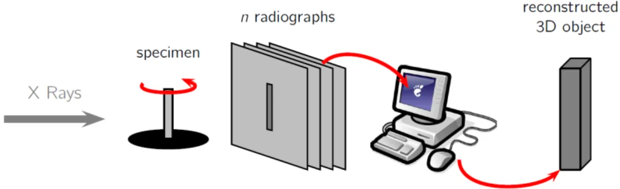
In micro CT scanning, the specimen is divided into a series of 2D slices which are irradiated from the edges with X-rays. Upon transversing through the slice, the X-rays are attenuated and the emergent X-rays with reduced intensities are captured by the detector array. From the detector measurements, the X-ray paths are calculated and the attenuation coefficients are derived. A 2D pixel map is created from these computations and each pixel is denoted by a threshold value which corresponds to the attenuation coefficient measured at a similar location within the specimen. As the attenuation coefficient correlates to the material density, the resultant 2D maps reveal the material phases within the specimen. The quality of the 2D maps is dependent on the scanning resolution which ranges from 1 to 50 mm. At high resolutions, intricate details are imaged, however more time is required for high resolution scanning and the resultant large data set poses a challenge for data storage and processing."
"
Figure 6 - MicroCT technique. In μCT to produce a three-dimensional CT image, a whole set of such two dimensional projections need to be acquired. These projections are usually taken in a setup in which the source and detector are at a fixed position and the object is rotated around its long axis. It can be shown that the number of projections taken over 180
degrees should be about twice the number of samples per projection to avoid aliasing artifacts. The two-dimensional projections can then be used to reconstruct a three-
dimensional image"
"
"
1.4.1 Scaffolds 3D Modeling!
"
Image thresholding is a crucial step that has to be executed prior to 3D modeling and it affects the subsequent analysis and visualization. In the conventional approach, the thresholding range is selected via histographics and visual estimation and the problem arises when the scaffold composes of multiple materials whose thresholding ranges overlap and this renders the digital separation of these materials a difficult task. Moreover, as polychromatic X-ray beams are used in microCT, the lower energy rays would be readily attenuated by the sample resulting in a high exposure at the center of the scaffold. This effect is known as beam hardening and as a result thresholding is no longer dependent solely on radiodensity but also on the specimen size. MicroCT analysis is not suitable for scaffolds containing metals as X-rays are heavily attenuated by these metals. The presence of metals results in dark and bright grainy artifacts which obscure important details in the scan images. As microCT is a relatively new technology, improved algorithms and setups are anticipated, thus resolving such imaging errors [53]."
"
"
"
Chapter 3 43 50 mm. At high resolutions, intricate details are imaged, however more time is required for high resolution scanning and the resultant large data set poses a challenge for data storage and processing.Figure 3-8: MicroCT technique. In µCT to produce a three-dimensional CT image, a
whole set of such two-dimensional projections need to be acquired. These projections are usually taken in a setup in which the source and detector are at a fixed position and the object is rotated around its long axis. It can be shown that the number of projections taken over 180 degrees should be about twice the number of samples per projection to avoid aliasing artifacts. The two-dimensional projections can then be used to reconstruct a three-dimensional image.
3D modeling programs such as Mimics (Materialize, Belgium), Velocity and Anatomics stack the 2D maps to create 3D models, without these programs, visualization and analysis would have been impossible. As computation is inherent in this technique, the selection of software and hardware facilities would influence the efficiency and effectiveness of this radiographical assessment, There are associated concerns despite of the numerous advantages of using micro CT. Image thresholding is a crucial step that has to be executed prior to 3D modeling and it affects the subsequent analysis and visualization. In the conventional approach, the thresholding range is selected via histographics and visual estimation and the problem arises when the scaffold composes of multiple materials whose thresholding ranges overlap and this renders the digital separation of these materials a difficult task. Moreover, as polychromatic X-ray beams are used in micro CT, the lower energy rays would be readily attenuated by the sample resulting in a high exposure at the center of the scaffold. This effect is known as beam hardening and as a result thresholding is no longer dependent solely on radiodensity but also on the specimen size. Micro CT analysis is not suitable for scaffolds containing metals as X-rays are heavily attenuated by
2. Materials and Methods!
"
2.1 Bioreactor Development!
"
In this thesis, a flow perfusion bioreactor was developed from a prototype (Figure 7 and Figure 8) made by Department of Science of Landspítali University Hospital and the Institute of Biomedical and Neural Engineering of Reykjavík University."
"
"
Figure 7 - The prototype consists out of one tube and three disks and a container "
"
Figure 8 - Bioreactor system with the scaffolds. The small holes which are in the disks form small tubes."
"
"
"
"
This device, however, had different aspects to be improved, such as:"
"
• Replacing the DC Motor!
"
The used DC Motor corresponded to the small engines that often found in small appliances or toys for children. These motors provided a functioning also at low voltages, reduced storage space and limited performance. The motor in this prototype (DC Motor 1) had the following characteristics:"
"
The replaced DC Motor (DC Motor 2) had better features instead. In particular it was controlled by a microcontroller that allowed to obtain a speed value between 0 and 255 svm, where 0 svm was about to 0 rpm and 255 svm was about to 5700 rpm. "
The DC Motor replacement was necessary because the DC Motor 1 did not provide sufficient continuity of working for all 4 weeks of cell culture. To investigate the stability were performed two types of tests:"
"
-
Operation test over time (2-3 days of test)"-
Operation test real-time (with digital multimeter) ""
The first test was carried out leaving the bioreactor in operation for 2-3 days, in the same operating conditions to be maintained during the cell culture." The second test was performed by the use of a digital multimeter that has allowed to detect in real time the voltages across the DC Motor, in order to understand at what speed (rpm), the engine would guarantee the best performance. "
For the DC Motor 1, the results were: for the first test, several blocks could be resolved only by restarting of operations; for the second test, a working speed had to be at least 220 svm."
For the DC Motor 2, the results were: for the first test, no blocking detectable; for the second test, a working speed equal to 90 svm, guaranteed good performance."
The following table shows the technical characteristics (Table 1) and the evaluation of the functioning (Table 2, Figure) of the DC Motor 2"
Table 1. Data for DC Motor 2"
"
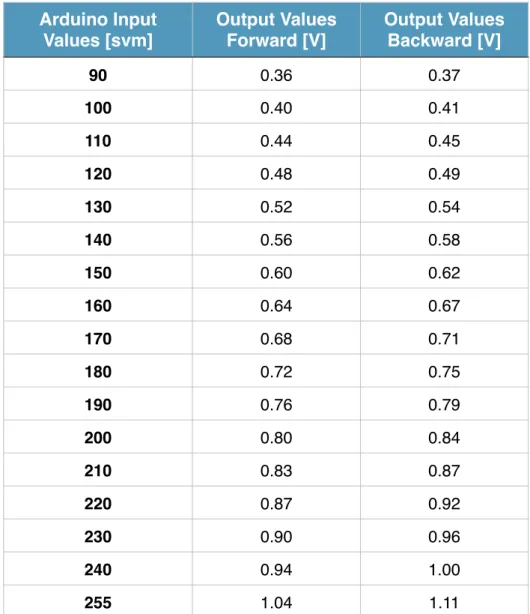
Table 2. Average output values for Forward and Backward Method measured with multimeter across the DC Motor 2."
Motor Data
Name Motrax xDrive 2025-22
Rated voltage [V] 1.5
Working voltage [V] 1.5-3.0
No load Revolutions [rpm] 8000
No load Current [A] 0.2
Max load revolutions [rpm] 5700
Max load current [A] 0.5
Max Power [W] 0.24 Max torque [Ncm] 0.04 Efficiency [%] 31.4 Weight [g] 18 Arduino Input Values [svm] Output Values Forward [V] Output Values Backward [V] 90 0.36 0.37 100 0.40 0.41 110 0.44 0.45 120 0.48 0.49 130 0.52 0.54 140 0.56 0.58 150 0.60 0.62 160 0.64 0.67 170 0.68 0.71 180 0.72 0.75 190 0.76 0.79 200 0.80 0.84 210 0.83 0.87 220 0.87 0.92 230 0.90 0.96 240 0.94 1.00 255 1.04 1.11
"
Figure 9 - The graph, obtained from the values present in the Table 2, show the stability of DC Motor 2 "
"
In particular, Table 2 shows that the DC Motor 2 has a greater stability, ensuring its operation for the entire period of cell culture, even at low speeds."
"
• Replacing the axis and the stabilization system!
"
The axis with propeller, necessary to the realization of the flow inside of the bioreactor and the stabilization system (Figure 10) were replaced with a steel shaft and a new case of support. This was necessary to avoid oscillations and then a non-uniform flow."
"
"
A B!
Figure 10 - A) Bioreactor before changes; B) Bioreactor after changes"
Voltage measurements
Me a su re d vo lt a g e [ V] 0 0,3 0,6 0,9 1,2 Arduino voltage [svm] 90 110 130 150 170 190 210 230 255 Forward Voltage [V] Backward Voltage [V]• Creating a plug between the bioreactor and control unit"
"
Finally, it was necessary to create a plug (shown in Figure 10.B) that would allow to connect and disconnect the bioreactor from the control unit quickly and easily. In particular this addition was very useful during the operations of exchange media; in fact it was possible to disconnect the control unit from the bioreactor and to extract only the bioreactor from the incubator without having to transport each time also inside the work hood."
"
"
2.1.1 Control Unit!
"
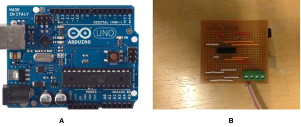
The control unit was the heart of the entire bioreactor and thanks to this was possible to configure each operation that the bioreactor had to play. It consisted of a microcontroller (Arduino UNO) powered via USB transformer and required wiring to create various connections (Figure 11)"
"
"
A B!
Figure 11 - A) Arduino UNO; B) Base with the various connections needed"
"
The Arduino Uno is a microcontroller board based on the ATmega328. It has 14 digital input/output pins (of which 6 can be used as PWM outputs), 6 analog inputs, a 16 MHz ceramic resonator, a USB connection, a power jack, an ICSP header, and a reset button. "
"
"
"
Its programming was carried out through two software:"
"
• Arduino ""
• Processing ""
“Arduino” is an application that uses the Java programming language and allows the implementation of the code realized directly on the microcontroller; in this study, this was useful when it was assumed that the operating characteristics of a device (e.g., speed, time, program, etc..) were set definitively. “Processing” is a software that exploits the bases of Arduino software and it allows the implementation of control interface; in this work, this was useful when it was necessary to configure and to control a device in real-time."
"
In this thesis, a code developed using the Arduino software was designed to allow the operation of the bioreactor during the 4 weeks of cell culture, this code implemented three operation methods (“Forward”, “Backward” and “Pulse”) at speeds of 170 svm."
In particular, the “Forward” and “Backward” methods allowed the propeller respectively a clockwise and couterclockwise, while the “Pulse” method allowed rotations very rapid (with a period of 150ms) alternately clockwise and counterclockwise. “Forward” and “Backward” methods were needed to allow the creation of two alternate streams, in order to improve the cell load above and internally to scaffolds. “Pulse” method was needed to maintain scaffolds in suspension and to create a light flow inside them."
A code, developed using the processing software, was realized for the creation of a user interface that would allow different operations in real-time during the use of the bioreactor."
"
"
"
"
"
2.1.2 Interface!
"
The user interface (Figure 12), as already mentioned above, allowed to operate on the device in real-time. Its implementation was primarily useful for testing different phases of the bioreactor"
"
"
Figure 12 - User interface, daily program"
"
The main available functions were:"
"
• Set a default program (daily)"
"
• Set a custom program"
"
Through the daily program, the bioreactor, worked alternating the phases of “Forward”, “Backward” and “Pulse”, for a time and speed defined by the user; “Forward” and “Backward” speeds were the same that were utilized for “Pulse” phase; it was possible to define a “Pause” between the phases; this feature was helpful to use the device for short time (e.g. during a normal working day)."
The custom program (Figure 13) allowed to setting the various phases in even greater detail, and this feature was helpful to use the device for long periods of time (e.g. 2-3 days or weeks)."
"
"
Figure 13 - User Interface, Custom Program"
"
Finally for both functions, it was possible to extract a "Command History" (Figure 14) that was useful, for example, to check if the bioreactor had actually carried out, on schedule, all the set phases and then to monitor if any anomaly was intervened."
"
! "
"
Figure 14 - Example of Command History"
"
"
"
2.2 Computational Fluid Dynamic Modeling
!"
The fluid dynamics is the part of mechanics that studies the motion of bodies within the fluids flowing. If a liquid flows in a pipe then it will define subject to an internal motion; if a body moves in a liquid this is called external motion. " In the field of fluid dynamics it is also necessary, given the numerous references, a brief definition of fluid that is defined as a substance whose molecules are so few adherents that can slide freely over each other, or they may move away from each other indefinitely. It is possible to divide fluids in two classes:"
AERIFORMS: when the matter, consisting of molecules, tends to occupy the entire available volume, compressible"
FLUIDS: liquid substances, having a proper mass and volume, assume the shape of the vessel that contains them, incompressible"
Therefore there are two possible situations:"
"
• Motion of fluids with the hypothesis of constant density (d), then incompressible fluid"
"
• Motion of bodies inside of the fluids, taking into account of the resultant of all forces acting on the body, to study the behavior of the concerned body (drag force)"
"
"
2.2.1 Viscosity!
"
To talk about motion of a fluid within a body, it is also necessary to introduce the concept of viscosity, before in a particular case and then generalizing the definition, starting to Newton's law. "
The viscosity is defined as the set of tangential forces between the surface and the fluid and between states of different fluid and it is opposed to the motion of these fluids respect to the surface, with velocity u (no constant)."
"
Figure 15 - Speed of a fluid in motion respect to a firm surface"
In the graph, it is possible to note that, given a fluid in relative motion, respect to the solid surface, if a normal to the surface is plotted, after a certain interval of time, it is possible to observe the situation of the motion itself, which represents the speed of a fluid in motion, respect to a firm surface."
The particle in contact with the solid surface has not moved, so it is in the case of adherence, in fact in all fluids, the particles in direct contact with the solid boundaries do not flow, respect to same boundary."
Newton's law, relative to the fluids in motion on solid surfaces, binds tensions that are created with the velocity gradient of the fluid, through the constant of viscosity μ"
! "
"
"
τ is directed in the opposite direction respect to motion, along the x direction on the normal surface to y (in descending toward) and it is measured in Pascal [Pa]."
"
It also defines the kinematic viscosity ratio"
" with μ=dynamic viscosity and ρ=fluid density"
The viscosity is not constant but it varies with temperature: in fact, increasing the temperature, the viscosity decreases, as it is possible to see in the following graphs"
"
Figure 16 - The viscosity decreases with increasing temperature"
"
"
"
"
"
"
"
Misura viscosità nei fluidi: Viscosimetro a rotazione
Dato un cilindro cavo, al cui interno ad una temperatura T = = costante è contenuto il liquido in esame, ed un secondo cilindro posto in rotazione intorno al suo asse da un momento motore ,che si misura in Newton*metro [N*m] , ad opera di un motorino elettrico. Tarando il cilindro interno con due tacche ,una per il livello 1 e l’altra per il livello 2 , dovrò misurare la differenza tra momento motore necessario a produrre la rotazione del cilindro interno quando il livello del liquido è in 1 e quando è in 2; la velocità di rotazione dovrà essere la medesima (fig.2).
2.2.2 Model!
"
It is supposed to consider the motion of a fluid inside a tube of radius r with velocity u. By convention, this situation is studied on a volume element of the fluid with the following characteristics"
!
"
Figure 17 - Forces acting on the volume fluid element"
"
L = length of considered volume element " D = diameter of the element"
r = D/2 = radius of the element "
τ = shear stress that curbs the fluid element in motion"
Considering an equation that expresses the balance of the forces acting on the fluid element, it is possible to write: "
! (1)"
"
p1, p2 = pressures acting on the fluid element"
A = area of the base circumference of the fluid element"
Now calculating"
! (2)"
"
And replacing the (2) in the (1) , it is possible to write:"
! "
"
! ""
! ""
! ""
! "The last obtained equation represents the velocity profile of the fluid in the tube. It is possible then to affirm:"
"
"
! "
"
"
Then:"
"
! ! " ! " ! " ! ""
Ultimately:" ! ""
! ""
"
"
"
"
"
"
"
"
"
2.2.3 Pressure in the Bioreactor!
"
In order to know the stresses that would have occurred on the cells, stimulating them mechanically, it was assumed to approximate the scaffold (small, light, very porous, made in titanium) as an element of the fluid with the following characteristics"
"
" " "
! "
where in particular the speed (w) was extrapolated, in first approximation, by measuring the distance covered by the scaffold and the number of times that this has covered it in one second."
"
"
A B!
Figure 18 - A) Simulation of the flow circulating inside the bioreactor; B) Detail of the flow, at the height of scaffolds area, where the pressure was calculating"
"
Then starting from"
"
"
"
"
"
The pressure, acting on each scaffold, was"
"
"
"
This parameter was very important in order to test if use of the bioreactor, improved or not the growth of the mineralized matrix. In fact, studies focused on the effects of fluid shear on osteoblastic differentiation have shown that shear stress affects osteogenic signal expression of mesenchymal stem cells [54-59]."
"
"
2.3 Stem Cell Culture!
"
In this work hES-MP (human Embryonic Stem Cells-Mesenchymal Progenitor) were used. Human embryonic derived mesenchymal progenitor cells are cells that belong to the cell line hES-MP002.5, that was developed by Cellartis in Gothenburg, Sweden, 2009. These cells have been described to have high resemblance to primary adult mesenchymal stem cells (MSC) in terms of proliferation and differentiation. Handling and culture of hES-MP cells and MSC is almost the same but hES-MP cells require gelatin coating of the growth surface prior to seeding."
To obtain a single cell suspension via trypsination of adherent cell cultures in monolayer, some equipment and procedures were necessary (see Appendix
A)."
"
"
2.3.1 Counting Cells!
"
Knowing the cell concentration was an important step for subsequent seeding cell. Cell concentration of viable cells in a solution was determined using hemocytometer."
Hemocytometer is a counting chamber made of glass especially designed for cell counting. The chamber has two counting grids that are each put