Dipartimento Farmaco-Biologico
(SSD: MED/46 SCIENZE TECNICHE DI MEDICINA DI LABORATORIO)
Dottorato di Ricerca in “Biochimica Cellulare ed
Attività dei Farmaci in Oncologia” (XXV ciclo)
DAX-1 at the connection between Androgen Receptor
and Aromatase: a novel mechanism in the inhibition of
estrogen-dependent cancer cell proliferation
Docente Tutor Dottoranda
Prof.ssa Marilena LANZINO Dott.ssa Pamela MARIS
Coordinatore
Prof. Diego SISCI
MATERIAL AND METHODS ... 7
Cell cultures, animals and treatments ... 7
Cell proliferation assays ... 7
Plasmids, transfections and luciferase reporter assays ... 8
RNA extraction and reverse transcription-PCR assays ... 9
Western Blotting analysis ... 9
Immunofluorescence ... 9
DNA affinity precipitation assay ... 10
Electrophoretic mobility shift assay ... 10
Aromatase activity assay ... 11
Radioimmunoassay (RIA) ... 11
Chromatin Immunoprecipitation ... 11
RNA silencing ... 12
Statistical analysis ... 13
RESULTS ... 14
Ligand-activated AR increases DAX-1 expression in MCF-7 breast cancer cells.14 Ligand-activated AR induces DAX-1 gene promoter activity ... 15
Identification of a functional Androgen Responsive Element (ARE) within the DAX-1 promoter ... 17
Androgen treatment inhibits aromatase expression through a DAX-1-mediated mechanism in breast cancer cells. ... 19
Androgens, by inducing DAX-1 expression, inhibit P450 aromatase in estrogen-dependent Leydig tumor cells, R2C ... 20
AR/DAX-1/Aromatase expression pattern in male Fisher rats ... 23
DISCUSSION ... 24
Catalano S., Morelli C. and Ando' S. Chenodeoxycholic acid through a TGR5-dependent CREB-signaling activation enhances Cyclin D1 expression and promotes human endometrial cancer cell proliferation. Cell Cycle. 2012 Jul 15;11(14):2699-710.
2. Puoci F., Morelli C., Cirillo G., Curcio M, Parisi O. I., Maris P., Sisci D. and Picci N. Anticancer Activity of a Quercetin-based Polymer Towards HeLa Cancer Cells. Anticancer Res. 2012 Jul; 32(7):2843-7.
3. Morelli C., Maris P., Sisci D., Perrotta E., Brunelli E., Perrotta I., Panno M.L., Tagarelli A., Versace C., Casula M.F., Testa F., Andò S., Nagya J.B. and Pasqua L. PEG-templated mesoporous silica nanoparticles exclusively target cancer cells. Nanoscale, 2011, 3, 3198.
4. Morelli C., Lanzino M., Garofalo C., Maris P., Brunelli E., Casaburi I., Catalano S., Bruno R., Sisci D., Andò S. Akt2 inhibiton enables the forkhead transcription factor FoxO3a to have a repressive role in Estrogen Receptor Alpha transcriptional activity in breast cancer cells. Mol. Cell Biol. 2010 Feb;30(3):857-70.
SUMMARY
In situ estrogen production by aromatase is a critical determinant for breast cancer growth and progression. Clinical and in vitro studies indicate that androgens have a protective role in mammary carcinogenesis. Here we demonstrated, in breast cancer cell lines, that ligand-activated androgen receptor (AR) induces the expression of the orphan nuclear receptor DAX-1 by a direct binding to a newly identified Androgen-Response-Element within the DAX-1 proximal promoter. In turn, androgen-induced DAX-1 is recruited, in association with the corepressor N-CoR, within the SF-1/LRH-1 containing region of the aromatase promoter, thereby repressing aromatase expression and activity. The molecular mechanism underlining the androgen-dependent modulation of DAX-1 and aromatase expression is reproducible in R2C rat Leydig tumor cells, highly expressing aromatase. Indeed, in vivo studies conducted in testes tissues from Fisher rats, spontaneously developing Leydig cell neoplasma, reveal an inverse relationship between AR/DAX-1 and aromatase levels.
In elucidating a novel mechanism by which androgens, through DAX-1, inhibit aromatase expression, these findings reinforce the theory of androgen- opposing estrogen-action opening new avenues for therapeutic intervention in endocrine-related cancers.
INTRODUCTION
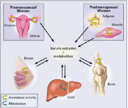
Sexual hormones, estrogens and androgens, determine biological response in a tissue- and gender-specific manner and play a pivotal role in endocrine-mediated tumorigenesis (1-5). Estrogens influence the pathological processes of several hormone-dependent cancers since they are thought to be the driving force for the formation of breast, endometrial, ovarian and testicular tumors (6,7). In breast, estrogen signaling plays a critical role in cell proliferation and tumor development (8), and estrogen receptor α (ER-α) positive tumors comprise ~ 70% of annually diagnosed breast cancer cases (9). Importantly, in addition to the estrogen supply from the ovary, biologically active estrogens, especially estradiol (E2), are locally produced in an intracrine mechanism within the breast cancer tissues and exert their actions on carcinoma cells. The puzzling hint was the observation that breast cancer incidence is higher in post-menopausal women, when the production of ovarian estrogens has ceased. It is now known that, in post-menopausal women, estrogen levels in breast tissue are 10-50 times elevated than in blood and significantly higher in malignant than in non-malignant tissues (10).
Figure 1. Schema illustrating sites of production of
biologically active estrogens in pre- and post-menopausal women.
A series of enzymes are involved in this intra-tumoral or in situ production of estrogens in breast carcinoma tissues but aromatase, a member of the cytochrome P450 family, is a key enzyme of estrogen production through conversion from circulating adrenal androgens in estrogen-dependent postmenopausal breast cancer (11).
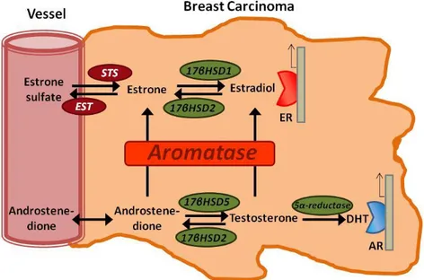
Figure 2. Scheme representing in situ production of sex steroids in human
breast carcinoma tissues. High concentrations of circulating inactive steroids, such as estrone sulfate and androstenedione are precursor substrates of local production of estrogens and/or androgens in breast carcinomas. STS; steroid sulfatase, EST; estrogen sulfotrasferase, and 17HSD; 17-hydroxysteriod dehydrogenase.
Consequently, substantial efforts have been devoted to explore ways to block estrogen activity in breast cancer cells, underpinning the widespread use of antiestrogens and aromatase inhibitors in the adjuvant treatment of breast cancer (12,13).
The balance between ER-α and androgen receptor (AR) signalling has been proposed as a critical determinant of growth in the normal and malignant mammary epithelium, supporting the prevalent theory of androgens opposing estrogens in the mammary gland.
Studies on non-human primate or rodent support the notions that androgens inhibit mammary epithelial growth by reducing E2-dependend cell proliferation (14-16). Several lines of evidence sustain the idea of similar effects in humans: 1) androgens, such as dihydrotestosterone, oppose the E2-induced proliferation
and induce apoptosis in explant cultures from normal breast tissues, in an AR-dependent manner (17); 2) genotypically male individuals suffering from complete androgen insensitivity develop morphologically normal female breast during puberty (18); 3) girls with congenital adrenal hyperplasia and androgen excess do not undergo pubertal thelarche (19); 4) reduced or impaired AR signalling has been implicated in the development of hereditary male breast cancers (20); and 5) women who have AR alleles that encode for short polyglutamine tracts in the AR protein, resulting in a higher AR transcriptional activity (21), have a reduced risk of developing breast cancer (22). These observations suggest that androgens do play a role in the regulation of mammary gland development and imply that androgen signaling in the breast might protect against cancer development and progression.
A significant number of primary well differentiated breast tumors express AR (23) whose presence and functional activity appear to be related to positive prognostic factors, including ER positivity, smaller tumor size, low tumor grade, improved response to hormone therapy and longer patient survival (24-37). Interestingly, several events involved in breast cancer genesis or progression have been shown to alter AR expression or function, conferring a growth advantage to cancer cells. Indeed, a trend towards a loss of AR has been shown in BRCA1-mutated breast tumors (38-40) as well as in HER2-positive breast cancers, generally associated with a worse outcome (37,41).
These findings are consistent with cell-based assays indicating that, in ER/AR-positive breast tumor cell lines, AR activation by the agonist dihydrotestosterone decreases ER-α transcriptional activity (36,42) and inhibits basal as well as estrogen-dependent cell proliferation (43-48). These effects occur by an AR-mediated mechanism that negatively regulates the transcriptional activity of the cyclin D1 gene promoter throughout the recruitment of a multiprotein repressor complex involving the participation of the orphan nuclear receptor DAX-1 (Dosage-sensitive sex reversal, Adrenal Hypoplasia Congenita (AHC) critical region on chromosome X, gene 1; NROB1) (47).
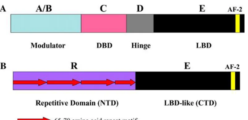
DAX-1 is an unusual orphan member of the nuclear receptor superfamily, lacking the classical zinc-finger DNA Binding Domain (49-52)
Figure 3. Comparison of functional domain structure of members
of the nuclear receptor superfamily (A) with DAX-1 (B).
Instead of directly binding to regulatory DNA sites, DAX-1 controls transcription mainly as a co-repressor by associating with nuclear receptors, (e.g., AR, ER and progesterone receptor), or other transcription factors (e.g., steroidogenic factor 1, SF-1 or Liver Receptor Homolog-1, LRH-1).
Figure 4. Mechanisms of DAX1-mediated repression of SF-1, ER, and LRH-1. (A) DAX1 binds the AF-2 domain of the nuclear receptors via its LXXLL motifs
and recruits corepressor proteins to target gene promoters. (B) Effects of intracellular levels of DAX1 and SF-1 on transcriptional repression. Increased SF1 levels relative to DAX1 favor transcriptional activation (left), and increased levels of DAX1 relative to SF-1 favor transcriptional repression (right).
DAX-1 has a restricted expression pattern to tissues directly involved in steroid hormone production and reproductive function, such as adrenal cortex, Leydig and Sertoli cells in the testis and theca and granulosa cells in the ovary (53-55).
Within these tissues, DAX-1 acts as a global anti-steroidogenic factor by working in pair with SF-1/LRH-1 and repressing the expression of multiple enzymes involved in the steroidogenic pathway. Major steroidogenic targets include cytochrome P450 (e.g. CYP19 aromatase), cholesterol transporters (e.g., steroidogenic acute regulator protein, StAR) and hydroxysteroid dehydrogenase (53,55,56). DAX-1 expression has also been reported in several types of cancers. In adrenocortical tumours DAX-1 presence is inversely correlated to the level of steroid production (57,58). DAX-1 expression in breast (34,59), ovarian (60), endometrial (61) and prostate cancers (62) has been additionally described even though not narrowly investigated. Moreover, there are few studies on how DAX-1 expression is regulated.
Here we report the identification of a novel AR-mediated mechanism controlling the expression of DAX-1. Based on our model, ligand-activated AR may negatively regulate in situ estrogen production through an androgen receptor-dependent activation of DAX-1 gene transcription in estrogen-related cancer cell lines, providing new clues for a better comprehension of the mechanisms underlying the inhibitory role exerted by androgens on estrogen-dependent cancer cell proliferation.
MATERIAL AND METHODS
Cell cultures, animals and treatments
Breast cancer epithelial cell line MCF-7 (American Type Culture Collection, ATCC, UK) and human embryonic kidney cell line HEK-293 were grown in DMEM (Gibco, USA) supplemented with 5% and 10% fetal bovine serum (FBS; Gibco, USA) respectively. Human ductal breast epithelial tumor cell line T47D (Interlab Cell Line Collection, ICLC, Italy) was maintained in RPMI (Gibco) supplemented with 10% FBS. Rat Leydig tumor cells (R2C) were cultured in Ham's F-10 supplemented with 15% horse serum (HS; Gibco,) and 2.5% FBS. Before each experiment, cells were synchronized in phenol red-free serum free media (PRF-SFM) for 24h. All the experiments were performed in PRF-media containing 2,5% charcoal-treated FBS (PRF–CT). Male Fischer 344 rats (a generous gift of Sigma-Tau), 6 (FRN), (n=3) and 24 (FRT) (n=4) months of age, were used for studies. Twenty-four-month-old animals presented spontaneously developed Leydig cell tumors, which were absent in younger animals. Testes of all animals were surgically removed by qualified, specialized animal care staff in accordance with the Guide for Care and Use of Laboratory Animals (NIH) and used for experiments. The following agents were used for treatments: Mibolerone (Mb, Perkin Elmer, USA), Bicalutamide (Casodex; Santa Cruz Biotechnology, USA) and Hydroxyflutamide (OH-Fl; Sigma, Italy).
Cell proliferation assays
MCF-7 and R2C cells were seeded on 12-well plates at a density of 105 cells/well and grown overnight. The following day, cells were synchronized in PRF-SFM for 24h to avoid growth differences among cells. After 24h, cells were exposed to the desired concentration of Mb or left untreated. The effects of Mb on cell proliferation were measured at different time points following initial exposure to treatments by counting cells using a Burker’s chamber, with cell viability determined by trypan blue dye exclusion or using the method of transcriptional and translational colorimetric assay (MTT) (47).
Plasmids, transfections and luciferase reporter assays
The following plasmids were used: pcDNA3-AR (AR) encoding full-length AR (63); CMV-P881 (AR(Cys574→Arg)) encoding the full-length AR carrying a mutation in the DNA-binding domain (DBD; Cys-574→Arg) (63); pGL3 vector containing human aromatase promoters II/1.3 ligated to a luciferase reporter gene (PII/1.3) (a gift from Dr. E. R. Simpson and Dr. C. D. Clyne, Prince Henry’s Institute of Medical Research, Clayton, Australia); pGL3 vector containing rat aromatase promoter II ligated to a luciferase reporter gene (PII) and the pGL3 vector containing rat aromatase promoter II mutated in SF-1 binding site ligated to a luciferase reporter gene (mutPII) (a gift from Dr. M.J. McPhaul , Southwestern Medical Center, Dallas, USA); the vector-based pSiAR plasmid, coding for small interfering RNA targeting the 5’-untranslated region of AR mRNA, and the scrambled control construct pSiCon (64). The Renilla reniformis luciferase expression vector used was pRL-Tk (Promega, USA). The deletions of SF-1/LHR-1 sequence in the human aromatase promoter PII/1.3 were generated by PCR. The resulting plasmid encoding the aromatase promoter II and 1.3 containing the desired deletions was designated mutPII/1.3 The desired deletions of sequences were confirmed by nucleotide sequence analysis. The luciferase expression vector Luc-promoter (Luc), encoding the DAX-1 promoter, was generated by PCR on genomic DNA. The following primer pairs were used to amplify a 1336bp fragment spamming from position -1370 to
-34 from the ATG of the DAX-1 gene:
5’-GAGGATGGGAGGGAGGGAAAAAGT-3’ (forward) and
5’-AGGGCAGGGGAAAAGAGGAAACAT-3’ (reverse). PCR primer pairs were selected analyzing the 5’-flanking region of DAX-1 gene. The PCR product was purified and the 1336bp fragment was inserted in the pCR 2.1 plasmid by using TOPO-TA cloning kit (Invitrogen, Milan, Italy) and sequenced. The fragment was cut with Kpn I and Xho I and cloned into the pGL3 basic vector (Promega, Italy). Cells were transfected using Fugene (Promega) according to the manufacturer’s instructions. pRL-Tk was used to assess transfection efficiency. Luciferase activity was measured using Dual Luciferase Assay System (Promega), normalized to renilla luciferase activity. For western blotting (WB) assays, cells were plated on 60-mm dishes and transfected with an appropriate amount of various plasmids, as indicated in figure legends.
RNA extraction and reverse transcription-PCR assays
Total RNA was isolated using TRIzol reagent (Life Technologies, USA) according to the manufacturer’s instructions and treated with DNase I (Ambion, USA). Two micrograms of total RNA were reverse transcribed with the High capacity cDNA Reverse Transcription Kit (Applied Biosystems, USA); cDNA was amplified by PCR to obtain products corresponding to cDNA fragments of DAX-1, P450 aromatase, GAPDH, or -Actin. RT-PCR was performed using the DreamTaq DNA Polymerase (Fermentas, USA) and GeneAmp PCR System 9600 thermocycler (Perkin Elmer). Primers are listed in Table 1.
Western Blotting analysis
MCF-7 and R2C cells or total tissue of FRNT and FRTT were lysed in 500µl of 50mM Tris-HCl, 150 mM NaCl, 1% Nonidet P-40, 0.5% sodium deoxycholate, 2 mM sodium fluoride, 2mM EDTA, 0.1% SDS, containing a mixture of protease inhibitors (aprotinin, phenylmethylsulfonyl fluoride, and sodium orthovanadate) for protein extraction. Nuclear extracts were prepared as previously described (65), Western Blotting (WB) analysis was performed as previously described (66). The following monoclonal (m) and polyclonal (p) antibodies (Ab) were used: anti-AR mAb (4i299), anti-DAX-1 pAb (K-17), anti-Lamin-B pAb (C-20), anti-GAPDH pAb (FL-335), anti--Actin mAb (AC-15), (Santa Cruz Biotechnology), and anti-human cytochrome P450 aromatase mAb (Serotech, USA).
Immunofluorescence
MCF-7 and R2C cells were plated on coverslips, serum starved, and then treated or left untreated with Mb 10-8 M for the appropriated time. After incubation, cells were fixed with 4% paraformaldehyde and permeabilized with 0.2% Triton X-100, and nonspecific sites were blocked with bovine serum albumin (BSA, Santa Cruz Biotechnology) (3% for 30 min). The blocked samples were incubated O/N with a mixture of primary antibody recognizing DAX-1(K-17) pAb (Santa Cruz Biotechnology), washed with phosphate-buffered
saline (PBS) (Gibco), and incubated with a mixture of fluorescein-conjugated goat anti-rabbit IgG (Santa Cruz Biotechnology) secondary Abs. The cellular localization of the two proteins was examined under a Leica TCS SP2 confocal laser scanning microscope at X100 magnification.
DNA affinity precipitation assay
DNA affinity precipitation assay was performed as previously described (63). The DNA motif probes were prepared by annealing a biotinylated sense oligonucleotide (for DAX-1-ARE, 5’-[Bio]-CCAAATATCACATGTTCTCAC-3’; for DAX-1-mutatedARE, 5’-[Bio]- CCAAATGGAACATGGGATCAC-3’) with the respective nonbiotinylated complementary oligonucleotide (for DAX-1-ARE,
5’-GTGAGAACATGTGATATTTGG-3’; for DAX-1-mutated ARE,
5’-GTGATCCCATGTTCCATTTGG-3’).
Electrophoretic mobility shift assay
Nuclear protein extracts were prepared as previously described (67). The double-stranded oligonucleotides used as probes were end labeled with [– 32P]
ATP and T4 polynucleotide kinase and purified using Sephadex G50 spin columns. The oligonucleotides used as probes and as cold competitors (Sigma Genosys, UK) were (nucleotide motifs of interest are underlined): probe:
CCAAATATCACATGTTCTCAC-3’; mutated probe:
5’-CCAAATGGAACATGGGATCAC-3’. Nuclear extracts (20 μg) were incubated with 50000 cpm. of labeled probe, under conditions previously reported (67) The mixture was incubated at room temperature for 20 min in the presence or absence of the unlabelled competitor oligonucleotide. Mouse anti-AR monoclonal antibody (441) or normal mouse IgG (Santa Cruz Biotechnology) were included in some of the reaction mixtures with an additional 12h incubation at 4°C before addition of labelled probe. The entire reaction mixture was electrophoresed through a 6% polyacrylamide gel for 3 h at 150 V.
Aromatase activity assay
The aromatase activity in subconfluent R2C cell culture medium was measured by tritiated water-release assay using 0.5 Amol/L [1h-3H(N)]androst-4-ene-3,17-dione (DuPont NEN) as a substrate (75). Incubations were done at 37°C for 3h under a 95%:5% air/CO2 atmosphere.
Radioimmunoassay (RIA)
Before the experiments, R2C cells were maintained overnight in Ham/F-10 (medium only). The estradiol content of medium recovered from each well was determined against standards prepared in low-serum medium using a RIA kit (DSL 43100; Diagnostic System Laboratories) according to manufacturer’s instructions.
Chromatin Immunoprecipitation
Chromatin Immunoprecipitation (ChIP) assay was performed as previously described (68). MCF-7, T47D, and R2C cells were grown in 100mm plates. Confluent cultures (90%) were shifted to SFM for 24 h and then treated with 10-8 M Mb for 2 h, or left untreated in 2,5% PRF–CT. Following treatment, the cells were washed twice with PBS and crosslinked with 1% formaldehyde at 37°C for 10 min. Next, the cells were washed twice with PBS at 4°C, collected and resuspended in 200 µl of lysis buffer (1% SDS, 10mM EDTA, 50mM Tris-HCl pH 8.1) and left on ice for 10 min. Then, the cells were sonicated four times for 10 s at 30% of maximal power (Fisher Sonic Dismembrator) and collected by centrifugation at 4°C for 10 min at 14.000 rpm. The supernatants were collected and diluted in 1.3 ml of IP buffer (0.01% SDS, 1.1% Triton X-100, 1.2mM EDTA, 16.7mM Tris-HCl pH 8.1, 16.7mM NaCl) followed by immunoclearing with 80 μl of sonicated salmon sperm DNA/protein A agarose (UBI) for 1 h at 4°C. The precleared chromatin was immunoprecipitated for 12 h with specific Abs: AR mAb (441), Polymerase II pAb (N-20), DAX-1 pAb (K-17), and anti-N-CoR pAb (H-303) (Santa Cruz Biotechnology). Normal rabbit IgG and normal mouse IgG were used instead of primary Abs as negative controls. After this,
60μl of salmon sperm DNA/protein A agarose was added and precipitation was continued for 2 h at 4°C. After pelleting, precipitates were washed sequentially for 5 min with the following buffers: Wash A (0.1% SDS, 1% Triton X-100, 2mM EDTA, 20mM Tris-HCl pH 8.1, 150mM NaCl), Wash B (0.1% SDS, 1% Triton X-100, 2mM EDTA, 20mM Tris-HCl pH 8.1, 500mM NaCl), and Wash C (0.25M LiCl, 1% NP-40, 1% sodium deoxycholate, 1mM EDTA, 10mM Tris-HCl pH 8.1), and then twice with TE buffer (10mM Tris, 1mM EDTA). The immune complexes were eluted with elution buffer (1% SDS, 0.1M NaHCO3). The eluates were reverse crosslinked by heating at 65°C for 12 h and digested with proteinase K (0.5 mg/ml) at 45°C for 1 h. DNA was obtained by phenol and phenol/chloroform extractions. A 2 μl portion of 10 mg/ml of yeast tRNA was added to each sample and DNA was precipitated with EtOH for 12 h at 4°C and then resuspended in 20 μl of TE buffer. Immunoprecipitated DNA was analysed by RT-PCR by using 4 μl of each sample for RT-PCR. Primers are listed in Table 1.
RNA silencing
For AR gene silencing experiments, MCF-7 cells were transfected using the vector-based pSiAR plasmid or the scrambled control construct pSiCon (64), as described in the ‘Plasmids, transfections and luciferase reporter assays’ paragraph. For DAX-1 gene silencing experiments, custom synthesized siRNA (Ambion, USA) annealed duplexes were used for effective depletion of DAX-1 mRNA. A scrambled siRNA (Ambion) that does not match with any human mRNA was used as a control for non-sequence-specific effects. Growing cells were switched to PRF-SFM for 24h, trypsinized and transfected in suspension with 80 pmol of siDAX-1 or 80 pmol of siScramble RNA in 60-mm dishes, using Lipofectamine 2000 (Life Technologies), following the manufactuer’s instructions. Cells were incubated with the siRNA-Lipofectamine 2000 complex at 37°C for 6 h and then switched to fresh PRF-CT and treated or not with Mb before analysis.
Statistical analysis
All data were expressed as the mean±SD of at least three independent experiments. Statistical significances were tested using Student’s t-test.
TABLE 1
Gene sequence Primer sequence Cycles AT(°C)
DAX-1 5’-CGGGCCACGGCGCTTCTGTA-3’(forward)
30 63
5'-TCCCGCCGCCTGGTGGTGAG-3’(reverse)
Human P450 Aromatase 5’-CAAGGTTATTTTGATGCATGG-3’(forward)
30 58
5’-TTCTAAGGTTTGCGCATGA-3’;(reverse)
Rat P450 Aromatase 5’-CAGCTATACTGAAGGAATCCACACTGT-3’(forward)
22 58
5’-AATCGTTTCAAAAGTGTAACCAGGA-3’(reverse)
GAPDH 5’-CCCACTCCTCCACCTTTGAC-3’(forward) 25 58
5’-TGTTGCTGTAGCCAAATTCGT-3’(reverse)
Rat -Actin 5’-AGGCATCCTGACCCTGAAGTAC-3’(forward) 18 58
5’-TCTTCATGAGGTAGTCTGTCAG-3’(reverse)
DAX-1 ARE promoter 5’-AATGCAGGAACAGAAAACCAAATA-3’ (forward) 33 62
5’-GGCAGCGAGCAGGATGTAAAAGTG-3’ (reverse)
Human P450Aromatase 5’-TGATGGAAGGCTCTGAGAAG-3’ (forward) 30 58
Promoter 5’-TAGCTCCTGTTGCTTCAGAGG-3’ (reverse)
Rat P450Aromatase
promoter 5’-ATGCACGTCACTCTACCCACTCAA-3’ (forward) 5’-TAGCACGCAAAGCAGTAGTTTGGC-3’ (reverse) 35 65
RESULTS
Ligand-activated AR increases DAX-1 expression in MCF-7 breast cancer cells.
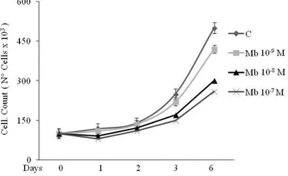
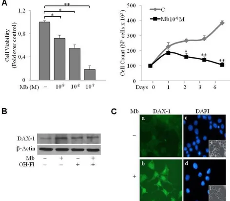
DAX-1 has been detected in breast cancer cell tissues (59,69) and cell lines (70). Our first aim was to investigate DAX-1 levels and cellular compartmentalization in human MCF-7 breast cancer cell line following androgen administration. Experiments were carried out using the synthetic AR agonist Mibolerone (Mb) that was proved to be as effective as dihydrotestosterone (DHT) (47) in inhibiting MCF-7 cell proliferation (Fig. 5).
Figure 5. Serum starved MCF-7 cells were grown in PRF–CT in
absence or presence of Mb as indicated. Cell proliferation was measured at 0, 1, 2, 3 and 6 days after initial exposure to Mb treatments by trypan blue dye exclusion test.
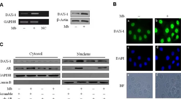
As shown in Fig.6, 24h Mb treatment increased both DAX-1 mRNA (Fig.6A, left panel) and protein cellular levels (Fig.6A, right panel). Immunofluorescence experiments, revealed that DAX-1 protein localizes in both cytoplasm and nucleus, even though a markedly stronger immunoreactivity could be observed in the nuclear compartment. Mb administration induced a significant increase of DAX-1 nuclear abundance (Fig. 6B). To ascertain whether activated AR could be involved in the positive regulation of DAX-1 levels a Short Hairpin plasmid was used to silence AR expression in MCF-7 cells. Western blotting analysis of
cytoplasmic and nuclear fractions further confirmed DAX-1 subcellular localization (Fig. 6C). Interestingly, reduction of the AR protein content resulted in the loss of the up-regulatory effect exerted by Mb on DAX-1 nuclear expression (Fig. 6C).
Figure 6. DAX-1 expression is modulated by ligand-activated AR in MCF-7 cells. (A) Mb increases DAX-1 expression. Cells were treated with 10-8 M Mb for 24h and DAX-1 mRNA (left
panel) or protein content (right panel) were detected by RT–PCR or WB analyses, respectively.
GAPDH mRNA, internal standard; NC, negative control. β-Actin protein, loading control. (B)
Subcellular localization of DAX-1. DAX-1 (a,b), DAPI (c,d) immunofluorescence was detected in
cells treated with 10-8 M Mb for 24h; BF, bright field (e,f); 100x optical magnification. (C) AR
silencing abrogates Mb-dependent DAX-1 modulation. Cytoplasmic and nuclear proteins
isolated from cells transfected with 0.5 μg pSiAR (sh-AR) or 0.5 μg pSiCon (scramble) and treated with 10-8 M Mb, were subjected to WB analysis. GAPDH and Lamin-B expression was assessed as control of protein loading and purity of lysate fractions. All the results are representative of three independent experiments.
Ligand-activated AR induces DAX-1 gene promoter activity
To determine whether the human DAX-1 gene is a target of the ligand-activated AR a 1.3kb (-1370/-34) sequence of the 5’-flanking region of the human DAX-1
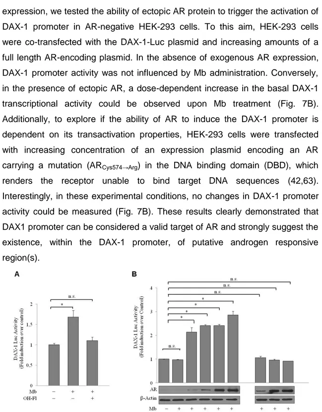
gene was cloned, inserted into a reporter plasmid (DAX-1-Luc plasmid) and used to perform transient transfection experiments on MCF-7 cells. As shown in Fig. 7A, Mb administration induced a significant enhancement of DAX-1 promoter activity. This effect was reversed by the addition of the AR antagonist, Hydroxyflutamide (OH-Fl), indicating that it was mediated by AR activation. To confirm the involvement of ligand-activated AR in the modulation of DAX-1 gene
expression, we tested the ability of ectopic AR protein to trigger the activation of DAX-1 promoter in AR-negative HEK-293 cells. To this aim, HEK-293 cells were co-transfected with the DAX-1-Luc plasmid and increasing amounts of a full length AR-encoding plasmid. In the absence of exogenous AR expression, DAX-1 promoter activity was not influenced by Mb administration. Conversely, in the presence of ectopic AR, a dose-dependent increase in the basal DAX-1 transcriptional activity could be observed upon Mb treatment (Fig. 7B). Additionally, to explore if the ability of AR to induce the DAX-1 promoter is dependent on its transactivation properties, HEK-293 cells were transfected with increasing concentration of an expression plasmid encoding an AR carrying a mutation (ARCys574→Arg) in the DNA binding domain (DBD), which renders the receptor unable to bind target DNA sequences (42,63). Interestingly, in these experimental conditions, no changes in DAX-1 promoter activity could be measured (Fig. 7B). These results clearly demonstrated that DAX1 promoter can be considered a valid target of AR and strongly suggest the existence, within the DAX-1 promoter, of putative androgen responsive region(s).
Figure 7. Mb-induced activation of DAX-1 gene promoter requires integrity of the AR DNA
binding domain. (A) Mb induces DAX-1 promoter activity. MCF-7 cells were transiently transfected with DAX-1-Luc plasmid (0.25 µg/well) and treated with 10-8 M Mb and/or 10-6M OH-Fl, for 24h. (B) DAX-1 promoter activation depends on a functional AR-DNA binding domain. HEK-293 cells were co-transfected with DAX-1-Luc-plasmid (0.25 µg/well) plus increasing amounts (µg) of pcDNA3-AR (AR) or CMVP881 (ARCys-574→Arg) as indicated, and treated with Mb
10-8M for 24h. Linear relation between transfected AR plasmid and expressed AR protein quantity was evaluated by WB analysis; -Actin protein, loading control. *, P<0.01; n.s., non significant.
Identification of a functional Androgen Responsive Element (ARE) within the DAX-1 promoter
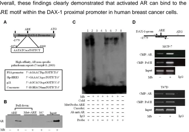
Sequence analysis revealed that the DAX-1 promoter contains a putative androgen response element (ARE) located at position -1074bp upstream of the translation starting codon (Fig. 8A, upper panel) which is homolougus to the AREs found in the promoters of several androgen responsive genes (71) (Fig. 8A, lower panel).
To test the hypothesis that AR can effectively bind to this putative ARE motif (DAX-1-ARE), a DNA affinity precipitation assay (DAPA) was performed by using a double-stranded oligonucleotide containing the core ARE sequence and 10-11 bp of flanking sequence on either side. Endogenous AR was found associated with the putative consensus oligonucleotide following Mb administration only, while AR binding was unnoticeable in nuclear cell lysates from untreated cells. The use of biotinylated mutant oligonucleotide, abolished AR binding, indicating that the in vitro DNA-AR protein binding is sequence specific (Fig. 8B).
To better demonstrate the specificity of the putative ARE site, an electrophoretic mobility shift assay (EMSA) was performed using the identified ARE motif as a DNA probe. Factors present in MCF-7 nuclear extracts and ARE probe formed a protein-DNA complex (Fig. 8C, lane 1) which was strongly enhanced by Mb treatment (Fig. 8C, lane 2). The specificity of the protein-DNA association was demonstrated by its disappearance when 100-fold molar excess of unlabeled wild-type probe was added to the reaction as competitor (Fig. 8C, lane 3) or when a mutated labeled probe was used (Fig. 8C, lane 4). Coadministration of the anti-androgen bicalutamide (casodex) markedly decreased the Mb-induced DNA binding complex indicating the involvement of the AR (Fig. 8C, lane 5). Moreover, the DNA-protein complex formation was reduced (immunodepletion) in the presence of a specific anti-AR monoclonal antibody, indicating that this antibody recognizes AR epitopes that interact or interfere with AR association to ARE (Fig. 8C, lane 6) and further confirming the presence of AR in the protein-DNA complex. As expected, protein-protein-DNA complex formation was unaffected by normal rabbit IgG (Fig. 8C, lane 7).
Chromatin immunoprecipitation (ChIP) analysis was next used to determine whether endogenous AR protein localizes to the native DAX-1 promoter in AR-positive MCF-7 and T47D breast cancer cells. AR occupancy of the DAX-1-ARE containing region of DAX-1 promoter was induced in a ligand-dependent manner, since AR recruitment was enhanced by Mb administration. The ability of AR to transactivate the DAX-1 gene is evidenced by the dynamic of RNA Pol II recruitment onto the DAX-1 promoter, that appears to be enhanced upon Mb treatment. Non-specific IgG antibody failed to precipitate any protein-DNA complexes (Fig. 8C).
Overall, these findings clearly demonstrated that activated AR can bind to the ARE motif within the DAX-1 proximal promoter in human breast cancer cells.
Figure 8. Activated AR binds to the ARE site within the DAX-1 promoter. A) Schematic
representation of the putative ARE site within the DAX-1 promoter (upper panel). List of known ARE sequences (lower panel). (B) Detection of AR/ARE binding. Nuclear extracts from MCF-7 cells treated with 10-8 M Mb were incubated with either wt- (ARE) or mutated (mut-ARE) biotinylated oligonucleotide and subjected to DAPA assay. The unbound fraction was loaded as negative control (NC); cell nuclear extracts were used as positive control (Input). (C) Evaluation
of AR/ARE binding specificity. Nuclear extracts from MCF-7 cells untreated (lane 1) or treated
with 10-8 M Mb (lane 2-7) were incubated with a specific labelled probe. 100-fold molar excess of unlabeled probe (lane 3); labeled mutated probe (lane 4); addition of 10-5 M Casodex (lane 5); pre-incubation with anti-AR antibody (lane 6) or IgG (lane 7); probe alone (lane 8). (D) AR
recruitment onto the DAX-1 promoter. Sheared chromatin from MCF-7 or T47D cells treated
with 10-8 M Mb for 2h, was precipitated using anti-AR or anti-RNA Pol II antibodies. IgG, control samples. The investigated sequence was detected by PCR using specific primers. DNA Inputs were amplified as loading controls. All the results are representative of three independent experiments.
Androgen treatment inhibits aromatase expression through a DAX-1-mediated mechanism in breast cancer cells.
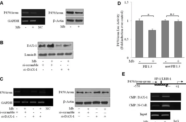
To comprehend the biological significance of the androgen-dependent up-regulation of DAX-1 expression, we explored the possibility that it might impact on in situ estrogen production since the aromatase gene CYP19 has been shown to represent a physiologic target gene for DAX-1 (56). To this aim the expression of P450 aromatase upon androgen treatment was examined in MCF-7 cells. Mb administration decreased the cellular content of the enzyme at both mRNA and protein levels (Fig. 9A).
To provide evidence of the participation of DAX-1 in the androgen-dependent modulation of aromatase expression, DAX-1 gene was silenced by siRNA technology as evidenced in Fig. 9B. Interestingly, DAX-1 gene silencing was able to reverse the down-regulatory effect induced by Mb administration on aromatase mRNA and protein levels (Fig. 9C), clearly demonstrating the involvement of DAX-1 in this process.
Previous studies, demonstrated that DAX-1 is able to repress SF-1/LRH-1-mediated transactivation of several steroidogenic genes, including P450 aromatase (56,72,73), by acting as an adaptor molecule and recruiting the steroid receptor corepressor N-CoR (72). Accordingly, the occurrence of a similar mechanism in our experimental system upon androgen treatment has been also investigated. To this aim, the ability of Mb to regulate aromatase gene transcriptional activity through the PII-1.3 proximal promoter was evaluated firstly by transient transfection-based reporter gene assays in MCF-7 cells. As shown in Fig. 9D, a significant reduction of promoter transcriptional activity was observed upon Mb administration. On the contrary, no significant changes could be observed in MCF-7-cells transfected with a PII-1.3 reporter plasmid bearing a SF-1/LRH-1 mutated binding site, underlying the importance of this motif in the regulation of aromatase expression by androgens in MCF-7 breast cancer cells.
Then, the ability of DAX-1 to associate to the SF-1/LRH-1 containing region of the PII-1.3 aromatase proximal promoter upon androgen treatment was examined by ChIP assay. DAX-1 occupancy of the SF-1/LRH-1 site containing sequence of the PII-1.3 promoter was induced in a ligand-dependent manner,
since DAX-1 recruitment was enhanced by Mb administration. Similarly, recruitment of the corepressor N-CoR was significantly increased following Mb treatment (Fig. 9E).
Figure 9. Mibolerone through DAX-1 inhibits P450aromatase expression and promoter activity
in MCF-7 cells. (A) Mb decreases aromatase expression. Cells were treated with 10-8 M Mb for 48h and aromatase mRNA (left panel) or protein content (right panel) were detected by RT– PCR or WB analyses, respectively. GAPDH mRNA, internal standard; NC, negative control. β-Actin protein, loading control. (B) DAX-1 gene knock-down. Nuclear extracts from cells transfected with siDAX-1 or scrambled control siRNA (si-scramble) and treated with 10-8 M Mb for 48h were subjected to WB analysis. Lamin B expression was assessed as protein loading control. (C) Aromatase expression in DAX-1-silenced cells. Cells were treated as above described and aromatase mRNA (left panel) or protein content (right panel) were detected by RT–PCR or WB analyses, respectively. GAPDH mRNA, internal standard; NC, negative control. β-Actin protein, loading control. All the results are representative of three independent experiments. (D) Mb modulates aromatase promoter activity. Cells were transiently transfected with wt- PII/1.3- or a SF-1/LRH-1 mutant PII/1.3- (mut PII/1.3) aromatase promoter reporter plasmids (0.25 µg/well) and treated with 10-8 M Mb for 24h. *, P<0.05; n.s., nonsignificant. (E)
“In vivo” DAX-1 recruitment onto the P450-aromatase promoter. Sheared chromatin from
MCF-7 cells treated with 10-8 M Mb for 2h, was precipitated using anti-DAX-1 or anti-N-CoR Abs. IgG, control samples. The investigated sequence was detected by PCR using specific primers. DNA Inputs were amplified as loading controls.
Androgens, by inducing DAX-1 expression, inhibit P450 aromatase in estrogen-dependent Leydig tumor cells, R2C
To investigated whether androgens might also play a role in the modulation of estrogen-dependent Leydig cell tumor growth, we used a rat Leydig tumor cell line R2C, characterized by a very low DAX-1 expression (74) but high aromatase content and activity responsible for high in situ estrogen production
which controls cell proliferation (75,76). The role of androgens on Leydig tumor cell proliferation was evaluated, firstly, by measuring the response of R2C cells to increasing concentrations of Mb for 6 days. As shown in Fig. 10A (left panel), Mb treatment significantly decreased R2C cell proliferation in a dose-dependent manner. The inhibitory effect of 10-8 M Mb was further confirmed, showing that it began 3 days after androgen administration and persisted thereafter (Fig. 10A, right panel).
Then, we aimed to evaluate, even in Leydigioma tumor cells, the existence of an androgen-dependent mechanism able to regulate DAX-1 cellular levels, as previously described in MCF-7 breast cancer cells. 72h Mb treatment induced an up-regulation of DAX-1 protein content (Fig. 10B). In addition, immunofluorescence analysis showed that immunoreactivity for DAX-1 was detectable in both cytoplasm and nucleus of untreated R2C cells and strongly enhanced in the nuclear compartment upon Mb administration (Fig. 10C).
Figure 10. Androgen-dependent modulation of DAX-1 in R2C cells. (A) Cell proliferation inhibition by Mb. MTT assay (left panel) or cell counting by trypan blue exclusion test (right panel) were performed on cells treated for 6 days with Mb as indicated. *, P<0,05; **, P<0,01. (B) Mb up-regulates DAX-1 expression. Nuclear extracts from cells treated with 10-8 M Mb and/or 10-6 M OH-Fl were subjected to WB analysis to detect DAX-1 protein expression. β-Actin protein, loading control. (C) Subcellular localization of DAX-1. DAX-1 (a, b), DAPI (c, d) immunofluorescence was detected in cells treated with 10-8 M Mb for 24h; small squares, bright field (e, f); x1000 optical magnification. Results are representative of three independent experiments.
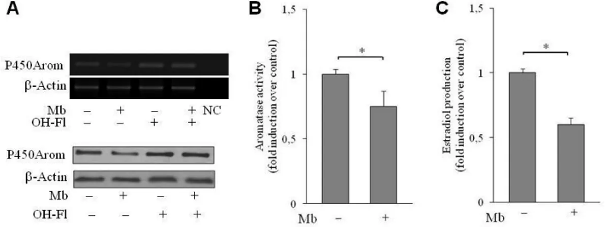
On the other hand, Mb treatment determined a decrease of aromatase mRNA and protein levels, which was completely abrogated by contemporary administration of the androgen antagonist Hydroxiflutamide (Fig. 11A).
Reduction of aromatase enzyme levels in R2C cells upon Mb treatment was paralleled by decreased aromatase enzymatic activity and estradiol production (Fig. 11B and C).
Figure 11. Androgen-dependent modulation of P450 aromatase in R2C cells (A) Modulation of aromatase expression by Mb. Cells were treated with 10-8 M Mb and/or 10-6 M OH-Fl and aromatase mRNA (upper panel) or protein content (lower panel) were detected by RT–PCR or WB analyses, respectively. β-Actin mRNA, internal standard; NC, negative control. β-Actin protein, loading control. Results are representative of three independent experiments.
Modulation of aromatase activity (B) and estradiol production (C) by Mb. Aromatase activity
and Estradiol production were assessed as described in materials and methods. The values represent the mean ± S.D. of three different experiments each performed with triplicate samples. (*, p < 0.01; **, p < 0,05).
To determine whether Mb treatment might negatively influence PII aromatase promoter transcriptional activity, transient transfection experiments were performed in R2C cells. As shown in Fig. 12A, Mb administration was able to determine a significant reduction of promoter activity. Disruption of the SF-1 binding site within the PII aromatase promoter resulted in the loss of the inhibitory effect exerted by Mb, evidencing that regulation by androngens requires the SF-1 motif. Additionally, ChIP assays indicated that DAX-1 accumulated at the SF-1 motif containing region of the PII aromatase promoter upon Mb treatment concomitantly with an equivalent increase in the occupancy of the corepressor N-CoR (Fig. 12B). These results underline the contribution of DAX-1 in the negative androgen-dependent modulation of aromatase gene.
Figure 12. Androgen-dependent modulation of P450 aromatase promoter in R2C cells (A) Mb modulates aromatase promoter activity. Cells were transiently transfected with wt- PII- or a SF-1
mutant PII- (mut PII) aromatase promoter reporter plasmids and treated with 10-8 M Mb for 24h. *, P<0.05; n.s., nonsignificant. (B) “In vivo” DAX-1 recruitment onto the P450 aromatase
promoter. Sheared chromatin from MCF-7 cells treated with 10-8 M Mb for 2h, was precipitated using anti-DAX-1 or anti-N-CoR antibodies. IgG, control samples. The investigated sequence was detected by PCR using specific primers. DNA Inputs were amplified as loading controls. Results are representative of three independent experiments.
AR/DAX-1/Aromatase expression pattern in male Fisher rats
To evaluate the biological significance of the up-regulation of DAX-1 and the inhibition of aromatase levels exerted by ligand-activated AR, the expression pattern of DAX-1, aromatase and AR was investigated in male Fisher rats. To this aim immunoblotting analysis was performed on testes tissues from younger (Fisher Rats Normal Testes: FRNT) and older (Fisher Rats Tumor Testes: FRTT) Fisher rats. The latter group have a high incidence of spontaneously developing Leydig cell tumors. Worthily, as shown in Fig. 13, DAX-1 protein was highly expressed in FRNT but scarcely detectable in FRTT. A similar expression pattern was displayed by AR. On the contrary, a strongly increased aromatase immunoreactivity could be observed in FRTT compared to younger FRNT.
Figure 13. Expression analysis of AR, DAX-1 and P450 aromatase in Fisher rats. Total proteins
extracted from R2C cells or from tissues of normal (FRNT) or tumoral (FRTT) Fisher rat testes were subjected to WB analysis to detect DAX-1, P450aromatase and AR expression levels. GAPDH levels were assessed as loading control. Single representative result.
DISCUSSION
In human hormone sensitive neoplasms such as breast and Leydig cell tumors, estrogens can be de novo produced (7,76-78), indicating an essential role of the androgen peripheral aromatization by P450 aromatase. Accordingly, aromatase inhibitors are currently established as the gold standard for the treatment for ER-positive breast cancers but resistance to the therapy still remains to be solved by other modes of suppression of intra-tumoral estrogen production (11). Hence, analysis of the modulation of intratumoral aromatase appears to be crucial not only for a better comprehension of the development and the biological behaviour of estrogen-dependent carcinomas, but also in the clinical management of cancer patients, since aromatase suppression by new agents may provide improvement in tumor endocrine therapy. Here we demonstrate a novel androgen receptor-mediated mechanism leading to the activation of the DAX-1 gene transcription which in turn negatively impacts on aromatase expression and activity in two distinct estrogen-related neoplasms, such as breast cancer and testicular Leydig cell tumor.
Orphan nuclear receptor DAX-1 has key roles in the development and the maintenance of reproductive function and hormone biosynthesis in mammals (53,79-82). However DAX-1 functions and regulation in sex steroid-dependent neoplasms, in which in situ steroid production and metabolism holds a crucial role, need to be further clarified. In node-negative breast cancer, DAX-1 expression correlates with positivity for ER, PR and AR, smaller tumor size (23,24,83) and excellent survival (84). Nevertheless, although several studies established DAX-1 as a crucial regulator of steroidogenesis, the precise molecular mechanism behind DAX-1 transcriptional control and/or its relationship to aromatase expression and activity in breast cancer cells still remains largely unknown. Our study identifies DAX-1 as an androgen-responsive gene, whose expression is positively modulated by ligand-activated AR. Indeed, in MCF-7 breast cancer cells, DAX-1 expression is up-regulated by Mb administration as evidenced by enhancement of its mRNA and protein levels, and increase of its promoter activity. These results are supported by previous observations in rat Sertoli cells indicating that DAX-1 expression is
regulated during spermatogenesis and peaks during the androgen-sensitive phase of the spermatogenic cycle (85). Activated AR has a fundamental role in the up-regulation of DAX-1 cellular levels since in AR-negative HEK-293 cells, induction of DAX-1 promoter activity can be evidenced only in the presence of exogenous AR expression and strictly depends on the integrity of the AR DNA binding domain, suggesting the existence, in the DAX-1 promoter, of putative androgen responsive region(s) which mediate androgen response. In the current study we identified an androgen response element (DAX-1-ARE: 5’-AATATCacaTGTTCT-3’) located between -1074bp to -1060bp upstream of the DAX-1 gene translation initiation codon which displays the conventional inverted repeats of the 5’-TGTTCT-3’ monomer-binding element found in the promoters of several androgen-responsive genes (71) such as prostate specific antigen (86), p21 (87) and slp-HRE3 (88). This DAX-1-ARE is functional, as demonstrated by transactivation studies, and capable to bind efficiently the AR, in a ligand-dependent manner. Furthermore, the in vivo interaction between AR and the DAX-1 promoter is supported by ChIP analysis showing that AR occupancy of the DAX-1-ARE containing promoter region is concomitant with an increase in RNA Pol II recruitment, consistent with the enhanced DAX-1 transcriptional activity. Thus, activated AR may directly regulate or modulate the expression of DAX-1 and hence it may lie upstream of DAX-1 in a regulatory cascade directing steroid hormones biosynthesis in various steroidogenic tissues. Indeed, DAX-1 plays significant roles in the regulation of steroidogenesis through a variety of mechanisms of transcriptional repression and interaction with many other factors. Interestingly, studies on Dax-1 deficient mice, displaying an increased aromatase expression, indicate that Cyp19 is a physiologic target for DAX-1 in Leydig cells (56). Consistent with these observations, we found that androgen-dependent up-regulation of DAX-1 significantly impacts on aromatase content in breast cancer cells as demonstrated by the evidence that silencing DAX-1 gene completely reversed the down-regulatory effect exerted by androgens on aromatase expression. Transcriptional control by DAX-1 occurs through its recruitment on the SF-1/LRH-1 binding site within the PII/1.3 aromatase proximal promoter in association with the transcriptional co-repressor N-CoR, leading to inhibition of aromatase gene transcription. The importance of our results is highlighted by
recent studies demonstrating that biologically active DHT is locally produced in breast carcinoma tissues (89) and increased by aromatase inhibitors treatment (90). Interestingly, intratumoral DHT levels are positively associated with AR and 5-α-reductase-1 expression but inversely correlated with tumor size, Ki-67 and aromatase expression (89). Thus in AR-positive breast carcinomas the use of aromatase inhibitors may be more effective by accumulation of the local DHT concentration which in turn, acting in an autocrine short loop through DAX-1, might further contribute to reduce aromatase levels (5). Therefore the molecular mechanism we identified might contribute to explain the negative influence of androgens on breast cancer cell proliferation, and well correlates with previous findings in endometrial carcinoma, where loss or decreased DAX-1 expression results in increased intratumoral steroids production and enhanced estrogen-dependent proliferation of cancer cells (61).
Estrogen dependence is also a feature of testicular tumors: excessive estrogen exposure has been shown to alter Leydig cell function, leading to hyperplasia, hypertrophy and Leydig cell adenomas (7,91). The above described androgen-dependent modulation of DAX-1 and aromatase expression, appears to be a more general mechanism since it can also be evidenced in R2C rat Leydig tumor cells in which growth and progression are strongly estrogen-dependent and consequent to a high aromatase expression (76). In the present study we demonstrated that androgen treatment in R2C cells decreases cell proliferation, enhances DAX-1 expression and reduces aromatase cellular levels. According to our findings, DAX-1 disrupted mouse model displays defective spermatogenesis and Leydig cell hyperplasia, possible due to aromatase up-regulation (56). The molecular mechanism responsible for the androgen-dependent inhibition of aromatase expression in R2C cells is transcriptional as well, involving the recruitment of DAX-1 within the SF-1 site containing region of the PII aromatase proximal promoter in association with N-CoR. Direct confirmation of the biological relevance of our findings comes from results obtained in testes tissues from younger (FRNT) and older (FRTT) Fisher rats. The latter group develops spontaneously Leydig cell neoplasma (92,93), a phenomenon not observed in young animals. Indeed, immunoblotting analysis reveals an inverse relationship between DAX-1 and aromatase expression. Specifically, FRNT are characterized by high levels of DAX-1 protein and very
low levels of aromatase enzyme. On the contrary, Leydig tumor development in FRTT is associated with an opposite expression pattern showing a barely detectable DAX-1 protein and a strongly increased aromatase immunoreactivity. Interestingly, DAX-1 expression pattern is paralleled by a similar mode of expression of the AR.
Collectively, our model for AR-mediated repression of aromatase in estrogen-related cancers (Fig. 14) proposes that ligand-activated AR directly binds to the DAX-1 promoter resulting in increased DAX-1 gene expression. In turn, DAX-1 represses aromatase through its recruitment within the SF-1/LRH-1 containing region of the aromatase promoter, causing reduced in situ estrogen production and inhibition of cell proliferation.
Our study identifies DAX-1 as an androgen target gene and provides new insights into the AR/DAX-1/aromatase interplay. Thus, targeting DAX-1 and AR signalling may represent an exploitable pharmacological tool in estrogen-related cancers, opening new avenues for therapeutic intervention.
Figure 14. Proposed model for AR/DAX-1/Aromatase interplay in estrogen-related cancer cells. (1) In the absence of androgens, P450 aromatase is regulated by several factors sustaining
gene transcription and aromatase protein expression leading to (2) enhanced in situ estrogen (E2) production responsible for cancer cell proliferation. In the presence of androgens, ligand-activated AR (3) binds to a specific ARE within the DAX-1 promoter gene triggering its activation. In turn, increased DAX-1 protein (4) is recruited onto the P450 aromatase proximal promoter and represses SF-1/LHR-1-mediated transcription, reducing locally produced estrogens thus affecting cancer cell proliferation (5).
REFERENCES
1. Pasqualini, J.R. and Chetrite, G.S. (2005) Recent insight on the control of enzymes involved in estrogen formation and transformation in human breast cancer. The Journal of steroid biochemistry and molecular biology, 93, 221-236. 2. Sasano, H., Nagasaki, S., Miki, Y. and Suzuki, T. (2009) New developments in
intracrinology of human breast cancer: estrogen sulfatase and sulfotransferase. Annals of the New York Academy of Sciences, 1155, 76-79.
3. Lopez-Otin, C. and Diamandis, E.P. (1998) Breast and prostate cancer: an analysis of common epidemiological, genetic, and biochemical features. Endocrine reviews, 19, 365-396.
4. Nakamura, Y., Suzuki, T., Nakabayashi, M., Endoh, M., Sakamoto, K., Mikami, Y., Moriya, T., Ito, A., Takahashi, S., Yamada, S. et al. (2005) In situ androgen producing enzymes in human prostate cancer. Endocrine-related cancer, 12, 101-107.
5. Takagi, K., Miki, Y., Nagasaki, S., Hirakawa, H., Onodera, Y., Akahira, J., Ishida, T., Watanabe, M., Kimijima, I., Hayashi, S. et al. (2010) Increased intratumoral androgens in human breast carcinoma following aromatase inhibitor exemestane treatment. Endocrine-related cancer, 17, 415-430.
6. Chen, G.G., Zeng, Q. and Tse, G.M. (2008) Estrogen and its receptors in cancer. Medicinal research reviews, 28, 954-974.
7. Fowler, K.A., Gill, K., Kirma, N., Dillehay, D.L. and Tekmal, R.R. (2000) Overexpression of aromatase leads to development of testicular leydig cell tumors : an in vivo model for hormone-mediated TesticularCancer. Am J Pathol, 156, 347-353.
8. Henderson, I.C. and Canellos, G.P. (1980) Cancer of the breast: the past decade (first of two parts). The New England journal of medicine, 302, 17-30.
9. Jemal, A., Siegel, R., Ward, E., Hao, Y., Xu, J. and Thun, M.J. (2009) Cancer statistics, 2009. CA: a cancer journal for clinicians, 59, 225-249.
10. Yager, J.D. and Davidson, N.E. (2006) Estrogen carcinogenesis in breast cancer. The New England journal of medicine, 354, 270-282.
11. Sasano, H., Miki, Y., Nagasaki, S. and Suzuki, T. (2009) In situ estrogen production and its regulation in human breast carcinoma: from endocrinology to intracrinology. Pathology international, 59, 777-789.
12. Jordan, V.C. and Brodie, A.M. (2007) Development and evolution of therapies targeted to the estrogen receptor for the treatment and prevention of breast cancer. Steroids, 72, 7-25.
13. Forbes, J.F., Cuzick, J., Buzdar, A., Howell, A., Tobias, J.S. and Baum, M. (2008) Effect of anastrozole and tamoxifen as adjuvant treatment for early-stage breast cancer: 100-month analysis of the ATAC trial. The lancet oncology, 9, 45-53.
14. Zhou, J., Ng, S., Adesanya-Famuiya, O., Anderson, K. and Bondy, C.A. (2000) Testosterone inhibits estrogen-induced mammary epithelial proliferation and suppresses estrogen receptor expression. FASEB journal : official publication of the Federation of American Societies for Experimental Biology, 14, 1725-1730.
15. Dimitrakakis, C., Zhou, J., Wang, J., Belanger, A., LaBrie, F., Cheng, C., Powell, D. and Bondy, C. (2003) A physiologic role for testosterone in limiting estrogenic stimulation of the breast. Menopause, 10, 292-298.
16. Yeh, S., Hu, Y.C., Wang, P.H., Xie, C., Xu, Q., Tsai, M.Y., Dong, Z., Wang, R.S., Lee, T.H. and Chang, C. (2003) Abnormal mammary gland development and growth retardation in female mice and MCF7 breast cancer cells lacking androgen receptor. The Journal of experimental medicine, 198, 1899-1908.
17. Eigeliene, N., Elo, T., Linhala, M., Hurme, S., Erkkola, R. and Harkonen, P. (2012) Androgens inhibit the stimulatory action of 17beta-estradiol on normal human breast tissue in explant cultures. The Journal of clinical endocrinology and metabolism, 97, E1116-1127.
18. Quigley, C.A., De Bellis, A., Marschke, K.B., el-Awady, M.K., Wilson, E.M. and French, F.S. (1995) Androgen receptor defects: historical, clinical, and molecular perspectives. Endocrine reviews, 16, 271-321.
19. New, M.I. (2004) An update of congenital adrenal hyperplasia. Annals of the New York Academy of Sciences, 1038, 14-43.
20. Lobaccaro, J.M., Lumbroso, S., Belon, C., Galtier-Dereure, F., Bringer, J., Lesimple, T., Heron, J.F., Pujol, H. and Sultan, C. (1993) Male breast cancer and the androgen receptor gene. Nature genetics, 5, 109-110.
21. Chamberlain, N.L., Driver, E.D. and Miesfeld, R.L. (1994) The length and location of CAG trinucleotide repeats in the androgen receptor N-terminal domain affect transactivation function. Nucleic acids research, 22, 3181-3186.
22. Giguere, Y., Dewailly, E., Brisson, J., Ayotte, P., Laflamme, N., Demers, A., Forest, V.I., Dodin, S., Robert, J. and Rousseau, F. (2001) Short polyglutamine tracts in the androgen receptor are protective against breast cancer in the general population. Cancer research, 61, 5869-5874.
23. Park, S., Koo, J., Park, H.S., Kim, J.H., Choi, S.Y., Lee, J.H., Park, B.W. and Lee, K.S. (2010) Expression of androgen receptors in primary breast cancer. Annals of oncology : official journal of the European Society for Medical Oncology / ESMO, 21, 488-492.
24. Moinfar, F., Okcu, M., Tsybrovskyy, O., Regitnig, P., Lax, S.F., Weybora, W., Ratschek, M., Tavassoli, F.A. and Denk, H. (2003) Androgen receptors frequently are expressed in breast carcinomas: potential relevance to new therapeutic strategies. Cancer, 98, 703-711.
25. Agoff, S.N., Swanson, P.E., Linden, H., Hawes, S.E. and Lawton, T.J. (2003) Androgen receptor expression in estrogen receptor-negative breast cancer. Immunohistochemical, clinical, and prognostic associations. Am J Clin Pathol, 120, 725-731.
26. Soreide, J.A., Lea, O.A., Varhaug, J.E., Skarstein, A. and Kvinnsland, S. (1992) Androgen receptors in operable breast cancer: relation to other steroid hormone receptors, correlations to prognostic factors and predictive value for effect of adjuvant tamoxifen treatment. European journal of surgical oncology : the journal of the European Society of Surgical Oncology and the British Association of Surgical Oncology, 18, 112-118.
27. Kimura, N., Mizokami, A., Oonuma, T., Sasano, H. and Nagura, H. (1993) Immunocytochemical localization of androgen receptor with polyclonal
antibody in paraffin-embedded human tissues. The journal of histochemistry and cytochemistry : official journal of the Histochemistry Society, 41, 671-678. 28. Sauter, E.R., Lininger, J., Magklara, A., Hewett, J.E. and Diamandis, E.P. (2004)
Association of kallikrein expression in nipple aspirate fluid with breast cancer risk. International journal of cancer. Journal international du cancer, 108, 588-591.
29. Sauter, E.R., Tichansky, D.S., Chervoneva, I. and Diamandis, E.P. (2002) Circulating testosterone and prostate-specific antigen in nipple aspirate fluid and tissue are associated with breast cancer. Environmental health perspectives, 110, 241-246.
30. Schippinger, W., Regitnig, P., Dandachi, N., Wernecke, K.D., Bauernhofer, T., Samonigg, H. and Moinfar, F. (2006) Evaluation of the prognostic significance of androgen receptor expression in metastatic breast cancer. Virchows Archiv : an international journal of pathology, 449, 24-30.
31. Rakha, E.A., El-Sayed, M.E., Green, A.R., Lee, A.H., Robertson, J.F. and Ellis, I.O. (2007) Prognostic markers in triple-negative breast cancer. Cancer, 109, 25-32. 32. Castellano, I., Allia, E., Accortanzo, V., Vandone, A.M., Chiusa, L., Arisio, R.,
Durando, A., Donadio, M., Bussolati, G., Coates, A.S. et al. (2010) Androgen receptor expression is a significant prognostic factor in estrogen receptor positive breast cancers. Breast cancer research and treatment, 124, 607-617. 33. Park, I.H., Ro, J., Park, S., Lim, H.S., Lee, K.S., Kang, H.S., Jung, S.Y. and Lee, S.
(2012) Lack of any association between functionally significant CYP2D6 polymorphisms and clinical outcomes in early breast cancer patients receiving adjuvant tamoxifen treatment. Breast cancer research and treatment, 131, 455-461.
34. Chae, B.J., Lee, A., Bae, J.S., Song, B.J. and Jung, S.S. (2011) Expression of nuclear receptor DAX-1 and androgen receptor in human breast cancer. Journal of surgical oncology, 103, 768-772.
35. Yu, Q., Niu, Y., Liu, N., Zhang, J.Z., Liu, T.J., Zhang, R.J., Wang, S.L., Ding, X.M. and Xiao, X.Q. (2011) Expression of androgen receptor in breast cancer and its significance as a prognostic factor. Annals of oncology : official journal of the European Society for Medical Oncology / ESMO, 22, 1288-1294.
36. Peters, A.A., Buchanan, G., Ricciardelli, C., Bianco-Miotto, T., Centenera, M.M., Harris, J.M., Jindal, S., Segara, D., Jia, L., Moore, N.L. et al. (2009) Androgen receptor inhibits estrogen receptor-alpha activity and is prognostic in breast cancer. Cancer research, 69, 6131-6140.
37. Lin Fde, M., Pincerato, K.M., Bacchi, C.E., Baracat, E.C. and Carvalho, F.M. (2012) Coordinated expression of oestrogen and androgen receptors in HER2-positive breast carcinomas: impact on proliferative activity. Journal of clinical pathology, 65, 64-68.
38. Berns, E.M., Dirkzwager-Kiel, M.J., Kuenen-Boumeester, V., Timmermans, M., Verhoog, L.C., van den Ouweland, A.M., Meijer-Heijboer, H., Klijn, J.G. and van der Kwast, T.H. (2003) Androgen pathway dysregulation in BRCA1-mutated breast tumors. Breast cancer research and treatment, 79, 121-127.
39. Haiman, C.A., Brown, M., Hankinson, S.E., Spiegelman, D., Colditz, G.A., Willett, W.C., Kantoff, P.W. and Hunter, D.J. (2002) The androgen receptor CAG repeat
polymorphism and risk of breast cancer in the Nurses' Health Study. Cancer research, 62, 1045-1049.
40. Kollara, A., Kahn, H.J., Marks, A. and Brown, T.J. (2001) Loss of androgen receptor associated protein 70 (ARA70) expression in a subset of HER2-positive breast cancers. Breast cancer research and treatment, 67, 245-253.
41. Ogawa, Y., Hai, E., Matsumoto, K., Ikeda, K., Tokunaga, S., Nagahara, H., Sakurai, K., Inoue, T. and Nishiguchi, Y. (2008) Androgen receptor expression in breast cancer: relationship with clinicopathological factors and biomarkers. International journal of clinical oncology / Japan Society of Clinical Oncology, 13, 431-435.
42. Lanzino, M., De Amicis, F., McPhaul, M.J., Marsico, S., Panno, M.L. and Ando, S. (2005) Endogenous coactivator ARA70 interacts with estrogen receptor alpha (ERalpha) and modulates the functional ERalpha/androgen receptor interplay in MCF-7 cells. J Biol Chem, 280, 20421-20430.
43. Ando, S., De Amicis, F., Rago, V., Carpino, A., Maggiolini, M., Panno, M.L. and Lanzino, M. (2002) Breast cancer: from estrogen to androgen receptor. Molecular and cellular endocrinology, 193, 121-128.
44. de Launoit, Y., Dauvois, S., Dufour, M., Simard, J. and Labrie, F. (1991) Inhibition of cell cycle kinetics and proliferation by the androgen 5 alpha-dihydrotestosterone and antiestrogen N,n-butyl-N-methyl-11-[16' alpha-chloro-3',17 beta-dihydroxy-estra-1',3',5'-(10')triene-7' alpha-yl] undecanamide in human breast cancer ZR-75-1 cells. Cancer research, 51, 2797-2802.
45. Lapointe, J. and Labrie, C. (2001) Role of the cyclin-dependent kinase inhibitor p27(Kip1) in androgen-induced inhibition of CAMA-1 breast cancer cell proliferation. Endocrinology, 142, 4331-4338.
46. Greeve, M.A., Allan, R.K., Harvey, J.M. and Bentel, J.M. (2004) Inhibition of MCF-7 breast cancer cell proliferation by 5alpha-dihydrotestosterone; a role for p21(Cip1/Waf1). Journal of molecular endocrinology, 32, 793-810.
47. Lanzino, M., Sisci, D., Morelli, C., Garofalo, C., Catalano, S., Casaburi, I., Capparelli, C., Giordano, C., Giordano, F., Maggiolini, M. et al. (2010) Inhibition of cyclin D1 expression by androgen receptor in breast cancer cells--identification of a novel androgen response element. Nucleic acids research, 38, 5351-5365.
48. Poulin, R., Baker, D. and Labrie, F. (1988) Androgens inhibit basal and estrogen-induced cell proliferation in the ZR-75-1 human breast cancer cell line. Breast cancer research and treatment, 12, 213-225.
49. Bardoni, B., Zanaria, E., Guioli, S., Floridia, G., Worley, K.C., Tonini, G., Ferrante, E., Chiumello, G., McCabe, E.R., Fraccaro, M. et al. (1994) A dosage sensitive locus at chromosome Xp21 is involved in male to female sex reversal. Nature genetics, 7, 497-501.
50. Iyer, A.K., McCabe, E.R. (2004) Molecular mechanisms of DAX1 action. Molecular Genetics and Metabolism, 83, 60–73.
51. Swain, A., Zanaria, E., Hacker, A., Lovell-Badge, R. and Camerino, G. (1996) Mouse Dax1 expression is consistent with a role in sex determination as well as in adrenal and hypothalamus function. Nature genetics, 12, 404-409.
52. Zanaria, E., Muscatelli, F., Bardoni, B., Strom, T.M., Guioli, S., Guo, W., Lalli, E., Moser, C., Walker, A.P., McCabe, E.R. et al. (1994) An unusual member of the