UNIVERSITA’ DEGLI STUDI DI MESSINA
TESI DI DOTTORATO DI RICERCA IN BIOLOGIA APPLICATA E
MEDICINA SPERIMENTALE
CURRICULUM MEDICINA SPERIMENTALE XXXII CICLO
2-pentadecyl-2-oxazoline the oxazoline of
Palmitoylethanolamide (PEA-OXA) a new
pharmacological strategy for neuropathic pain
Candidato:
Enrico GUGLIANDOLO
Relatore:
Ch.mo Prof.
Salvatore CUZZOCREA
Coordinatore:
Ch.ma Prof.ssa Maria Assunta Lo Gullo
Table of contest
1 INTRODUCTION ... 5
1.1 Neuropathic pain ... 5
1.2 Pathophysiology of Neuropathic Pain ... 6
2 Endocannabinoid system and PEA in neuropathic pain ... 14
2.1 Endocannabinoid system in neuropathic pain ... 14
2.2 Palmytohilethanolamide (PEA) and 2-pentadecyl-2-oxazoline the oxazoline of Palmitoylethanolamide (PEA-OXA) ... 17
3. AIM OF THE STUDY ... 21
4. Matherials and Methods ... 22
4.1 Animal ... 22 4.2 Surgery ... 22 4.3 Experimental groups ... 23 4.4 Histological analysis ... 24 4.7 Tunel staining ... 26 Behavioral testing ... 26
4.8 Thermal hyperalgesia (paw withdrawal test) ... 26
4.9 Mechanical allodynia Dynamic Aesthesiometer ... 27
4.10 Beam Walking ... 27
4.11 Functional studies ... 28
4.12 Materials ... 29
4. 13 Statistical evaluation ... 29
5. Results ... 30
5.1 Effect of PEA-OXA on histological changes after sciatic nerve crush ... 30
5.2 Effect of PEA-OXA on mast cells density and Pain behavioral ... 32
5.3 Effect of PEA-OXA on inflammatory response after sciatic nerve crush ... 34
5.4 Effect of PEA-OXA on astrocytes and microglia activation after sciatic nerve crush ... 36
5.5 Effetti dell’Adelmidrol sull’espressione di nitrotirosina, PAR e sulla perossidazione lipidica . 38 5.6 Effect of PEA-OXA on NGF expression and functional recovery after sciatic nerve crush ... 40
6. DISCUSSION ... 42
Abstract
Background: Animal models of sciatic nerve injury are commonly used to study neuropathic pain as
well as axon regeneration. Inflammation/immune response at the site of nerve lesion is known to be an essential trigger of the pathological changes that have a critical impact on nerve repair and regeneration, moreover the damage to peripheral nerve can cause a loss of sensory function and produces a persistent neuropathic pain. N-acylethanolamines (NAEs) involve a family of lipid molecules existent in animal and plant, of which N-Palmitoylethanolamide (PEA) that arouses great attention owing to its anti-inflammatory, analgesic and neuroprotective activities. The modulation of specific amidases for NAEs (and in particular NAE-hydrolysing acid amidase NAAA, which is more selective for PEA) could be a condition to preserve its levels. Here we investigated, in a mice model of sciatic nerve crush, the effect of 2-Pentadecyl-2-oxazoline (PEA-OXA) the oxazoline of PEA, that reportedly modulates activity of NAAA.
Methods: In this experimental model the mice, following the sciatic nerve crush, were treated daily
with OXA at a dose of 10 mg \ Kg for 14 days. Therefore, we evaluated the effects of PEA-OXA on the degree of injury, on the inhibition of neuropathic pain, and on the inflammatory process, as in the improvement of reparative processes and therefore in the restoration of locomotor function.
Results: Ours results showed that PEA-OXA (10mg/kg) treatment, daily, for 14 days after sciatic
nerve crush, have an anti-inflammatory and neuroprotective effect, and moreover have an analgesic protective effect on hypersensitivity, and improve the functional recovery after nerve crush.
Conclusions: Therefore, treatment with PEA-OXA as a whole has shown a protective effect, which
makes it a powerful candidate for the treatment of peripheral nerve injury and neuropathic pain.
List of abbreviations:
NAAA: NAE-hydrolysing acid amidase PEA: N-Palmitoylethanolamide
NAEs: N-acylethanolamines
PEA-OXA: 2-pentadecyl-2-oxazoline of Palmitoylethanolamide SFI: sciatic functional index (SFI)
GFAP: Glial fibrillary acidic protein
Iba1: ionized calcium binding adaptor molecule 1 NGF: nerve growth factor
Ikb-a: kappa-B inhibitor alpha Nf-kb: Nuclear factor kappa-B TNF-a: Tumor Necrosis Factor alpha IL-1b: Interleukin 1 beta
1 INTRODUCTION
1.1 Neuropathic pain
Pain generally can be divided into nociceptive, inflammatory and neuropathic pain, even if, there is no clear distinction between the latter. Nociceptive pain is produced by nociceptors activation in response to a nociceptor stimulus signalling or tissue damage. After tissue damage, the damaged cells and inflammatory cells recruited to the site release several mediators including cytokines and chemokines leading to a reduction of the nociceptors activation threshold and thus making the damaged area more sensible to any stimuli. The pain generated during this process is called inflammatory pain (Woolf, 2010). Neuropathic pain differs from the other types of pain because it does not have a physiological role. Neuropathic pain is one of the most debilitating and disabling chronic pain states, often generated by a multi-factor causes such as, diabetes, anti-cancer therapies, nerve injury due to traumatic injury, infection, autoimmune diseases, surgery, diabetes mellitus or bone compression in cancer, alone on in conjunction. Therefore, it is different from the other types of pain, because it involves the participation of several systems and should be viewed as a multi-factorial disease that needs a new pharmacological strategy to achieve the desired pain relief, also because the conventional analgesics agents are not very effective in the neuropathic pain treatment (Maia, 2017). Commons symptoms of neuropathic pain may also include excruciating pain, pins and needles, difficulty correctly sensing, and numbness and this kind of pain is usually described as a burning sensation and affected. Typically, the neuropathic pain may be constant, or it may occur randomly. Chronic pain is also often accompanied also by short-term memory deficit, and\or depression (Hart, Martelli, & Zasler, 2000). Current pharmacological therapeutic strategies include the use of antidepressants, anticonvulsants, topical lidocaine, non-steroidal anti-inflammatory drugs (NSAIDs) as well as the use of opioids. But these therapies are often limited by different side effects both at the CNS level and not, such as gastrointestinal disorders, risk of renal and hepatic injury, and also somnolence and dizziness, and for the latter, the potential for drug dependence (Finnerup et al.,
2015; Gahr, Freudenmann, Hiemke, Kolle, & Schonfeldt-Lecuona, 2013). Moreover, numerous clinical studies and meta-analyses highlighted how the current pharmacological strategies for the control of neuropathic pain are often ineffective or at least have a low impact in improving the quality of life of these patients (Costigan, Scholz, & Woolf, 2009; Finnerup et al., 2015; Finnerup, Sindrup, & Jensen, 2010). All these studies indicate the need for novel therapies to treat neuropathic pain that is demonstrated by the analysis of analgesia success. Peripheral nerve injuries and diseases often lead to pain persisting beyond the resolution of damage, indicating an active disease-promoting process, which may result in chronic pain. This is regarded as a maladaptive mechanism resulting from neuroinflammation that originally serves to promote regeneration and healing. The impact of nerve damage is a change in nerve function both at the site of the injury and areas around it.
1.2 Pathophysiology of Neuropathic Pain
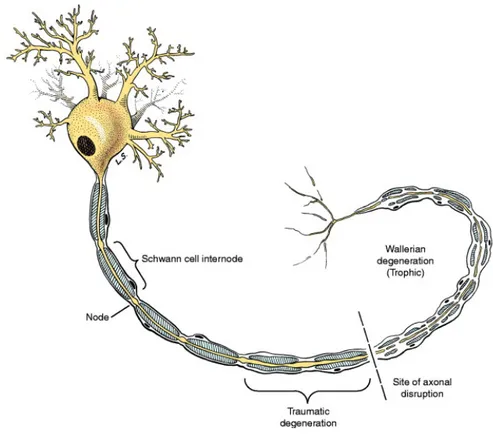
Several studies have shown that neuropathic pain often caused by a lesion or a disease that affects the somatosensory system and involves the nervous system and is supported by a cascade of inflammatory processes. In fact, diffusible inflammatory mediators released both by resident and infiltrating immune cells, and by immune-like glial cells, can directly activate or sensitize nociceptors, thus causing an aberrant activity in the nociceptive system, causing symptoms of neuropathic pain. Injury to peripheral nerves, are often due to compression, cutting and through a variety of trauma, or ischemic and metabolic disorder, injury to nerves initiates a cascade of events, that includes the degeneration of the distal part of the nerve, the increase in infiltration of inflammatory cells such as macrophages, events that are part of a complex mechanism defined as Wallerian degeneration (Koeppen, 2004). The immune response and the consequent inflammatory process play a key role in the pathogenesis of chronic pain, in fact one of the five cardinal signs of inflammation is pain, that in the presence of excessive imbalance in favour of inflammatory processes, the pain can become persistent and become chronic even, after the injury be healed. In
fact, a goal of modern medicine is to find an effective treatment in the management of chronic neuropathic pain (Gureje, Von Korff, Simon, & Gater, 1998).
Figura 1.1- Wallerian degeneration of an LMN
(From: Veterinary Neuroanatomy and Clinical Neurology; Third Edition, 2009. de Lahunta Et al.)
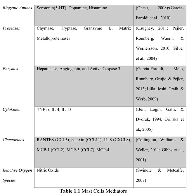
Primary peripheral nerve injury results from compression, focal contusion, traction or transection of the nerve. This process is followed by secondary ischemic injury, which is dominated by inflammation (also referred to as degeneration). Cytokines, and in particular, TNF-α, play a major role in this inflammatory response and also the glial cells and neurons participate in this cascade. Generally, the degeneration is followed by regeneration and recovery process. Glial cells have been shown to activate microglial cells and astrocytes in the regional inflammation area (Scholz & Woolf, 2007). A key role on this inflammatory cascade is played by the inflammatory cytokines, among the first mediators and activators of the inflammatory response to be involved. In particular, TNF-α is released by the Schwann cells, mast cells, endothelial cells, and fibroblasts after injury and plays a key role in inducing the upregulation of other inflammatory cytokines. Several studies reported how
the suppression of inflammatory cytokines after experimental peripheral nerve injury was shown to have a positive effect on neurological recovery (Ozbek et al., 2017). In response to disease and/or trauma a key event is the neural-immune interactions (Steinman, 2004). Immune activation and the subsequent release of immune mediators in the peripheral nervous system have important effects on nerve degeneration and regeneration. A variety of peripheral immune cells, including mast cells, macrophages, and lymphocytes, reside in peripheral nerves and/or are recruited to sites of peripheral nerve after injury (Watkins, Hutchinson, Milligan, & Maier, 2007) . In particular mast cells are a powerful immunological cell, they are distributed in several tissues in different part of the body. And they plays a key role in a great variety of different inflammatory pathological condition (Akin, 2017) (T. Zhang, Finn, Barlow, & Walsh, 2016). In fact since their discovery by Paul Ehrlich in 1878 they have been identified for their granule-laden cytoplasm (Ribatti & Crivellato, 2009). Mast cells in their granules have a huge variety of pro-inflammatory mediators as show in Table 1.1 and the process of degranulation of mast cells is a fundamental step in the inflammatory response and tissue hypersensitization (da Silva, Jamur, & Oliver, 2014). For example considering the sciatic nerve, mast cells are present in the connective tissue epineurial, and within the endoneurial nerve fibres (Olsson, 1968), they also reside in meninges, within the dural layer associated with vessels and meningeal pain receptors (Kaur, Singh, & Jaggi, 2017). Several studies pointed out the role of mast cells in neuropathic pain (Demir et al., 2013; Jaggi, Kaur, Bali, & Singh, 2017; Skaper, 2016).Therefore it is evident that an effective pharmacological strategy against neuropathic pain must take into consideration and include a stabilizing effect on this type of immune cell. But also, other important cell types and meditators are involved in the complex pathophysiology of neuropathic pain and should be consider as a pharmacological target in neuropathic pain management. One of the neuronal mechanisms participating in neuropathic pain development is synaptic plasticity, which contribute to potentiated sensory responses after injury (Zhuo, Wu, & Wu, 2011). The synaptic connectivity is modulated by several neuron-derived neurotransmitters but under certain condition is also modulated by immune mediators released from the CNS-resident microglia and astrocytes as well as from
infiltrating immune cells such as macrophage and mast cells. This neuro-immune interaction is an essential phenomenon that underlies the pathology of neuropathy.
MEDIATORS REFERENCES
Biogenic Amines Serotonin(5-HT), Dopamine, Histamine (Ohtsu, 2008);(Garcia-Faroldi et al., 2010) Proteases Chymase, Tryptase, Granzyme B, Matrix
Metalloproteinases
(Caughey, 2011; Pejler, Ronnberg, Waern, & Wernersson, 2010; Silver et al., 2004)
Enzymes Heparanase, Angiogenin, and Active Caspase 3 (Garcia-Faroldi, Melo, Ronnberg, Grujic, & Pejler, 2013; Lilla, Joshi, Craik, & Werb, 2009)
Cytokines TNF-a, IL-4, IL-15 (Beil, Login, Galli, & Dvorak, 1994; Orinska et al., 2005)
Chemokines RANTES (CCL5), eotaxin (CCL11), IL-8 (CXCL8), MCP-1 (CCL2), MCP-3 (CCL7), MCP-4
(Collington, Williams, & Weller, 2011; Gibbs et al., 2001)
Reactive Oxygen Species
Nitric Oxide (Swindle & Metcalfe, 2007)
Table 1.1 Mast Cells Mediators
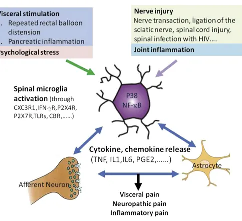
Astrocyte and microglia are immune competent cells (Bilbo, Smith, & Schwarz, 2012) and thus have an active role in regulate the neuronal function such as neurotransmitter release, synaptic connectivity and cell viability (Araque et al., 2014; Kettenmann, Kirchhoff, & Verkhratsky, 2013; Salter & Beggs, 2014). Microglia cell are resident macrophages in the CNS approximately 10% of CNS cells, and they rapidly respond to an injury by proliferating, changing shape and producing pro-inflammatory
cytokines (Taves, Berta, Chen, & Ji, 2013). After a peripherical nerve injury, the microglial activated especially in the spinal cord significantly increase the expressions of CD11b (typically macrophage and microglia marker) and Ionized calcium binding adaptor molecule 1 (Iba-1) (Suter, Wen, Decosterd, & Ji, 2007) several studies have shown a significant increase in microglial markers and microglial proliferation after sciatic nerve partial ligation, constriction or spared nerve injury (Echeverry, Shi, & Zhang, 2008; Suter et al., 2007). An possible mechanism proposed in microglia evoked pain is the activation of elevated phosphorylation of p38 mitogen-activated protein kinase (MAPK) after a peripherical injury, this phosphorylation of p38 in microglia has been seen to be correlated with the increase of pro-inflammatory cytokines IL-1b, IL-6 and TNF-a levels and also with an increase in activation of Nuclear transcription Factor kB (NF-kB) (Wen, Tan, Cheng, Liu, & Ji, 2011). These increase in IL-1b and TNF-a in dorsal horn neurons is well known as an increase in neuron hyperactivity, effect responsible for central sensitization and pain hypersensitivity (Kawasaki, Zhang, Cheng, & Ji, 2008; Ren & Torres, 2009).
Another glial cell-type that contribute in neuropathic pain are astrocyte cells. Astrocytes have a characteristic star shape with long, slender processes and have a strategic position between the vasculature and neurons where they monitor and modify neuronal activity and transmitter release, they also have contact with synapses, approximately 100,000 synapses for one astrocyte which makes their impact on synaptic transmission significant. A recent study suggest a cross-talk between astrocyte and microglia during a response to inflammatory stimuli (Bian et al., 2014; Miyoshi, Obata, Kondo, Okamura, & Noguchi, 2008) and as seen for microglia cells, after a nerve injury also for the astrocyte cells it is possible to highlight an activation and involvement of this cellular population. After nerve injury is possible to detect in spinal cord an significant increase of a typical marker of activated astrocyte which is the glial fibrillary acidic protein (GFAP), and moreover the activation in astrocyte results in NF-kB activation and the subsequent release of pro-inflammatory cytokines IL-1b, IL-6 and TNF-a (Miyoshi et al., 2008) that as seen previously sensitize the dorsal horn neurones in spinal cord. Thus, appear evident the intricate mechanism that underline the neuropathic pain development. Therefore, the process described so far highlight the role of peripheral and immune cell in response to nerve injury, but once activated the inflammatory response driven so far by molecular mediators like Nf-kB, IL-1b and TNF-a that produces different several changes and the involvement of different pathways.
Figure 1.3 The neuron-astrocyte and microglia interaction in neuroinflammation (Neal &
Richardson, 2018)
As seen previously in response to a nerve injury the activation of NF-kB and the release of proinflammatory cytokines such as IL-1b and TNF-a that produce a hyper sensibilization both at the site of injury and in the spinal cord dorsal horn, this up regulated neuronal activity is also confirmed by the high levels of c-fos protein a marker of neuronal activity (Bullitt, 1990), these protein has been seen increase in response to a nociceptive stimuli (Nemoto et al., 2016) and high level c-fos protein is thus find under neuropathic pain condition (Nemoto et al., 2016; M. Q. Zhang et al., 2017), these process have a clinical manifestation in mechanical allodynia and thermal hyperalgesia, condition observed in both experimental and human neuropathic pain (Jensen & Finnerup, 2014). Thermal hyperalgesia is a condition of altered perception of temperature and means an increased response to a painful stimulus (Jensen & Finnerup, 2014) instead, mechanical allodynia is a painful sensation caused by innocuous stimuli like light touch (Lolignier, Eijkelkamp, & Wood, 2015; Salvat, Yalcin, Muller, & Barrot, 2018). As seen previously the activation of MAPK phosphorylation in spinal cord is a key step in the inflammatory response, but are responsible also of regulation of neural apoptosis (Tsuda, Mizokoshi, Shigemoto-Mogami, Koizumi, & Inoue, 2004) process also involved in pathogenesis of neuropathic pain (Cruz & Cruz, 2007) (Cao, Wang, Ren, & Zang, 2015). After a
nerve injury, high levels of marker of programmed cell dead such as caspase-3, that is responsible for the proteolytic cleavage of many key proteins. (Eldadah & Faden, 2000). Caspase-3 high levels at peripherical levels has been correlated with an impairment in nerve regeneration and with a delayed nerve repair (Saito, Kanje, & Dahlin, 2009) and at spinal cord level caspase-3 has been reported to be correlated with neuropathic pain states (Joseph & Levine, 2004) (Scholz et al., 2005). This imbalance towards the apoptosis process under neuropathic pain condition is also revelated by an increase in the ratio of BAX\Bcl2 pro- and anti-apoptotic protein respectively (Kaeidi et al., 2011) (Amin, Hajhashemi, Abnous, & Hosseinzadeh, 2014) .
2 Endocannabinoid system and PEA in neuropathic pain
2.1 Endocannabinoid system in neuropathic pain
Thus, elucidation of the regulatory components involved in the ensuing inflammatory reaction to neural injury holds great promise in the development of effective treatment strategies to maximize immune elements critical for repair, while simultaneously suppressing aspects of the immune response responsible for further damage to offset disease or injury progression. After peripheral nerve injury, axonal regeneration does not always allow for adequate functional recovery, and therefore the patients do not recover normal motor control and fine sensibility (Allodi, Udina, & Navarro, 2012) Beyond the immune-inflammatory response described above, there are several systems and pathways that balance the inflammatory action and provide for the “extinguishing” of the inflammation and therefore the return to the basal condition of homeostasis. Therefore, more and more interest is turned to the study and identification of these endogenous anti-inflammatory mediators, and an increasing attention and turned to the study of pharmacological strategies that aim at the strengthening of these pathways. Of all systems participating in neuropathic pain control a particular interest is turned to the endocannabinoid system, both for its other expression both in neurons and in immune cells, interests confirmed also by several preclinical studies both for its other expression both in neurons and in immune cells, interests confirmed also by several preclinical studies (Maldonado, Banos, & Cabanero, 2016). The use of preparations of the Cannabis sativa plant as analgesics has been known for centuries, but only in the 1960s that the major active constituent (Δ9-tetrahydrocannabinol) was
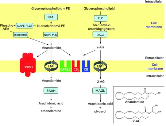
characterized (Mechoulam & Gaoni, 1967) and only in 1990s the molecular targets were discovered, the cannabinoid receptor CB1 And CB2 (Devane, Dysarz, Johnson, Melvin, & Howlett, 1988). This discovery suggest that endogenous molecules are present in the mammalian body that could stimulate or inhibit these receptors. And so the endogenous mediators of this system have been identified, which are lipid molecules of which the most important are the arachidonoylethanolamide also called anandamide and 2-arachidonoyl glycerol (2-AG) (Howlett et al., 2002)
Figure 2.1.1 Biosynthesis and breakdown of the two best-studied endocannabinoids, anandamide
and 2-arachydonoylglycerol (2-AG).
This lipidic molecules are produced on demand (Varma, Carlson, Ledent, & Alger, 2001) from cell membrane phospholipid precursors (Di Marzo, Bisogno, Sugiura, Melck, & De Petrocellis, 1998). And several studies showed how these mediators levels are increase in experimental model of neuropathic pain, and has been demonstrated the effect of CB1\CB2 agonist in modulation of neuropathic pain (Masocha, 2018). As show in figure 2.1 different enzymes are involved in endocannabinoid biosynthesis [N-acyl-phosphatidylethanolamine-specific phospholipase D (NAPE-PLD),α/β-hydrolase domain type-4 (Abdh4),glycerophosphodiesterase-1 (GDE1), protein tyrosine phosphataseN22 (PTPN22), for AEA; and diacylglycerol lipase-αor -β(DAGLαand DAGLβfor 2-AG] or degradation [fatty acid amide hydrolase-1(FAAH) for AEA; and monoacylglycerol lipase (MAGL),α/β-Hydrolase Domain Containing Protein 6 and 12 (ABDH6 and 12), and FAAH-1 for2-AG . These components are expressed almost ubiquitously throughout nociceptive pathways, and
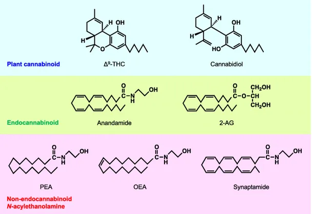
thus targeting the system via exogenous cannabinoid ligands or enhancement of endogenous signaling can regulate nociceptive signaling at multiple sites. More and more interest are directed towards mediators who are able to resolve the inflammatory process, in particular different lipid mediators have been reached able to "extinguish the inflammatory process". and then start the inflammation resolution processes (Buckley, Gilroy, Serhan, Stockinger, & Tak, 2013). Therefore, in the context of these studies aimed at the characterization of the endocannaboid system, it emerged that. In addition to anandamide also some ethanolamides of polyunsaturated fatty acids such as dihomo-γ-linolenic acid (C20: 3 ω6), mead acid (C20: 3 ω9), and adrenic acid (C22: 4), showed the ability to bind cannabinoid receptors (Mechoulam et al., 1996). Interestingly, it has been seen that the N-acetyl ethanolamides of saturated or mono unsaturated fatty acids, show no binding activity with cannabinoid receptors, but show important biological activities through different receptors. Of this group of substances belong, N-arachidonoylethanolamide (an endocannabinoid), and its congeners N-stearoyl ethanolamine (C:18:0), N-oleoyl ethanolamine (C18:1), and N-palmitoyl ethanolamine ( C16:0, PEA or Palmitoylethanolamide) that are more abundant than anandamide in most animal tissues (Pacher, Batkai, & Kunos, 2006). Biosynthetic enzymes for N-acylethanolamines so far reported do not show selectivity for anandamide over other N-acylethanolamine species. Thus, anandamide could be concomitantly produced as a kind of by-product of non-endocannabinoid N-acylethanolamines.
Figure 2.1.2 Chemical structures of plant cannabinoids, endocannabinoids and N-acetyl
ethanolamides(K. Tsuboi, Uyama, Okamoto, & Ueda, 2018)
2.2 Palmytohilethanolamide (PEA) and 2-pentadecyl-2-oxazoline
the oxazoline of Palmitoylethanolamide (PEA-OXA)
A molecule of particular interest is the Palmytohilethanolamide (PEA). PEA is a well food
component since 1957, research on PEA has been conducted for more than 50 years, and over 350 papers are referenced in PubMed describing the physiological properties of this endogenous modulator and its pharmacological and therapeutically profile (Keppel Hesselink, de Boer, & Witkamp, 2013). The major focus of PEA research, since the work of the Nobel laureate Levi-Montalcini in 1993, has been neuropathic pain states and mast cell related disorders. It exerts a multitude of physiological functions related to metabolic and cellular homeostasis. The anti-inflammatory effect of PEA has been described in literature since 1939 (Coburn & Moore, 1939). Recently several studies have highlighted how the administration of exogenous PEA is a good
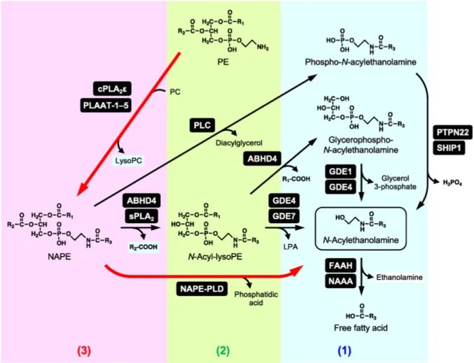
pharmacological strategy against inflammatory and neurodegenerative processes (di Marzo & Skaper, 2013) (Skaper et al., 2015), moreover, the pea has shown that it possesses analgesic properties in different models of neuropathic pain (Guida et al., 2015) (Di Cesare Mannelli et al., 2015), which increases its interest as a new therapeutic approach, also considering its good safety profile (Keppel Hesselink & Kopsky, 2015) (Bertolino et al., 2017). Actually, in the USA and Europe, PEA is currently marketed as a nutraceutical, a food supplement, or a food for medical purposes, depending on the country, which is effective for chronic pain represented by neuropathic pain. PEA is also a constituent of cream marketed for dry, irritated, and reactive skin. In animal tissues, PEA is synthesized through common pathways for N-acylethanolamides and anandamide, starting from glycerophospholipids. Fatty acid ethanolamides are degraded by the N-acylethanolamine acid amidase (NAAA) and fatty acid amide hydrolase (FAAH). FAAH is a membrane-bound serine hydrolase, belonging to the amidase signature family, FAAH hydrolyzes various N-acylethanolamines with a higher reactivity toward anandamide. FAAH is ubiquitously present in various tissues, and FAAH-deficient mice exhibit increased tissue levels of various N-acylethanolamines including anandamide, suggesting the central role of this enzyme in the degradation of N-acylethanolamines (Sultan, Nagarkatti, & Nagarkatti, 2019). NAAA is a lysosomal enzyme hydrolyzing N-acylethanolamines and in contrast with FAAH, that has as main substrate anandamide, the NAAA has as main substrate PEA.
Figure 2.2.1 Metabolism of N-acylethanolamine (K. Tsuboi, Uyama, Okamoto, & Ueda, 2018)
NAA is present in various tissues with an abundant expression in macrophages (Kazuhito Tsuboi, Zhao, et al., 2007). It has been seen that NAAA-deficient animals show an important suppression of the inflammatory response, Thus, NAAA inhibitors may have the therapeutic potential as novel anti-inflammatory drugs (Oscar Sasso et al., 2018). And it has been seen how the inhibition of these enzyme increase the levels of PEA(Kazuhito Tsuboi, Takezaki, & Ueda, 2007) (Starowicz et al., 2013). However, the pharmacological blockage of these enzymes can lead to disorders from the metabolic point of view, since the substrates such as PEA are produced on demand, so it has been seen that from the point of view of the pharmacological approach it is more useful to be modular and not blocking the activity of these enzymes (Skaper et al., 2015).
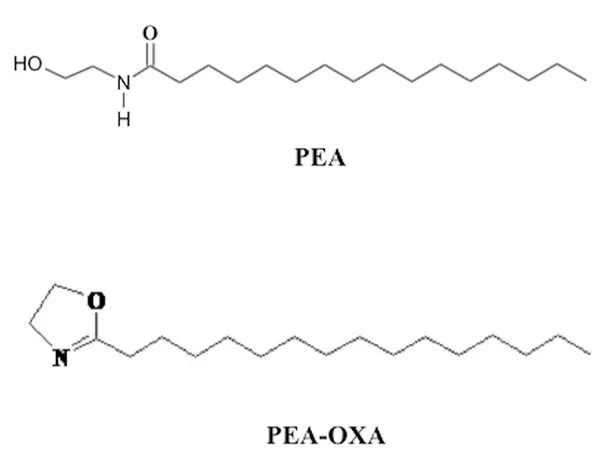
This has been seen to be possible thanks to a modification in the structure of the PEA obtaining the oxazoline, PEA 2-pentadecyl-2-oxazoline of Palmitoylethanolamide (PEA-OXA) (Petrosino et al., 2017), that has been identified in several natural compounds such as coffee, and it has been observed that its effectiveness in reducing inflammation and hyperalgesia is considerably significantly greater compared to PEA (Impellizzeri et al., 2016). The effects of PEA-OXA, on the inhibition of neuro-inflammatory processes as well as in the reduction of oxidative stress damage, have been seen in a mouse model of traumatic brain injury and spinal cord injury (Impellizzeri et al., 2017), and more recently the effects of PEA-OXA in reducing neuroinflammation, oxidative stress associated with vascular dementia in an experimental model of repeated bilateral common carotid arteries occlusion have been evaluated (Impellizzeri et al., 2019).
Figure 2.2.2 Structure of Palmytohilethanolamide (PEA) and 2-pentadecyl-2-oxazoline of
3. AIM OF THE STUDY
Therefore, the aim of this study was to evaluate, for the first time, the effects of PEA-OXA in an experimental mouse model of neuropathic pain induced by sciatic nerve compression. Thus, to evaluate the molecular mechanism of action of this new molecule that combines the already known properties of the PEA, with those of the modulation of its main enzyme delegated to degradation NAAA. And finally, to evaluate the effects of treatment with PEA-OXA, its analgesic component and the effects on the neuroinflammatory process induced in an experimental model of neuropathic pain induced by sciatic nerve compression.
4. Materials and Methods
4.1 Animal
CD1 mice (male 25–30 g; Envigo, Milan, Italy) were placed in a controlled location (room kept at 22 ± 1˚C with a 12-h dark/light cycles) and provided with standard rodent water and chow ad libitum. The animals were familiarized to their setting for one week. The University of Messina Review Board for the care of animals approved the research. All animal experiments observe the regulations in Italy (D.M. 116192) as well as the EU regulations (O.J. of E.C. L 358/1 12/18/1986).
4.2 Surgery
For nerve crush it was conducted with minor changes to what was seen previously (Morrison et al., 2015) ,mice were anesthetized with 2% isoflurane/oxygen, lateral thigh shaved, and a 1 cm incision in the skin made over the lateral femur. The muscle layers were split with blunt scissors, the sciatic nerve localized isolated and crushed just before the bifurcation with an ultra-fine, smooth, straight haemostat (tip width 0.6 mm/ Fine Science Tools) for 30 seconds.
Figure 4.1- A) The normal sciatic nerve; B) Sciatic nerve after crush - injury
4.3 Experimental groups
Mice were randomly divided into the following groups (n = 10 for each group)
Vehicle: mice were subjected to sciatic nerve crush as described above and vehicle (carboxymethylcellulose (CMC) 2.5% p/p in water), was orally administered for 14 days.
PEA\OXA: mice were subjected to sciatic nerve crush as described above and PEA-OXA (10 mg/kg) (dissolved in carboxymethylcellulose (CMC) 2.5% p/p in water) was orally administered daily for 14 days.
SHAM: were subjected to the surgical procedures as above group except that sciatic nerve crush was not applied, and vehicle was administered at 1 after
Sham + PEA-OXA: were subjected to the surgical procedures as above group except that sciatic nerve crush was not applied, and PEA-OXA (10 mg/kg) (dissolved in carboxymethylcellulose (CMC) 2.5% p/p in water) was orally administered daily for 14 days.
As described below mice, (N=10 from each group for each parameter;) were sacrificed 14 days after sciatic nerve crush.
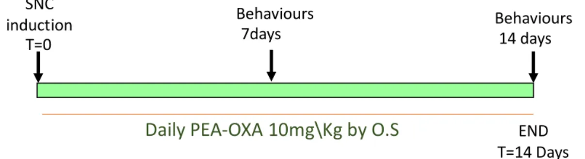
Figure 4.1- General outline of experimental design SNC
induction
T=0 7days
Behaviours Behaviours
14 days
Daily PEA-OXA 10mg\Kg by O.S
END4.4 Histological analysis
At the end of the 14 days, for histological examination standard haematoxylin and eosin (H&E) staining was performed as seen previously (Gugliandolo et al., 2018), sciatic nerve tissue specimens were then observed under an optical microscope (Leica QWin V3, Cambridge, UK). sciatic nerves, scores of 0, 1, 2, 3, and 4 indicate 0, 1–25, 26–50, 51–75, and .75% infiltration, respectively. Identification of Mast Cells was performed in sciatic nerve tissue specimens sections by blue toluidine staining as described previously (Smith et al., 2018).
4.5 Western Blot Analysis
Western blot analysis was performed on the lumbar portion of the spinal cord, the samples
was homogenized in lysis buffer. Cytosolic and nuclear extracts were prepared as described previously (15). The filters were probed with specific Abs: anti- NF-κB p-65 (1:1000; Santa Cruz Biotechnology) or IκB-α (1:1000; Santa Cruz Biotechnology), or anti TNF-a (1:1000; Santa Cruz Biotechnology), or anti IL-1b (1:1000; Santa Cruz Biotechnology), or anti Bax (1:500; Santa Cruz Biotechnology) anti-Bcl-2 (1:500; Santa Cruz Biotechnology), or anti Caspase-3 (1:500; Santa Cruz Biotechnology), or anti c-fos (1:500; Santa Cruz Biotechnology), or anti NGF (1:1000; Santa Cruz Biotechnology), or anti NAAA (1:500 Sigma–Aldrich Corp), or anti b-III-tubulin (1:1000 cell signalling) in 1 × PBS, 5% w/v non-fat dried milk, 0.1% Tween-20 at 4 °C, overnight. To ascertain that blots were loaded with equal amounts of proteins they were also incubated in the presence of the antibody against β-actin protein (cytosolic fraction 1:500; Santa Cruz Biotechnology) or lamin A/C (nuclear fraction 1:500 Sigma–Aldrich Corp.). Signals were detected with enhanced chemiluminescence (ECL) detection system reagent according to the manufacturer's instructions (Thermo, USA). The relative expression of the protein bands was quantified by densitometry with BIORAD ChemiDocTM XRS+software and standardized to β-actin and lamin A/C levels. Images of
blot signals (8 bit/600 dpi resolution) were imported to analysis software (Image Quant TL,v2003). The blot was stripped with glycine 2% and reprobed several times to optimize detection of proteins and to visualize other proteins without the need for multiple gels and transfers.
4.6 Immunofluorescence
After deparaffinization and rehydration, detection of b-III-tubulin was carried out after boiling the tissue sections in 0.1 M citrate buffer for 1 min as described previously (17). Non-specific adsorption was minimized by incubating in 2% (vol/vol) normal goat serum in PBS for 20 min. Sections were incubated with b-III-tubulin primary antibodies (1:400 cell signalling) in a humidified chamber overnight at 37◦C. Sections were then incubated with secondary antibody: fluorescein isothiocyanate-conjugated anti- mouse Alexa Fluor-488 (1:2000, Molecular Probes, Monza, Italy) or Texas Red-conjugated anti-rabbit Alexa Fluor-594 (1:1000, Molecular Probes) for 1 h at 37◦C. For nuclear staining, 2 μg/ml 4′, 6′ -diamidino-2-phenylindole (DAPI; Hoechst, Frankfurt, Germany) in PBS was added. Sections were observed using a Leica DM2000 microscope (Leica, Milan, Italy. Optical sections of fluorescence specimens were obtained using a HeNe laser (543 nm), an ultraviolet laser (361–365 nm) and an argon laser (458nm) at a one-mi, 2s scanning speed with up to eight averages; 1.5 μm sections were obtained using a pinhole of 250. The same settings were used for all images obtained from the other samples that had been processed in parallel. Digital images were cropped, and figure montages prepared using Adobe Photoshop 7.0 (Adobe Systems; Palo Alto, California, United States). The co-localization of images was examined with Image J software (National Institutes of Health) as described previously (Zhou, Masliah, & Spector, 2011).
4.7 Tunel staining
TUNEL staining protocol was according to a Roche protocol. Paraffin-embedded sections were dewaxed in xylene and rehydrated in graded ethanol series to water, permeabilized with citrate buffer 0,1 M and then incubated in TUNEL reaction mixture for 60 min at 37°C in the dark. The tissue was then rinsed in PBS 3 times for 5 min and then observed using an excitation wavelength in the range of 520-560 nm (maximum 540; green) and in the range of 570-620 nm (maximum 580 nm; red).
Behavioural testing
4.8 Thermal hyperalgesia (paw withdrawal test)
To assess hind paw heat sensitivity, test was conducted using a plantar test device (plantar test; Ugo Basile, Italy) (Hargreaves, Dubner, Brown, Flores, & Joris, 1988) as seen previously (Choi et al., 2007). Animals were allowed to freely move within an open-topped transparent plastic box on a glass floor 20 min before the test. A mobile radiant heat source was then placed under the glass floor and focused onto the hind paw. Paw withdrawal latencies were measured with a cut-off time of 15 seconds to prevent tissue damage. The heat stimulation was repeated three times with a 10-min interval to obtain the mean latency of paw withdrawal. Results are expressed as paw-withdrawal latency(s).
4.9 Mechanical allodynia Dynamic Aesthesiometer
Mechanical allodynia was evaluated using the Dynamic Plantar Aesthesiometer (Ugo Basile). This equipment employs a single non-flexible filament (0.5-mm diameter) to apply an increasing force to the plantar surface of the mouse hind paw. Animals were placed in a cage with a wire mesh floor and allowed to acclimatize before testing. The filament was applied to the plantar area of the hind paw and it began to exert an increasing upward force, reaching a maximum of 30 g in 10 s, until the paw was withdrawn. The withdrawal threshold was defined as the force, in grams, at which the mouse withdrew its paw. Withdrawal were determined three times, and the reported value is the mean of the three evaluations.
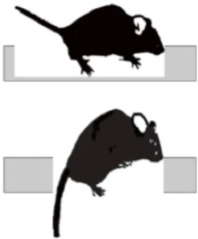
4.10 Beam Walking
Coordination and balance were assessed, with minor changes to what has been seen previously (Taylor, Holdeman, Weltmer, Ryals, & Wright, 2005; Taylor et al., 2001; Zhu, Li, Wang, Jia, & Xu, 2016), by measuring the ability of mice to traverse a wooden beam (1 m x 26 mm), in order to reach a dark, enclosed safety platform containing food and bedding. Mice were trained over two consecutive days (3 trials per day) by placing them at the starting point and allowing them to traverse a 70 cm section of the beam. Once trained, mice were tested using three consecutive trials. Observers were blinded to the study and a video camera was used to record three trials. The time taken for the mouse to cross the full length of the beam to the goal box was recorded for a maximum time of 120 s. If the mouse fall before reaching the goal box, the time was recorded as 120 s. The time spent in a frozen posture and number of right paw slips were also noted. Data from three trials were averaged for each and the difference between the experimental group were analysed by two-way ANOVA followed by Fisher PLSD post hoc tests.
Figure 4.2- Representative images of on the top side, a normal performance, and in the lower side a hid paw slip
4.11 Functional studies
The sciatic functional index (SFI) was evaluated, as previously described, briefly, 7 and 14 days after sciatic nerve crush. The paw prints were recorded by moistening the back legs of each animal with blue ink and making them walk without assistance along a 6 × 44 cm corridor, on white sheets of paper. the animals pawprints were recorded, and two measurements were taken: (i) the print length (PL), corresponding to the distance from the heel to the third toe; and (ii) the toe spread (TS), corresponding to the distance from the first to fifth toe. Both measurements were taken from injured (E, for experimental) as well as no injured (N, for normal) sides, and the SFI was calculated according to the formula:
𝑆𝐹𝐼 = 118.9 )𝐸𝑇𝑆 − 𝑁𝑇𝑆
𝑁𝑇𝑆 . − 51.2 )
𝐸𝑃𝐿 − 𝑁𝑃𝐿
The SFI value varies from 0 to −100, with 0 corresponding to normal function and −100 corresponding to complete dysfunction.
Figure 4.3- Representative image of paw prints from sham mice in blue, and after sciatic nerve
crush in red
4.12 Materials
PEA-OXA was kindly supplied by Epitech Group SpA (Saccolongo, Italy). Unless otherwise stated, all compounds were obtained from Sigma-Aldrich. All solutions used for in vivo infusions were prepared using non-pyrogenic saline (0.9% NaCl; Baxter Healthcare Ltd., Thetford, Norfolk, UK).
4.13 Statistical evaluation
All values are expressed as mean ± standard error of the mean (S.E.M.) of N observations. For in vivo studies N represents the number of mice used. In experiments involving histology, immunohistochemistry and immunofluorescence the figures shown are representative of at least three experiments performed on different days on tissue sections collected from all animals in each group. The results were analysed by one-way ANOVA followed by a Bonferroni post-hoc test for multiple comparisons. A P-value of less than 0.05 was considered significant.
5. Results
5.1 Effect of PEA-OXA on histological changes after sciatic nerve
crush
Sciatic nerve from the sham group showed a normal structure of sciatic nerve (Figure 5.1 A), 14 days after sciatic nerve crush the sciatic nerve from vehicle group exhibited several areas of oedema with an abundant presence of infiltrate and degraded myelin sheets as show in figure 5.1 B, PEA-OXA treatment significantly reduce the presence of oedema and infiltrate (Figure 5.1 C) compared with vehicle group as showed by the Nerve histological score figure 5.1 D. After, as a marker of an efficient nerve repair process we evaluated the expression of b-III-tubulin, both in the spinal cord and in the injured nerve. The immunofluorescent staining b-III-tubulin on longitudinal sections of the sciatic nerve Figure 5.1 E, shows that following crushing of the sciatic nerve and treatment with PEA-OXA there is a significant increase in expression of b-III-tubulin as show in (Figure 1 E), compared to the vehicle group, (Figure 5.1 panels E). Also, western blot analysis on the lumbar portion of spinal cord, showed a significant reduction in b-III-tubulin expression 14 day after sciatic nerve crush, instead PEA-OXA treatment daily for 14 days after sciatic nerve crush, significantly increase b-III-tubulin
expression compared to vehicle group, as show in Figure 5.1 F,
Figura 5.1- Panels A, show the sciatic nerve structure from sham mice. Panels B, nerve from vehicle
group exhibited, 14 days after sciatic nerve crush, the several areas of oedema with an abundant presence of infiltrate and degraded myelin sheets. Panels C, PEA-OXA treatment significantly reduce the presence of oedema and infiltrate, as indicated by nerve histological score panels D. Panels E, showed the immunofluorescent staining for b-III-tubulin on longitudinal sections of the sciatic nerve. following crushing of the sciatic nerve and treatment with PEA-OXA there is a significant increase in expression of b-III-tubulin, compared to the vehicle group. Panels F, F1 showed the western blot analysis of the lumbar portion of the spinal cord showed the expression and relative densitometric analysis for tubulin. The PEA-OXA treatment was able to significantly increase the b-III-tubulin compared with vehicle group. Each data is expressed as Mean ± SEM from N = 10 Mice for each group. *** p < 0.001 vs corresponding sham group. ## p< 0,01 vs vehicle group. ### p < 0.001 vs. vehicle group.
5.2 Effect of PEA-OXA on mast cells density and Pain behavioural
We subsequently assessed whether treatment with PEA-OXA had effects on the number of mast cells, that play a key role in the inflammatory process and in particular in the development of hyperalgesia. As show in figure 5.2, B, 14 days after sciatic nerve crush there is a significant increase in mast cell number, compared with uninjured nerve from sham group figure 5.2 A and D. Daily treatment with PEA-OXA significantly reduce the presence of mast cell compared to vehicle group as showed by figure 5.2 C and D. As show in figure 5.2 Panels E, mice from vehicle group present a significant increase in the response to thermal stimuli, from day 7 to 14 after nerve crush when compared with basal response, also show in figure 5.2 panels E mice treated with PEA-OXA show a significant reduction of their pain threshold compared to vehicle group. Figure 5.2 panel F show, the sciatic nerve crush produced a reduction in the nociceptive threshold to the mechanical stimuli from day 7 to 14 in the operated paw. Instead daily treatment with PEA-OXA at a dose of 10 mg \ Kg significantly increased the thresholds to mechanical stimuli. Therefore, as a marker of the activation of neurons by algesic stimuli we evaluated the expression of c-fos, western blot analysis on the lumbar portion of spinal cord, showed that 14 days after the sciatic nerve crush there is a significantly increase in c-fos expressions in mice from vehicle group. Instead treatment with PEA-OXA was able to significantly reduce the c-fos expression as show in figure 5.2 panels G,G1. As a mechanism of action of PEA-OXA has been proposed, the combination of the beneficial effects of PEA with those of the inhibition of NAAA, the main enzyme responsible for the degradation of PEA(Petrosino et al., 2017). therefore, we evaluated the expression of NAAA at the spinal level, as shown in Figure 5.2 H,H1. 14 days after the crush to sciatic nerve there was a significant increase in the expression of NAAA whereas daily treatment with PEA-OXA at the dose of 10 mg \ Kg significantly reduced the expression of NAAA induced by the sciatic nerve crush.
Figura 5.2- The blue toluidine staining for highlight the Mast cells, panels A for sham group. Panels
B showed that 14 days after sciatic nerve crush there is a significant increase in mast cell number in sciatic nerve from vehicle group. Panels C show how daily treatment with PEA-OXA significantly reduce the presence of mast cell compared to vehicle group as showed by Panels D. Panels E and F showed the analgesic effect of daily treatment PEA-OXA 10 mg\Kg, on thermal hyperalgesia (Plantar test) and mechanical allodynia respectively. Panels G,G1, showed the c-fos expressions by western blot analysis in the lumbar portion of the spinal cord. Panel H,H1, western blot analysis on the lumbar portion of the spinal cord for NAAA expressions. Each data is expressed as Mean ± SEM from N = 10 Mice for each group. *** p < 0.001 vs corresponding sham group. #p < 0.05 vs. vehicle group. ## p< 0,01 vs vehicle group ### p < 0.001 vs. vehicle group.
5.3 Effect of PEA-OXA on inflammatory response after sciatic
nerve crush
As shown by our results, crush to sciatic nerve produce an important inflammatory response, which is a key event both for the development of neuropathic pain and for neurodegenerative processes. In particular, as showed in figure 3 panels A,A1 and B,B1, western blot analysis of the lumbar portion of the spinal cord showed that, 14 days after sciatic nerve crush, in the vehicle group there is an significantly increase in Ikb-a degradation and consequently an increase in Nf-kb translocation to nucleus, this is a key event both for the inflammatory response and for changes in the transcription of different genes. Compared to vehicle group, PEA-OXA treatment significantly reduce the Ikb-a degradation and Nf-kb translocation (Fig 3 A,A1. B,B1). Moreover as show in figure 3 panels C,C1 and D,D1, crush to sciatic nerve produce a significantly increase in proinflammatory cytokines TNF-a TNF-and IL-1b, insteTNF-ad dTNF-aily treTNF-atment with PEA-OXA for 14 dTNF-ays TNF-after sciTNF-atic nerve crush, significantly reduce the expression of TNF-a and IL-1b.
Figura 5.3-Western blot analysis of the lumbar portion of the spinal cord showed the expression and
relative densitometric analysis for: Ikb-a panels A,A1; panels B,B1 for Nf-kb; panels C,C1 for TNF-a TNF-and pTNF-anels D,D1 for IL-1b. ETNF-ach dTNF-atTNF-a is expressed TNF-as MeTNF-an ± SEM from N = 10 Mice for eTNF-ach group. *** p < 0.001 vs corresponding sham. ## p< 0,01 vs vehicle group ###p < 0.001 vs. vehicle group.
5.4 Effect of PEA-OXA on astrocytes and microglia activation after
sciatic nerve crush
Astrocytes and microglia activation play a critical role in neuroinflammation, and in neuropathic pain state. When compared to the sham group (figure 5.4 panels A, for GFAP and panels E for Iba-1, see yellow arrows), immunofluorescence evaluation of Glial fibrillary acidic protein (GFAP) and ionized calcium binding adaptor molecule 1 (Iba1) revealed a significant increasing in GFAP and Iba-1 positive cell as show in figure 5.4 B and F respectively. Treatment with PEA-OXA at dose of 10 mg\Kg post crush to sciatic nerve, significantly reduce the number of positive cells for GFAP and Iba-1 figure 5.4 C and G respectively, compared to the vehicle group. As sow by western blot analysis figure 5.4 D,D1 and H,H1 respectively, also at the spinal cord level we found an increase in GFAP and Iba-1, 14 days after sciatic nerve crush. Instead daily treatment with PEA-OXA significantly reduce the GFAP and Iba-1 expression, (Figure 5.4 D,D1 and H,H1 respectively).
Figura 5.4-The immunofluorescent staining on longitudinal sections of the sciatic nerve for GFAP and Iba-1, showed a significantly increase in GFAP and Iba-1 expression in the vehicle group (panel B and F respectively) compared to sham mice, panel A and E respectively. PEA-OXA treatment significantly reduce the expression for GFAP and Iba-1 compared to vehicle group, panel C and G respectively. The western blot analysis of the lumbar portion of the spinal cord showed the expression and relative densitometric analysis for GFAP and Iba-1, panels D,D1 and H,H1 respectively. Each data is expressed as Mean ± SEM from N = 10 Mice for each group. *** p < 0.01vs corresponding sham. ###p < 0.001 vs. vehicle group
5.5 Effect of PEA-OXA on apoptotic pathway after sciatic nerve
crush
Western blot analysis on the lumbar portion of spinal cord, showed an increase in activation of the apoptotic process. in particular 14 days after sciatic nerve crush, in mice from vehicle group there is a significant increase in expression for the pro-apoptotic protease caspase-3, instead the treatment with PEA-OXA significantly reduce the expression of Caspase-3, as showed in figure 5.5 by panel A,A1. Crush of sciatic nerve also produce an alteration on expression of a proapoptotic protein Bax, and Bcl-2, an antiapoptotic protein as show in figure 5.5 B,B1 and C,C1 respectively. In fact, compared with sham mice, in the vehicle group there is a significant increase in Bax expression and consequently reduction in Bcl-2 expression, instead PEA-OXA treatment significantly reduce the expression of BAX and increase the Bcl-2 expression (Fig. 5.5 B,B1 and C,C1 respectively). Moreover, TUNEL assay is used to identify cells undergoing apoptosis in sciatic nerve tissue. As show in figure 5.5 D, vehicle group showed a significant increase in number of cells undergoing apoptosis as indicated by intense staining, compared with sham group. Treatment with PEA-OXA (10mg\kg) significantly decrease the apoptotic process in sciatic nerve tissue.
Figure 5.5-Western blot analysis of the lumbar portion of the spinal cord showed the expression and relative densitometric analysis for: Caspase-3 panel A,A1; Bax panel B,B1; Bcl-2 panel C,C1. Panels D showed the TUNEL staining on longitudinal section of sciatic nerve tissue. Each data is expressed as Mean ± SEM from N = 10 Mice for each group. *** p < 0.001 vs corresponding sham group. ## p< 0,01 vs vehicle group. ### p < 0.001 vs. vehicle group.
5.6 Effect of PEA-OXA on NGF expression and functional recovery
after sciatic nerve crush
Subsequently we evaluated the effects of treatment with PEA-OXA on the improvement of reparative processes, therefore we evaluated the expression at the spinal level of the neurotrophic factor NGF, which plays a key role in neuronal survival. western blot analysis showed that following the sciatic nerve crush in the vehicle group there is a significant reduction in the expression of NGF instead the treatment with PEA-OXA had a protective effect in increasing the expression of NGF compared to the vehicle group as show in figure 5.6 panels A, A1. Subsequently we evaluated the sciatic functional index SFI, which is an index of functional nerve recovery. As show in figure 5.6 panels B, the mice from vehicle group show a significantly increase in SFI value, instead the treatment with PEA-OXA was able to improve a fictional recover as indicated by a reduction in SFI compared to vehicle group. We also used the beam walk test to assess motor coordination improvement after sciatic nerve crush, the test consists in evaluating the ability of mice to cross a wooden beam, as shown in Figure 5.6 panel B, the mice of the vehicle group used a longer time to cross the beam after crushing the sciatic nerve, compared to the PEA-OXA group. Moreover, the treatment with PEA-OXA for 14 days reduced the number of paw slips and freezing time (Fig.5.6 panel A and C respectively) during the crossing of the beam respect to the vehicle group, indicating a better recovery in the motor coordination for the PEA-OXA group following the crushing of the sciatic nerve.
Figura 5.6- Panels A,A1 showed the western blot analysis of the lumbar portion of the spinal cord
showed the expression and relative densitometric analysis for NGF, the PEA-OXA treatment was able to significantly increase the NGF compared with vehicle group. As an index of functional nerve recovery, we evaluated the sciatic functional index SFI panel B, the SFI value varies from 0 to −100, with 0 corresponding to normal function and −100 corresponding to complete dysfunction. Beam walk test to assess motor coordination improvement after sciatic nerve crush, the test consists in evaluating the ability of mice to cross a wooden beam, the PEA-OXA treatment significantly improve the motor coordination when compared with vehicle group. in fact, the mice of the vehicle group, showed a greater number of sliding of the paw (C), a greater time of crossing of the beam (D) and a greater freeze time spent during the crossing (E). Each data is expressed as Mean ± SEM from N = 10 Mice for each group. *** p < 0.001 vs corresponding sham group. ## p< 0,01 vs vehicle group. ### p < 0.001 vs. vehicle group.
6. DISCUSSION
Damage to peripheral nerves often produces a condition of neuropathic pain, characterized by an increase in painful sensitivity, such as hyperalgesia and allodynia. Furthermore, damage to the nerves produces immunological and neuronal changes and also in the expression of genes and proteins even at the level of the spinal cord (Moalem & Tracey, 2006). Moreover, the compression of the peripheral nerves is often also associated with the loss of motor function, mainly due to an insufficient regeneration of the nerve. The therapeutic approach to this type of pathology, and in particular to neuropathic pain, is a clinical problem (Karsidag et al., 2008). In this study we used a sciatic nerve crush model, one of the most used models for studying cellular and molecular mechanisms in peripheral nerves (Tos et al., 2009), to study the effects of PEA-OXA on pain inhibition and pathological processes after crush to the sciatic nerve. PEA-OXA is the oxazoline (2-pentadecyl-2-oxazoline) of Palmitoylethanolamide (PEA), a fatty acid amide belonging to the family of N-acylethanolamines (NAEs). Numerous studies show that PEA is an important endogenous mediator, in controlling the inflammatory and analgesic phenomena (Di Cesare Mannelli et al., 2015; Esposito & Cuzzocrea, 2013) with neuroprotective effect (Skaper et al., 2015). In particular, a recent clinical study has highlighted the role of PEA as a valid therapeutic strategy for the treatment of nerve compression pain: such as the treatment of sciatic nerve pain and carpal tunnel syndrome, in addition clinical trials have shown that no adverse effects are reported for PEA, making PEA a valid and safe alternative to opioids and co-analgesics in the treatment of neuropathic pain (Keppel Hesselink & Kopsky, 2015). Recently, it has been seen that the pharmacological inhibition of NAAA produces a marked analgesic and anti-inflammatory effect (O. Sasso et al., 2013), therefore, treatment with PEA-OXA combines the known effects of PEA with those of NAAA inhibition, which has among other
effects, the increase of endogenous endocannabinoid levels (Petrosino et al., 2017). So, in this study, the histological analysis of sciatic nerve showed that 14 days after the sciatic nerve crush, there is a significantly presence of oedema, infiltration and degradation of the myelin layer. Instead, compared with vehicle the PEA-OXA treatment at dose of 10mg\Kg daily for 14 days following the sciatic nerve crush, has had a beneficial effect, in fact the histological analysis shows a significant reduction of oedema, infiltrated and therefore a beneficial effect in axonal regeneration. As an indicator of a efficiency reparative process, we evaluated the expression of b-III-tubulin both at the spinal level and in the sciatic nerve (Paula F Moskowitz & Oblinger, 1995) (P. F. Moskowitz, Smith, Pickett, Frankfurter, & Oblinger, 1993), our results showed a significant reduction in the expression of b-III-tubulin both at the spinal level and in the sciatic nerve, following the sciatic nerve crush. Instead, when compared to the vehicle group, daily treatment with PEA-OXA at a dose of 10mg \ Kg has significantly increased the expression of b-III-tubulin both at the spinal level and the damaged sciatic nerve. Subsequently we assessed the presence of mast cells following the damage and if the treatment with PEA-OXA was able to have a protective effect. In fact, numerous evidences suggest that mast cells play a key role in the development of the inflammatory process and in the generation of neuropathic pain following the peripheral nerves injury (Zuo, Perkins, Tracey, & Geczy, 2003). It has been seen that mast cells accumulate near the area of the injured nerve (Zochodne, Nguyen, & Sharkey, 1994), where they are responsible for the release of various pro-inflammatory mediators such as TNF-a and IL-1b (Metcalfe, Baram, & Mekori, 1997), these factors are also responsible for the recruitment of leukocytes. In addition, the activated mast cells are responsible for the release of substances that sensitize the nociceptors and contribute to hyperalgesia among these in particular histamine (Zuo et al., 2003), in fact blockers of histamine receptors alleviate or they inhibit neuropathic pain (Raffa, 2001). Our results show an increase in mast cells 14 days after sciatic nerve crush, instead daily treatment with PEA-OXA at a dose of 10mg \ Kg significantly reduced the number of mast cells in the injured nerve. In fact, we have found that the sciatic nerve crush produces a significant diminution of the nociceptive threshold to mechanical and thermal stimuli. Daily
treatment for 14 days with PEA-OXA at a dose of 10mg \ Kg was able to significantly increase the pain threshold to mechanical and thermal stimuli. Another way to monitor the activation of the nociceptive pathway is by measuring the levels of c-fos (Kudo, Takahashi, & Suzuki, 2014), in fact c-fos is recognized as a marker of activity of neurons excited by algesic stimuli, and has been seen to increase chased to damage to the peripheral nerves together with the mechanic allodynia and thermal hyperalgesia (Bester, Beggs, & Woolf, 2000). In fact, at the level of the lumbar portion of the spinal cord we found a significant increase in the expression of c-fos, 14 days after the sciatic nerve crush, while treatment with PEA-OXA significantly reduced the expression of c-fos, consistently with the decrease of the mechanical allodynia and thermal hyperalgesia that we observed. In addition, the mechanism of the analgesic effect of PEA-OXA, involves the combination of the already known analgesic effects of PEA with those of the inhibition of the main enzyme responsible for its degeneration NAAA (Petrosino et al., 2017), it has also been seen that the inhibition of NAAA produce an analgesic effect (Yang et al., 2015). In fact, we found at the spinal level a significant increase in the expression of NAAA following sciatic nerve crush, instead the daily treatment for 14 days with PEA-OXA at a dose of 10 mg \ Kg significantly reduced the expression of NAAA compared with the vehicle group. We subsequently assessed whether sciatic nerve damage could produce changes in protein and gene expression even at the spinal level. In particular, by western blot analysis of the lumbar portion of the spinal cord, we first evaluated the involvement of the inflammatory process, nerve damage has been shown to produce an important inflammatory response, and inhibition of this process is a strategy for the treatment of neuropathic pain (Ellis & Bennett, 2013). Our results showed that 14 days after sciatic nerve crush, at the spinal cord level of the mice from the vehicle group, there was a significant increase in Ikb-a degradation and therefore a consequent increase in Nf-kB translocation, the activation of the Nf-kb pathway is a pivotal event in a whole series of changes, as in gene expression, and in the increase of the expression of different proteins (such as cytokines and chemokines) neurotransmitters and therefore in painful hypersensitivity (Yin et al., 2015). Ours results showed an significative increase in Ikb-a degradation and consequently
Nf-kB translocation into the nucleus, 14 days after sciatic nerve crush, instead PEA-OXA treatment exerts an significantly protective effect, furthermore, at the lumbar spinal level, from the vehicle group we found a significant increase in the expressions of the pro-inflammatory cytokines TNF-a and IL-b while PEA-OXA treatment daily for 14 days following the sciatic nerve crush significantly reduce the expressions of pro-inflammatory cytokines TNF-a and IL-1b. A typical sign of damage to the central nervous system is the increase in reactive astrocytes and of microglia as indicated by an increase in GFAP and Iba-1 expression (Kernie, Erwin, & Parada, 2001; Zhao, Liang, Xu, Yan, & Zhang, 2016). In addition, several evidences show how microglia and astrocyte are activated, at spinal level, in the neuropathic pain state (Tsuda, Masuda, Tozaki-Saitoh, & Inoue, 2013) (Nakagawa & Kaneko, 2010). Our results show that both at the damaged nerve level and at the spinal cord level, there is a significant increase in the expression of GFAP and Iba-1, in the vehicle group compared to sham mice. The treatment with PEA-OXA has significantly reduced, compared to the vehicle group, the expression of GFAP and Iba-1 both at the level of the sciatic nerve and at the spinal level. after the damage to the peripheral nerves follow different events such as the inflammatory response that we have observed, the Wallerian degeneration and neuronal apoptosis, in particular the neuronal apoptosis plays a key role as an excessive activation of this process can compromise the efficiency of the reparative processes (T. Zhang et al., 2014). Therefore we evaluated the expression of caspase-3 as a marker of the process of programmed cell death (apoptosis) (Stenberg, Kanje, Dolezal, & Dahlin, 2012). At the spinal level we found that 14 days after sciatic nerve crush there was a significant increase in caspase-3 expression, and instead daily PEA-OXA treatment significantly reduced caspase-3 expression. furthermore, to indicate an increase in the apoptotic process we found in the vehicle group an increase in the ratio between Bax \ Bcl-2 , two factors involved in the regulation of this process (Renno, Al-Maghrebi, Rao, & Khraishah, 2015), also in this case the PEA-OXA treatment had a protective effect. Moreover, through TUNEL staining we evaluated the apoptotic process in the sciatic nerve tissue. 14 days after sciatic nerve crush, the vehicle group showed a significant increase in the intensity of TUNEL staining. instead, when compared to the
vehicle, treatment with PEA \ OXA 10mg \ Kg significantly reduced the number of apoptotic cells. In opposition to the apoptotic process there are the reparative processes of neuronal growth, in this type of processes a key role is played by the growth factors like the nerve growth factor (NGF) (Gumus et al., 2015). therefore, consistent with what was observed for the apoptotic process, at the spinal level we found a significant reduction in NGF expression 14 days after the sciatic nerve crush, while daily treatment with PEA-OXA significantly increased the expression of NGF when compared to the vehicle group. Finally, we evaluated the events described so far from a macroscopic point of view, in fact we evaluated the recovery of motor functions, by evaluating the ability of mice to cross a wooden beam (beam walking test) and the sciatic functional index (SFI). the calculation of SFI is often used to evaluate Functional recovery following sciatic nerve injury (George, Myckatyn, Jensen, Hunter, & Mackinnon, 2003). The SFI is scaled such that -100 represents a complete nerve injury and 0 represents normal function. our results show how when compared to the vehicle, treatment with PEA-OXA significantly improves the functional recovery following sciatic nerve injury both from 7 and 14 days. we observed both 7 and 14 days after the sciatic nerve crush that the mice of the vehicle group took a significantly longer time to cross the beam and also with greater difficulty as indicated by a greater number of foot fall from the beam. instead, treatment with daily PEA-OXA at a dose of 10 mg \ Kg produced a significant improvement in locomotor ability.
7. CONCLUSIONS
Then in conclusion our results showed how in this sciatic nerve crush model, treatment with PEA-OXA at a dose of 10 mg \ Kg daily for 14 days following the injury, was able to reduce the degree of injury the inflammatory process is histologic, also by reducing the number of mast cells, as well as the degeneration of the nerve structure, inhibiting an excessive activation of the apoptotic process and promoting instead the activation of reparative processes, in addition the treatment with PEA-OXA had a marked analgesic effect, inhibiting the mechanical allodynia and thermal hyperalgesia, then overall the processes so far described have produced a marked improvement of the locomotor function of the group treated with PEA-OXA compared to the vehicle group.