UNIVERSITÀ DEGLI STUDI DI CATANIA
Dipartimento di Scienze Biomediche e Biotecnologiche
Microbiology Section
DOTTORATO DI RICERCA INTERNAZIONALE IN SCIENZE MICROBIOLOGICHE E BIOCHIMICHE
Ciclo XXVIII
Dott. ssa Virginia Fuochi
Amensalistic activity of Lactobacillus spp.,
isolated from human samples
_______________________
PhD THESIS
_______________________
Co-ordinator:
Prof.ssa Adriana Garozzo
Tutor:
Prof. Pio Maria Furneri
Lasciatevi circondare solo da coloro
che pur non avendo vissuto le vostre esperienze,
riescono a percepire e comprendere appieno
le vostre gioie e le vostre paure.
Perché non è questione di età, sesso, forza
e nemmeno intelligenza,
è questione di empatia.
Siate empatici, sarete unici e meravigliosi.
ABSTRACT
Introduction
Lactobacillus are a bacterial genus belonging to LAB (Lactic Acid Bacteria) and they
are among the most common probiotics. Recent guidelines on probiotics, issued by the Italian Ministry of Health, state that, on the basis of the available literature, the amount sufficient to obtain a temporary colonization of the intestine by a probiotic strain is at least 109 living cells.
A microorganism can be defined as a probiotic strain if it is of human origin, if survive to the gastrointestinal tract, resisting the acidity of the stomach and the action of bile, and it should have immunostimulant activity. In addition, the strain should be able to adhere to the mucosa causing no toxicity, and to produce substances with antibacterial activity against some pathogens.
Aims of the study and results
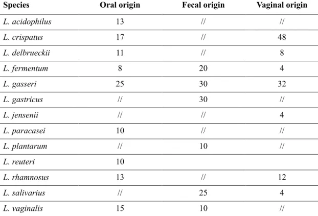
The aim of the work was the isolation and identification of lactobacilli of human origin. It was also deepened the study of their amensalistic properties, with particular attention to the resistance to gastrointestinal transit and their antagonism against pathogenic microorganisms.
Three hundred fifty-nine lactobacilli strains were isolated from swabs of healthy people and identified using molecular techniques based on the study of 16S rDNA. The identification of some strains was confirmed by further analysis DHPLC V1 and V3 of 16S rDNA. The strains were subjected to the evaluation of the resistance to bile salts and low pH, to the production of hydrogen peroxide and more particularly, it has been evaluated the ability to produce substances with antibacterial activity.
Finally, the attention was focused on the characterization of an active supernatant produced by an oral strain. The isolation of the substance provided chromatographic procedures such as SEC (Size Exclusion Chromatography using Sephadex 50) and SPE (Reverse Phase Chromatography using C18 column). The results were shown that the active fraction has a low molecular weight and for its chemical-physical characteristics is not a common bacteriocin, for this reason are on going further chromatographic studies using columns with increasing polarity (C4, phenyl, cyano, and amino).
Future outlooks
Future outlooks are focused on the identification of the molecule in question, by MALDI-TOF and ESI-TOF and then optimizing the whole process to standardize the entire method. In this way, the opportunity to bring to light new molecules will be possible, with the ultimate goal of being able to take advantage from these antibacterial substances.
ABSTRACT
Introduzione
I lattobacilli appartengono ai LAB (Lactic Acid Bacteria) e sono tra i più comuni microrganismi probiotici. Le recenti linee guida rilasciate dal Ministero della Salute, affermano che, sulla base della letteratura disponibile, la quantità sufficiente per ottenere una colonizzazione temporanea da parte di un ceppo probiotico è di almeno 109 cellule viventi. Un microrganismo per essere definito tale, deve essere di origine umana, avere attività immunostimolante e sopravvivere al tratto gastrointestinale, resistendo all'acidità dello stomaco e all'azione della bile. Inoltre, il ceppo deve essere in grado di aderire alla mucosa non causando tossicità, e di produrre sostanze ad attività antibatterica contro alcuni patogeni.
Obiettivi della ricerca e risultati
Scopo del lavoro è stato l'isolamento e l’identificazione di lattobacilli di origine umana. Inoltre, è stato approfondito lo studio delle proprietà amensalistiche, con particolare attenzione alla resistenza al transito gastro-intestinale e all’antagonismo nei confronti dei microorganismi patogeni.
Trecentocinquantanove ceppi di lattobacilli sono stati isolati da tamponi ottenuti da persone sane e identificati tramite tecniche molecolari basate sulla 16S rDNA. Per alcuni ceppi selezionati l’identificazione è stata ulteriormente confermata tramite la tecnica DHPLC applicata agli amplificati delle regioni V1 e V3 della 16S rDNA. In tutti ceppi è stata la valutata la resistenza ai sali biliari e al pH acido, la produzione di perossido di idrogeno, e infine è stata valutata la capacità di produrre sostanze ad attività antibatterica.
Particolare attenzione è stata posta alla caratterizzazione delle componenti “antibiotiche” di un surnatante prodotto da un ceppo di origine orale. L’isolamento della “sostanza inibitrice” è stato condotto mediante tecniche cromatografiche quali SEC (Size Exclusion Chromatography mediante Sephadex 50) e SPE (Reverse Phase Chromatography mediante colonna C18). I risultati hanno dimostrato che la sostanza contenuta nella “frazione attiva” possiede un peso molecolare basso e per le sue caratteristiche chimico-fisiche non è, molto probabilmente, una batteriocina, per questo
motivo sono in avanzamento ulteriori studi cromatografici con colonne a polarità crescente (C4, fenile, ciano e ammino).
Prospettive future
I progetti futuri sono focalizzati sull’identificazione della molecola in questione, tramite analisi MALDI-TOF ed ESI-TOF, e successivamente sull’ottimizzazione del processo di produzione e purificazione. In tal modo si avrà l’opportunità di portare alla luce nuove molecole con l’obiettivo finale di poter sfruttare l’attività antibatterica di tali sostanze.
I
Contents
1 The genus Lactobacillus ... 1
1.1 Nutritional Characteristics and Metabolism of the genus Lactobacillus ... 2
1.1.1 Obligately Homofermentative Lactobacilli... 3
1.1.2 Facoltatively Heterofermentative Lactobacilli. ... 3
1.1.3 Obligately Heterofermentative Lactobacilli. ... 4
1.2 Habitat in Human Organism ... 7
1.2.1 Oral Cavity... 7
1.2.2 Gastro-Intestinal (GI) Tract ... 8
1.2.3 Vagina ... 10
2 Isolation by Classic Cultural Techniques ... 12
2.1 Evaluation of Special Characteristics ... 14
2.1.1 Detection of bile salt hydrolysis ... 14
2.1.2 Detection of H2O2 production ... 14
2.1.3 Detection of lactic acid production ... 15
3 Taxonomy and the Importance of Phylogenetic Classification ... 16
4 Identification by Culture-Independent Techniques ... 26
4.1 Terminal Restriction Fragment Length Polymorphism (T-RFLP) ... 26
4.2 Amplified Restriction Length Polymorphism (AFLP) ... 27
4.3 Amplified Ribosomal Restriction Fragment Analysis (ARDRA) ... 27
4.4 Randomly Amplified Polymorphic DNA (RAPD) ... 28
4.5 Denaturing Gradient Gel Electrophoresis (DGGE) ... 28
II
4.7 Denaturing High Pressure Liquid Chromatography (DHPLC): a new
culture-independent technique ... 29
5 16S rRNA as a Sequencing Gene, the Reason Why ... 33
6 Probiotic Properties ... 37
7 Bacteriocins ... 42
7.1 Classification ... 44
7.2 Biosynthesis and Mode of Action... 46
7.3 Bacteriocins Applications ... 51
8 Introducing Proteomics ... 53
8.1 Size-Exclusion Chromatography (SEC) ... 54
8.2 Solid Phase Extraction (SPE) ... 56
8.3 Mass Spectrometry (MS) ... 59
9 Materials and Methods ... 60
9.1 Bacterial strains and culture conditions ... 60
9.2 Isolation from oral and fecal specimens ... 60
9.3 Isolation from vaginal specimens ... 62
9.4 Susceptibility testing ... 64
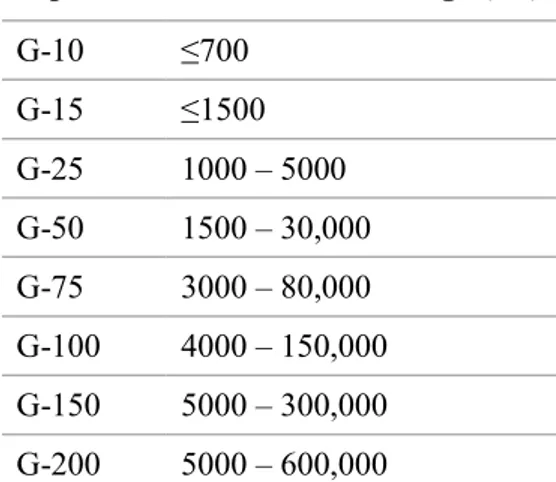
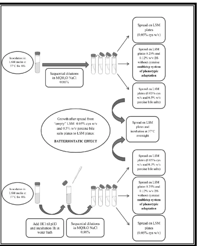
9.5 Evaluation of resistance to low pH and bile salts ... 65
9.6 Evaluation of the capacity of Lactobacillus strains to produce hydrogen peroxide... 68
9.7 DNA Extraction ... 69
9.8 DNA Quantitation Using Spectrophotometer ... 70
III
9.10 Two-steps multiplex PCRs: 16S-ITS-23S and the flanking region of 23S rDNA 74
9.11 tuf gene amplification ... 77
9.12 Nested PCR-amplifications of bacterial V1-V3: DHPLC (WAVE® System) analysis. ... 79
9.12.1 DHPLC collection protocol ... 81
9.12.2 Mutation analysis ... 82
9.13 Detection of inhibitory activity by agar-well diffusion assay ... 83
9.13.1 Sensitivity to different pH values ... 85
9.14 Partial chemical characterization of the antimicrobial fraction produced by an oral origin strain ... 86
9.14.1 Size Exclusion Chromatography ... 86
9.14.2 Reverse Phase Chromatography ... 87
9.14.3 1D SDS-PAGE Analysis ... 87
9.14.4 Evaluation of cytotoxicity by the producer strain on the BV2 cell line ... 88
10 Results ... 89
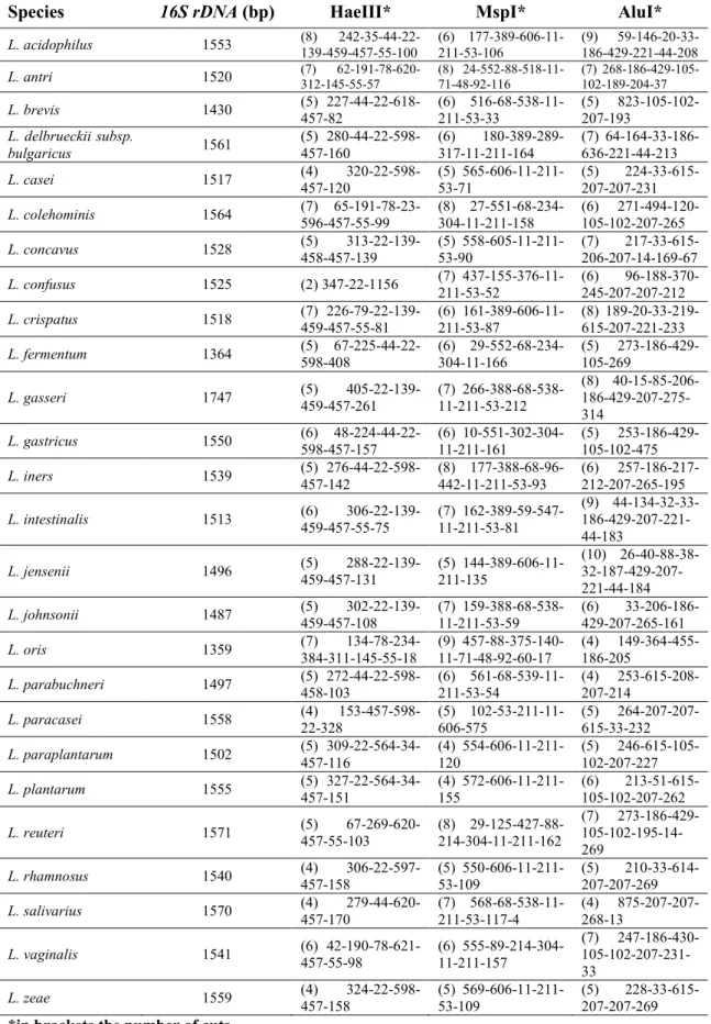
10.1 Molecular identification of Lactobacillus spp. ... 89
10.2 Discrimination of 14 type strains of Lactobacillus spp. by Denaturing High Performance Liquid Chromatography (DHPLC): pilot study ... 93
10.2.1 V1-V3 Analysis ... 95
10.2.2 V1 Species Discrimination ... 101
10.2.3 DHPLC analysis application to Lactobacillus strains ... 104
10.3 Susceptibility testing ...109
IV
10.5 Evaluation of the capacity of Lactobacillus strains to produce hydrogen
peroxide ...115
10.6 Detection of inhibitory activity by agar-well diffusion assay ...117
10.6.1 Sensitivity to different pH values ... 125
10.7 Partial chemical characterization of the antimicrobial fraction produced by an oral origin strain ...126
10.7.1 SEC (Size-Exclusion Chromatography) ... 126
10.7.2 Reverse Phase Chromatography ... 126
10.7.3 1D SDS-PAGE Analysis ... 128
10.7.4 Evaluation of cytotoxicity by the producer strain on BV2 cell line ... 128
11 Discussion & Future outlooks ... 132
11.1 Identification ...132
11.2 Susceptibility test ...133
11.3 Amensalistic activity ...134
11.4 Partial chemical characterization of the antimicrobial fraction produced by an oral origin strain ...136
12 Funding ... 138
13 Acknowledgements ... 138
1
1 The genus Lactobacillus
The genus Lactobacillus comprises a homogenous group of organisms with morphological, biochemical and physiological characteristics that are very similar; some species, initially included in the Lactobacillus genus, have been separated by creating two new genera, Weissella and Carnobacterium (Collins et al., 1987; 1993). With this separation Lactobacillus can be called a non-pathogenic bacterial genus. However, sometimes there are exceptions such as L. psittaci, which was isolated from an air bag of a dead parrot, but our knowledge about the infectivity or the habitats of this species is very poor (Lawson et al., 2001). Lactobacilli are part of our daily life, first of all, they are members of the human microbiome, and they are also indispensable fermentation agents of food and feed (Dworkin & Falkow, 2006; Vos et al., 1984-1989, 2009).
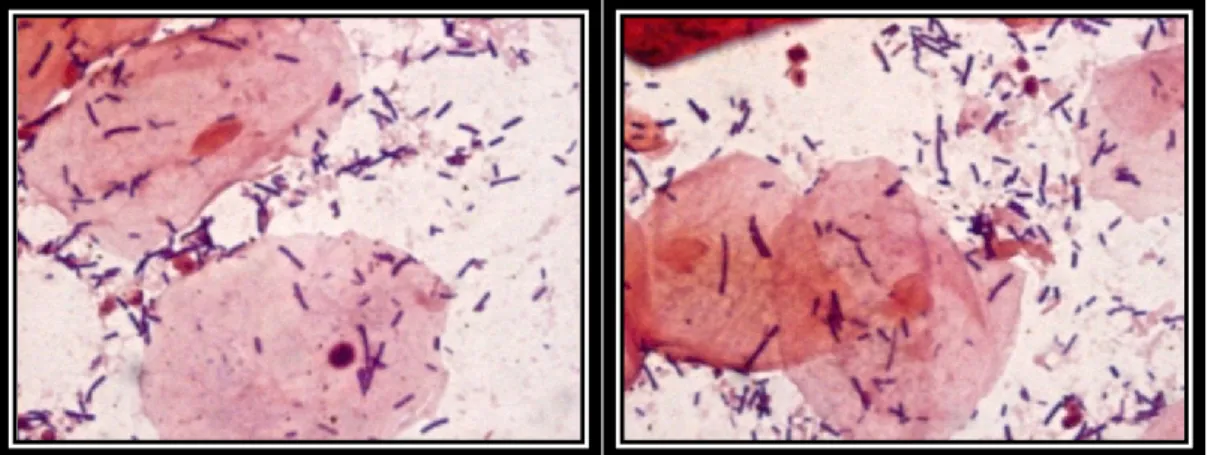
The cellular morphology of lactobacilli is variable, they resemble rods but they may differ in length among various species. For example, some heterofermentative species are short and stocky (coccobacillus) and they could be confused with Leuconostoc, on the other hand, some anaerobic homofermentative species are more similar to bifidobacteria. Sometimes they are curved, as in L. curvatus, or they have a spiral shape such as L. delbrueckii subsp. bulgaricus. In addition, within each species, the length varies according to the stage of development, the soil composition and the oxygen concentration (Dworkin & Falkow, 2006).
2
1.1 Nutritional Characteristics and Metabolism
of the genus Lactobacillus
Lactobacilli are Gram-positive bacteria, non-spore forming, motionless, aero tolerant but whose optimal growth is achieved in microaerobic conditions (CO2≈5%) or at low oxygen concentrations.
Most lactobacilli are mesophilic and they grow in an optimal range of temperature between 20-40°C; few species grow below 15°C or above 40°C with a maximum limit of 55 °C. Thanks to their metabolism lactobacilli cause a lowering of the pH values and this represents one of the factors with which they inhibit the growth of other bacteria favoring their establishment in many habitats. They are devoid of catalase, in fact, they consume oxygen by the action of flavoprotein (NADH-oxidase), and suppress the accumulation of H2O2 through the enzymatic activity of NADH-peroxidase. In addition, they possess nitrate reductase and cytochrome oxidase, and their metabolism is fermentative producing large quantities of lactic acid. However, these microorganisms are demanding and they are adapted to living on complex substrates using not only carbohydrates as a source of energy, but also amino acids, nucleotides, vitamins and ions (Kandler et al. 1986; Elli et al., 2000). The nutritional requirements change from one species to another and sometimes even from strain to strain, for example, biotin and vitamin B12 are required by a few strains, while acids such as pantothenic acid and nicotinic acid are required by all the species. Since the demands of nutrients are different, usually media for laboratory cultures contain carbohydrates, peptone, meat and yeast extracts with the addition of tomato juice, manganese acetate or Tween80, as in the most used medium; MRS (De Man, Rogosa, & Sharpe, 1960), besides, the optimum pH for growth is between 4.5-6.4, although there are exceptions, for example,
Lactobacillus suebicus that grows even at extremely low pH (pH 2; Kleynmans, Heinzl, & Hamme, 1989).
Metabolically speaking, we can divide the genus Lactobacillus into three groups: (A) Obligately homofermentative lactobacilli, (B) Facultatively heterofermentative lactobacilli, and (C) Obligately heterofermentative lactobacilli (Vandamme et al., 1996).
3
1.1.1 Obligately Homofermentative Lactobacilli.
They degrade hexoses to lactic acid by the Embden-Meyerhof-Parnas pathway, but they cannot metabolize pentoses and gluconate.
The Embden-Meyerhof-Parnas pathway is the most common way for the degradation of glucose to pyruvate and it works in both cases, with or without oxygen. This pathway can be divided into two stages: in the first one, a molecule of six carbon atoms (glucose) is phosphorylated twice, and is converted into fructose 1,6-diphosphate, in this step two molecules of ATP are consumed. In the second phase, the enzyme fructose 1,6-diphosphatealdolase results in the cleavage of fructose 1,6-diphosphate into two halves, one half is dihydroxyacetone phosphate. Since dihydroxyacetone phosphate can be easily modified changing it into glyceraldehyde-3-phosphate, the two halves of fructose 1,6-bisphosphate are both used in this second phase. The two molecules of glyceraldehyde 3-phosphate will become, in a process of five steps, two molecules of pyruvate. The Embden-Meyerhof-Parnas pathway leads from the degradation of one molecule of glucose to the formation of two molecules of pyruvate (Fig. 1-2) (Willey,
Sherwood, & Woolverton, 2008). Finally, the two molecules of pyruvate are reduced by lactic fermentation to two molecules of lactate (dissociated form of the lactic acid).
1.1.2
Facoltatively Heterofermentative Lactobacilli
.
These lactobacilli convert the hexoses to lactic acid by the Embden-Meyerhof-Parnas pathway. Instead, if glucose is scarce they exploit the Warburg-Dickens-Horecker pathway, also known as the pentose phosphate cycle; it consists in the oxidation of glucose-6-phosphate to 6-phosphogluconate (with the reduction of one molecule of NAD); 6-phosphogluconate is subsequently decarboxylated producing a sugar at five atoms of carbon. Then, this sugar is converted into glyceraldehyde-3-phosphate and acetyl phosphate. Glyceraldehyde is converted to lactic acid with the same reactions of the second part of glycolysis while the acetyl-phosphate is converted into ethanol (Fig. 1-3) (Willey, Sherwood, & Woolverton, 2008).
4
1.1.3
Obligately Heterofermentative Lactobacilli
.
Species belonging to this group only exploit the phosphogluconate pathway thanks to a phosphoketolase (Fig. 1-4) (Willey, Sherwood, & Woolverton, 2008).
The most important species of Lactobacillus are reported in Table 1.1, divided by type of metabolism.
Figure 1-2.The Embden-Meyerhof-Parnas pathway in obligately homofermentative Lactobacillus spp. These species have only 1,6-bisphosphatealdolase, thus they cannot metabolize pentose and gluconate (Nelson, Lehninger, & Cox, 2008).
5
Figure 1-3. Facultatively heterofermentative Lactobacillus spp. have a 1,6-bisphosphatealdolase (Embden-Meyerhof-Parnas pathway) and an inducible phosphoketolase with pentoses acting as inducers (Warburg-Dickens pathway) (Nelson, Lehninger, & Cox, 2008).
Figure 1-4. Obligately heterofermentative Lactobacillus spp. only Warburg-Dickens pathway using a phosphoketolase enzyme (Nelson, Lehninger, & Cox, 2008)
6
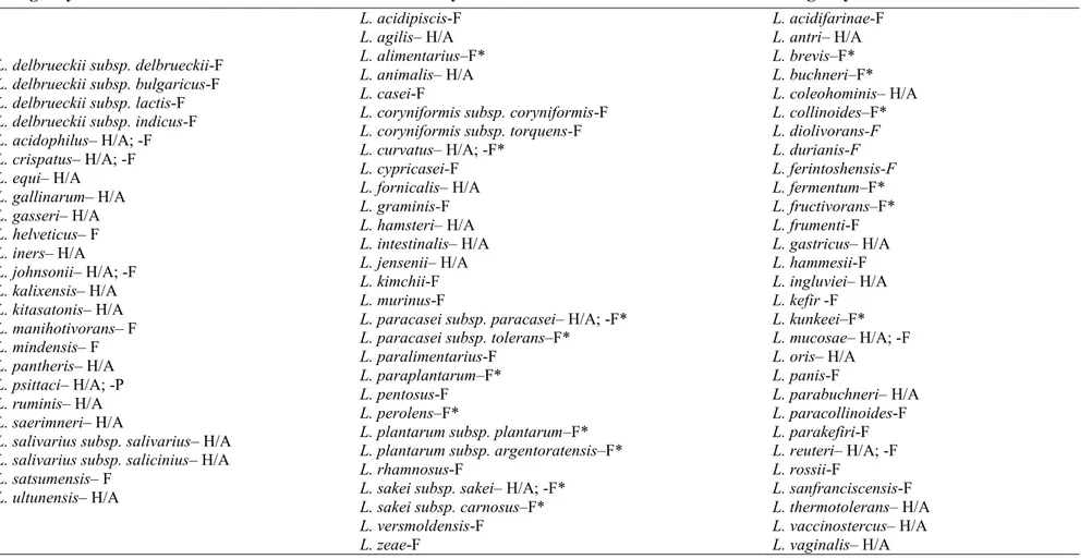
Table 1.1. List of the species of the genus Lactobacillus divided by type of metabolism (Vos, et al., 2009).
H/A: isolation from human or animal isolation; F/F*: involved in food fermentation/also in spoilage; P: pathogen
Obligately homofermentative Facultatively heterofermentative Obligately heterofermentative
L. delbrueckii subsp. delbrueckii-F L. delbrueckii subsp. bulgaricus-F L. delbrueckii subsp. lactis-F L. delbrueckii subsp. indicus-F L. acidophilus– H/A; -F L. crispatus– H/A; -F L. equi– H/A L. gallinarum– H/A L. gasseri– H/A L. helveticus– F L. iners– H/A L. johnsonii– H/A; -F L. kalixensis– H/A L. kitasatonis– H/A L. manihotivorans– F L. mindensis– F L. pantheris– H/A L. psittaci– H/A; -P L. ruminis– H/A L. saerimneri– H/A
L. salivarius subsp. salivarius– H/A L. salivarius subsp. salicinius– H/A L. satsumensis– F L. ultunensis– H/A L. acidipiscis-F L. agilis– H/A L. alimentarius–F* L. animalis– H/A L. casei-F
L. coryniformis subsp. coryniformis-F L. coryniformis subsp. torquens-F L. curvatus– H/A; -F* L. cypricasei-F L. fornicalis– H/A L. graminis-F L. hamsteri– H/A L. intestinalis– H/A L. jensenii– H/A L. kimchii-F L. murinus-F
L. paracasei subsp. paracasei– H/A; -F* L. paracasei subsp. tolerans–F*
L. paralimentarius-F L. paraplantarum–F* L. pentosus-F L. perolens–F*
L. plantarum subsp. plantarum–F* L. plantarum subsp. argentoratensis–F* L. rhamnosus-F
L. sakei subsp. sakei– H/A; -F* L. sakei subsp. carnosus–F* L. versmoldensis-F L. zeae-F L. acidifarinae-F L. antri– H/A L. brevis–F* L. buchneri–F* L. coleohominis– H/A L. collinoides–F* L. diolivorans-F L. durianis-F L. ferintoshensis-F L. fermentum–F* L. fructivorans–F* L. frumenti-F L. gastricus– H/A L. hammesii-F L. ingluviei– H/A L. kefir -F L. kunkeei–F* L. mucosae– H/A; -F L. oris– H/A L. panis-F L. parabuchneri– H/A L. paracollinoides-F L. parakefiri-F L. reuteri– H/A; -F L. rossii-F L. sanfranciscensis-F L. thermotolerans– H/A L. vaccinostercus– H/A L. vaginalis– H/A
7
1.2 Habitat in Human Organism
1.2.1 Oral Cavity
The oral cavity is a system constantly exposed to external factors because of respiration and nutrition. Here reside at least 700 microbial species (Moore & Moore, 1994; Aas et al., 2005), the composition and density is determined by kind of nutrition as well as sex, age, health status, composition of saliva and oral hygiene (Tannock, 1999; Slot & Chen, 1999).
As for the genus Lactobacillus, they make up about 1% of the bacteria in saliva, 0.1% of bacteria found on the epithelium of the cheek and tongue, and 0.005% of the intragingival plaque. The predominant species found both in childhood and adulthood are L. casei, L. rhamnosus and L. fermentum. To date, we know that many of the strains previously classified as L. casei might, instead, belong to the species L. paracasei (Ventura et al., 2003). Lactobacillus brevis strains isolated from human saliva (Hayward & Davis, 1956; Hayward, 1957) were then classified into a new species, L.
oris (Farrow & Collins, 1988). Strangely, L. salivarius was isolated with a low
incidence. In general, in the oral cavity lactobacilli are limited in number also because they are in an area that we can define “transient” (MacFarlane & Samaranayake, 2014). As suggested by many articles, dental caries is caused by the metabolism of the bacteria such as Streptococci, Neisseria, Actinomyces, and Capnocytophaga that adhere to tooth enamel causing damage (Tannock, 1999). The progression of the disease is promoted mainly by Streptococcus mutans, but when the tooth has deteriorated, lactobacilli multiply inside. As already mentioned, Lactobacilli produce lactic acid by the metabolism of carbohydrates and thus alter the ecological conditions, resulting in selection of more acidophilic bacteria such as L. casei, L. paracasei and L. rhamnosus that will take possession of the niche. This helps us to understand that the high density of lactobacilli in the cases of caries is the result of the disease rather than the cause (Aas et al., 2005; Alaluusua et al, 1987; Wijeyeweera & Kleinberg, 1989).
8
1.2.2 Gastro-Intestinal (GI) Tract
The entire gastrointestinal tract is home to a substantial amount of bacteria. There are more than 400 bacterial species in the intestine but quantity and composition change from section to section: pH varies from values of 5.5 at proximal levels to values of 6.9 at the level of the distal colon, the temperature can change between 37-40°C; also, the constitution of the mucous membranes, the diet of the subject and not least, health, have always to be considered. All this involves a considerable variety of strains, even the anaerobic conditions are important, in fact, near the mucous there is a high oxygen tension which promotes the growth of facultative anaerobic bacteria (Dworkin & Falkow, 2006).
As for the lactobacilli, in the human stomach they are in low quantities, about <103cfu/ml and there are only species adapted to the extremely acidic environment such as L. gastricus and L. antri, or L. kalixensis and L. ultunensis (Roos, Engstrand, &
Jonsson, 2005).
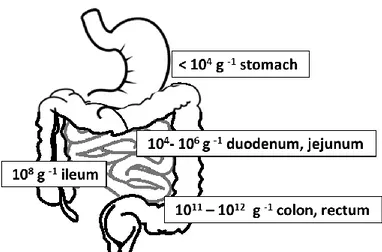
The number of bacteria in the intestines increases from ≈104cfu/ml in the duodenum, up to 108-109cfu/ml in the ileum with predominantly lactobacilli and enterococci (Fig. 1-5). The colon is the most colonized part of the human intestine and most of the knowledge acquired on the intestinal microbial populations is from stool analysis. Human feces contain >1011cfu/g, which constitute about 50% of the solids within the colon (Tannock, 1995; Vos et al., 2009) but the percentage of lactobacilli isolated is only a low fraction (Tannock et al., 2000); these latter, in fact, are found in greater amounts in the proximal section of the colon while they are considerably reduced at the level of the distal colon, and then in the stool (Marteau et al., 2001).
9
Figure 1-5. Numbers in individual sections depict the amount of bacteria per gram of intestinal content typically found in healthy individuals (Leser & Mølbak, 2009).
By molecular analysis it was found that L. acidophilus, L. fermentum and L. salivarius,
L. gasseri, L. crispatus and L. reuteri are the dominant species (Mitsuoka, 1992; Reuter,
2001) while L. paracasei, L. rhamnosus, L. delbrueckii, L. brevis and L. plantarum are quite transient whose stay varies depending on the diet, age and health of the individual (Heilig et al., 2002).
The amount of lactobacilli decreases in cases of gastrointestinal diseases. Studies of patients with Crohn's disease and ulcerative colitis have shown a prevalence of unclassified strains of the phyla Bacteroidetes and Verrucomicrobia. In addition, an increase of enterobacteriaceae with particular attention to the presence of a non-commensal Escherichia coli has been found.
Finally, Clostridium perfringens was detected, too. The profiles of the analysis of biopsy, aspirations and fecal samples taken from each patient are similar. Instead, the results of stool samples of patients with pouchitis (inflammation of the ileal pouch) or familial adenomatous polyposis show profiles very different from each other, but even in these cases the diseases are associated with the presence of non-commensal bacteria in human gut; the administration of antibiotics brings an improvement of the pathologies in question (Tannock, 2008; 2010).
10
1.2.3 Vagina
The vagina is a habitat in which, under normal conditions, there is a balance between different bacterial species in a commensal symbiosis with the host. Lactobacilli account for 50-90% of the vaginal aerobic microbiota in most Caucasian women (Zhou et al., 2007; Yamamoto et al., 2009) and they are present in concentrations of 107-108cfu/g of vaginal fluid, where glycogen is considered the maximum source of fermentable carbohydrates to be exploited for nourishment. Different ethnic women are at the heart of the most important differences observed in the composition of endogenous microbiota. Similar to the gut, there are many reasons why entire populations can differ from one another, variables can be included such as hygiene, sexual activity, number of sexual partners, diet, use of antibiotics or vaginal douches, cervical dysplasia, specific sampling site, age, and many other issues relating to socio-economic status (Marrazzo et al., 2002; Zhou et al., 2007; Yamamoto et al., 2009; Martínez-Peña et al., 2013; Pendharkar et al., 2013).
L. crispatus, L. gasseri, and L. jensenii were identified as predominant species in
non-cultural analysis of samples, taken from healthy, sexually active women, while L.
rhamnosus, L. paracasei, L. fermentum, L. plantarum, L. vaginalis, and L. iners have
occasionally been identified (Antonio, Hawes, & Hillier, 1999; Falsen et al., 1999; Vásquez et al., 2002), finally, L. coleohominis was isolated only on a few occasions (Nikolaitchouk et al., 2001).
On the other hand, subsequent studies showed that the vaginal pH is already low at birth (Cook &Sobel, 1990; Boskey et al, 1999; Gorodeski et al., 2005), and therefore not correlated with the presence of lactobacilli (Boskey et al., 1999) but because of hormonal activity (Gorodeski et al., 2005), for these reasons the bacterial contribution for the vaginal pH is still debated. To create further confusion about the influence of the vaginal microbiota (mainly consisting of lactobacilli) on the pH was caused by Zhou et al. (2007), where the identification of eight different ecotypes of vaginal microbiome would seem to agree with Gorodeski et al. (2005) more than with Boskey et al. (1999). In cases of vaginal diseases the amount of lactobacilli decreases or, in cases where it remains predominant, the bacterial composition changes (Kuebrichet al., 2004; Vitali et al., 2007; Zhou et al., 2007).
11
The most common vaginal disorders are linked to bacterial vaginosis (BV), vulvovaginal candidiasis (VVC), and aerobic vaginitis (AV). Among these infections, the most common genital infection during reproductive age, which causes vaginal discharge characterized by bad odor, is BV. An illness caused by anaerobic bacteria such as Mobiluncus, Gardnerella vaginalis, and Mycoplasma hominis, which does not cause inflammation (Priestly, 1997; Beigi et al., 2004; Klebanoff et al., 2004).VVC, however, is often caused by Candida albicans, a fungus and member of the vaginal microbiota in women of childbearing age, which is responsible for more than 90% of symptomatic cases of vaginitis. Nevertheless, in recent years, the incidence of vulvovaginitis can diasis due to non-C.albicans species has increased (Sobel et al., 1998; Wilson, 2005).
Finally, AV is pathology similar to BV but differs for different reasons: at the microbiological level there is an increase of Gram-positive cocci, such as Streptococcus
agalactiae, staphylococci and enterococci and Gram-negative bacteria such as
Enterobacteriaceae (in particular E. coli). The lactobacilli component of the microbiota decreases until it disappears completely (Donders et al., 2002; Kuebrich et al., 2004). From a clinical point of view, the infection is characterized by the presence of toxic leukocytes and parabasal cells, yellow secretion, foul odor (KOH negative test), elevated vaginal pH, redness, itching, burning and different degrees of dyspareunia (Donders et al., 2002; 2007; Kuebrich et al., 2004). Clinical studies have shown that L.
rhamnosus GR-1 and L. reuteri RC-14 can reduce the risk of BV (Reid et al., 2001;
2003), not only by increasing the effectiveness of the antibiotic by oral administration of a probiotic, but they can also cure BV by direct intravaginal administration. Saunders et al. (2007) demonstrated that strains of Lactobacillus spp. have the ability to destroy and remove biofilms formed by Gardnerella spp., and they potentially reduce the need for antibiotics.
12
2 Isolation by Classic Cultural Techniques
The methods for lactobacilli isolation must consider, first of all, the nutritional needs complex, the pH (values between 4.5 and 6.4 will foster a better growth) and environmental conditions (microaerobicor sometimes strictly anaerobic conditions). Therefore, it is advisable to incubate the plates in microaerobic atmosphere using generating kits for H2 and CO2. Media should contain growth factors such as yeast extract, vitamins, peptone, manganese, acetate and Tween®80 considering any supplements as needed such as meat extract, tomato juice, malt extract or ethanol. These conditions encourage a higher similarity to the environment from which they must be isolated (Dworkin & Falkow, 2006; Vos et al., 2009).Sources where Lactobacilli are the only present microorganisms are rare. When lactobacilli are the predominant bacteria, a non-selective media, such as MRS (De Man, Rogosa, & Sharpe, 1960), can be used. If the microbial population is more complex, you can use the medium proposed by Rogosa SL et al. in 1951, to isolate the lactobacilli from human samples (Rogosa, Mitchell, & Wiseman, 1951), but it should be considered that even other lactic acid bacteria (Leuconostoc, pediococci, enterococci, Weissella or Bifidobacterium) and yeasts could grow. The latter can be inhibited with the addition of cycloheximide (0.4 g/L) (Haley, Trandel, & Coyle, 1980).
Nowadays, to isolate the lactobacilli from human samples more specifically, according to the kind of sample (vaginal or oro-fecal) we can prepare a more selective media. A useful media is LAMVAB (Lactobacillus Anaerobic MRS with Vancomycin and Bromocresol Green), highly selective because of low pH and vancomycin (20 mg/L) which inhibit competitive fecal microbiota such as enterobactiaceae, Clostridium spp., other Gram-negative bacteria, and also Bifidobacterium spp. and all other lactic acid bacteria. This medium contains cysteine–HCl to enhance anaerobic conditions and bromocresol green as an indicator, indeed, the colonies grow green or blue, and the medium loses the color (becoming yellowish) due to the production of acid (Hartemink, Domenech, & Rombouts, 1997).
13
Moreover, to isolate the lactobacilli from vaginal specimens, media containing blood is needed, in these cases the swabs are directly streaked onto MRS agar and Columbia agar with 5% horse blood.In general, colonies grown on agar are small (2-5 mm diameter), convex, smooth, translucent or opaque, whose pigmentation depends on the medium used. In addition, many strains can show a weak proteolytic activity (caused by enzymes released from the cell) and lipolytic activity (caused by intracellular lipases). The edges are well-defined and the growth does not cause clear zones around the colonies. Finally, growth in broth appears homogeneous, smooth and whitish.
Generally, Lactobacilli develop a characteristic smell, more or less strong, caused by the production of lactic acid.
14
2.1 Evaluation of Special Characteristics
2.1.1 Detection of bile salt hydrolysis
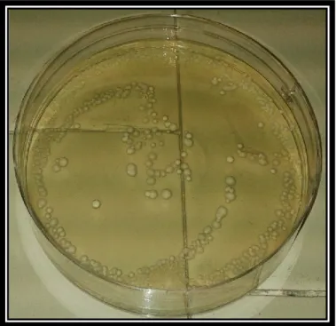
The test is performed with the growth of Lactobacillus strains, in anaerobic conditions, on SL or MRS media by adding taurocholic acid, taurochenodeoxycholic acid, or taurodeoxycholic acid. Hydrolysis of bile salts is manifested by the formation of halos around white colonies, these latter may appear opaque and granular (Fig. 2-1). This test can provide a quick way to determine the functional activity of Lactobacillus species and a rapid identification of hydrolase-deficient strains (Dashkevicz & Feighner, 1989).
Figure 2-1. Lactobacillus gastricus isolated from a human rectal sample grown on MRS containing taurocholic acid.
2.1.2 Detection of H
2O
2production
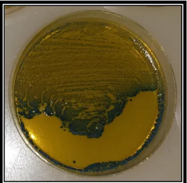
This test, for the detection of H2O2, exploits horseradish peroxidase, which is isolated from horseradish roots. It is a single chain polypeptide containing four disulfide bridges and 18% carbohydrates (galactose, arabinose, xylose, fucose etc.). It is used as a component of an MRS medium containing 3,3′,5,5′-tetramethylbenzidine (TMB); this latter acts as a chromogenic substrate of peroxidase. The peroxidase generates O2 from the H2O2 produced by the lactobacilli, and then the TMB stains the colonies because it
15
reacts with oxygen The colonies that produce H2O2 appear dark green/blue (Fig. 2-2), while H2O2 not-producers appear colorless (Otero & Nader-Macías, 2006; Pascual et al, 2006).Figure 2-2. Lactobacillus salivarius isolated from a human oral sample grown on MRS containing horseradish peroxidase and TMB.
2.1.3 Detection of lactic acid production
There are various essays for such determination. One of these uses the following reactions: L-lactic acid is oxidized to pyruvate by NAD in the presence of L-lactate dehydrogenase (LDH-L). However, the equilibrium of this reaction is in favor of L-lactic acid, but it is possible to facilitate the balance shifting towards the formation of pyruvate and NADH with a second reaction catalyzed by the enzyme glutamate-pyruvate transaminase (GPT) in the presence of L-glutamate. The increase in NADH is determined by its absorbance at 334, 340 or 365nm (Noll, 1966; Bergmeyer, 2012).
16
3 Taxonomy and the Importance of
Phylogenetic Classification
Ταξι: taxis, "sort", and νομο: nomos, "norm" or "rule." Taxonomy is the science that deals with the classification, or the description, of an entity in a system, in order to facilitate the study of living beings. Each species has a scientific binary name of Latin or Greek origin that identifies it uniquely.
What about the genus Lactobacillus? The genus Lactobacillus belongs, together with the genera Paralactobacillus and Pediococcus, to the phylum Firmicutes, class Bacilli, order Lactobacillales, family Lactobacillaceae (Garrity et al., 2004). The phylogenetic structure of the family Lactobacillaceae considers Lactococcus lactis and Streptococcus
thermophilus as limit groups.
The NCBI taxonomy database currently recognizes more than 200 species of
Lactobacillus; therefore it is the most populous genus of the order Lactobacillales.
Indeed, only this year seven new species have been described: Lactobacillus
formosensis (Chang et al., 2015), Lactobacillus plajomi and Lactobacillus modestisalitolerans (Miyashita et al., 2015), Lactobacillus mixtipabuli (Tohno et al.,
2015), Lactobacillus insicii (Ehrmann et al., 2015), Lactobacillus herbarum (Mao, Chen, & Horvath, 2015) and Lactobacillus wasatchensis (Oberg et al., 2015) (Table 3.1).
Historically, from Sharpe et al. (1979) to Kandler & Weiss (1986), morphological characteristics, fermentation of carbohydrates, nutritional requirements, the temperature dependence of growth and the properties of agglutination, were the principles of grouping of bacterial taxonomy.
The subdivision of the genus Lactobacillus was made on the basis of fermentation in three groups, but the terms about the different kind of fermentation have been described by many authors and in a different way and, for this reason, could be misleading. The metabolic terminology accepted is the following: obligately homofermentative Lactobacilli, which ferment hexoses almost exclusively to lactic acid by the Embden-Meyerhof-Parnas (EMP) pathway, but they lack phosphoketolase, therefore they are not able to ferment pentose and gluconate; facultatively heterofermentative Lactobacilli
17
possess the enzyme aldolase (EMP pathway) and are also able to degrade pentoses and gluconate through an inducible phosphoketolase; obligately heterofermentative Lactobacilli degrade hexoses and pentoses by the phosphogluconate pathway. These metabolic processes have already been described in the previous chapter (Hammes & Vogel, 1995).A good method to identify bacteria is a polyphasic approach, combining phenotypic analysis to genetic analysis. The phylogenetic approach has revolutionized the study of bacteria and thus also the method of classification that has become more efficient and correct. The comparison of molecular sequences is able to precisely estimate the phylogenetic relationships among organisms.
18
Table 3.1 Species of Lactobacillus at the time of writing (November 2015)
Lactobacillus acetotolerans Lactobacillus crispatus
Lactobacillus acidifarinae Lactobacillus crustorum
Lactobacillus acidipiscis Lactobacillus curieae
Lactobacillus acidophilus Lactobacillus curvatus
Lactobacillus agilis Lactobacillus delbrueckii
Lactobacillus algidus L. delbrueckii subsp. bulgaricus
Lactobacillus alimentarius L. delbrueckii subsp. delbrueckii
Lactobacillus alvei L. delbrueckii subsp. indicus
Lactobacillus alvi L. delbrueckii subsp. jakobsenii
Lactobacillus amylolyticus L. delbrueckii subsp. lactis
Lactobacillus amylophilus L. delbrueckii subsp. sunkii
Lactobacillus amylotrophicus Lactobacillus dextrinicus
Lactobacillus amylovorus Lactobacillus diolivorans
Lactobacillus animalis Lactobacillus equi
Lactobacillus animata Lactobacillus equicursoris
Lactobacillus antri Lactobacillus equigenerosi
Lactobacillus apinorum Lactobacillus fabifermentans
Lactobacillus apis Lactobacillus faecis
Lactobacillus apodemi Lactobacillus faeni
Lactobacillus aquaticus Lactobacillus farciminis
Lactobacillus aviarius Lactobacillus farraginis
L. aviarius subsp. araffinosus Lactobacillus fermentum
L. aviarius subsp. aviarius Lactobacillus floricola
Lactobacillus backii Lactobacillus florum
Lactobacillus bifermentans Lactobacillus formosensis
Lactobacillus bombi Lactobacillus fornicalis
Lactobacillus bombicola Lactobacillus fructivorans
Lactobacillus brantae Lactobacillus frumenti
Lactobacillus brevis Lactobacillus fuchuensis
Lactobacillus brevis subsp. coagulans Lactobacillus furfuricola Lactobacillus brevis subsp. gravesensis Lactobacillus futsaii
Lactobacillus brevisimilis Lactobacillus gallinarum
Lactobacillus buchneri Lactobacillus gasseri
Lactobacillus cacaonum Lactobacillus gastricus
Lactobacillus camelliae Lactobacillus ghanensis
Lactobacillus capillatus Lactobacillus gigeriorum
Lactobacillus casei Lactobacillus ginsenosidimutans
Lactobacillus paracasei Lactobacillus gorillae
L. paracasei subsp. paracasei Lactobacillus graminis
L. paracasei subsp. tolerans Lactobacillus guizhouensis
Lactobacillus zeae Lactobacillus halophilus
Lactobacillus catenefornis Lactobacillus hammesii
Lactobacillus ceti Lactobacillus hamsteri
Lactobacillus coleohominis Lactobacillus harbinensis
Lactobacillus collinoides Lactobacillus hayakitensis
Lactobacillus composti Lactobacillus heilongjiangensis
Lactobacillus concavus Lactobacillus helsingborgensis
Lactobacillus coryniformis Lactobacillus helveticus
L. coryniformis subsp. coryniformis L. helveticus subsp. jugurti L. coryniformis subsp. torquens Lactobacillus herbarum
19
Lactobacillus heterohiochii Lactobacillus odoratitofui
Lactobacillus hilgardii Lactobacillus oeni
Lactobacillus hokkaidonensis Lactobacillus oligofermentans
Lactobacillus hominis Lactobacillus oris
Lactobacillus homohiochii Lactobacillus oryzae
Lactobacillus hordei Lactobacillus otakiensis
Lactobacillus iatae Lactobacillus ozensis
Lactobacillus iners Lactobacillus panis
Lactobacillus ingluviei Lactobacillus pantheris
Lactobacillus insectis Lactobacillus parabrevis
Lactobacillus insicii Lactobacillus parabuchneri
Lactobacillus intermedius Lactobacillus paracollinoides
Lactobacillus intestinalis Lactobacillus parafarraginis
Lactobacillus iwatensis Lactobacillus parakefiri
Lactobacillus japonicus Lactobacillus paralimentarius
Lactobacillus jensenii Lactobacillus paraplantarum
Lactobacillus johnsonii Lactobacillus pasteurii
Lactobacillus kalixensis Lactobacillus paucivorans
Lactobacillus kefiranofaciens Lactobacillus pentosus
L. kefiranofaciens subsp. kefiranofaciens Lactobacillus perolens L. kefiranofaciens subsp. kefirgranum Lactobacillus plantarum
Lactobacillus kefiri L. plantarum subsp. argentoratensis
Lactobacillus kimbladii L. plantarum subsp. plantarum
Lactobacillus kimchicus Lactobacillus pobuzihii
Lactobacillus kimchiensis Lactobacillus pontis
Lactobacillus kisonensis Lactobacillus porcinae
Lactobacillus kitasatonis Lactobacillus psittaci
Lactobacillus koreensis Lactobacillus rapi
Lactobacillus kullabergensis Lactobacillus rennanquilfy
Lactobacillus kunkeei Lactobacillus reuteri
Lactobacillus larvae Lactobacillus rhamnosus
Lactobacillus leichmannii Lactobacillus rodentium
Lactobacillus letivazi Lactobacillus rogosae
Lactobacillus lindneri Lactobacillus rossiae
Lactobacillus malefermentans Lactobacillus ruminis
Lactobacillus mali Lactobacillus saerimneri
Lactobacillus manihotivorans Lactobacillus sakei
Lactobacillus mellifer L. sakei subsp. carnosus
Lactobacillus mellis L. sakei subsp. sakei
Lactobacillus melliventris Lactobacillus salivarius
Lactobacillus mindensis Lactobacillus sanfranciscensis
Lactobacillus mixtipabuli Lactobacillus saniviri
Lactobacillus mobilis Lactobacillus satsumensis
Lactobacillus mucosae Lactobacillus secaliphilus
Lactobacillus mudanjiangensis Lactobacillus selangorensis
Lactobacillus murinus Lactobacillus senioris
Lactobacillus nagelii Lactobacillus senmaizukei
Lactobacillus namurensis Lactobacillus sharpeae
Lactobacillus nantensis Lactobacillus shenzhenensis
Lactobacillus nasuensis Lactobacillus sicerae
20
Lactobacillus nodensis Lactobacillus siliginis
Lactobacillus similis Lactobacillus uvarum
Lactobacillus songhuajiangensis Lactobacillus vaccinostercus
Lactobacillus spicheri Lactobacillus vaginalis
Lactobacillus sucicola Lactobacillus vermiforme
Lactobacillus suebicus Lactobacillus versmoldensis
Lactobacillus sunkii Lactobacillus vini
Lactobacillus taiwanensis Lactobacillus wasatchensis
Lactobacillus thailandensis Lactobacillus xiangfangensis
Lactobacillus tucceti Lactobacillus yonginensis
Lactobacillus ultunensis Lactobacillus zymae
Nowadays, in fact, the identification is no longer based only on metabolism, but above all on the study of genomic analysis. One of the fastest methods for the identification of
Lactobacillus spp. is the sequencing of the 16S rRNA gene, but remembering that
closely related species cannot be discriminated, such as L. plantarum, L. pentosus and
L. paraplantarum (99.7 to 99. 9%) or L. casei, L. paracasei, L. rhamnosus, L. kimchii
and L. paralimentarius (99.9%), L. mindensis and L. farciminis (99.9%), or also, L.
animalis and L. murinus (99. 7%).
The first phylogenetic analysis of lactobacilli was carried out by Collins et al. (1991), and the genus Lactobacillus was divided into 3 groups: L. delbrueckii group, L.
casei-Pediococcus group, and Leuconostoc group, which contained some Lactobacillus. In
1995, Schleifer & Ludwig attributed to the L delbrueckii group the name L. acidophilus, and they divided the L. casei-Pediococcus group into four sub-clusters (L. salivarius group, L. reuteri group, L. buchneri group and L. plantarum group). Instead, all lactobacilli of the Leuconostoc group are now reclassified as species of the genera
Leuconostoc or Weissella.
The recent identification of a large number of species has led to revisions of the genus, allowing the reorganization of these groups in a better and more flexible way. Subsequently, Hammes & Hertel (2006), through the study of the 16S rRNA gene, divided the genus Lactobacillus into 7 different groups: Lactobacillus buchneri group,
Lactobacillus casei group, Lactobacillus delbrueckii group, Lactobacillus plantarum
group, Lactobacillus reuteri group, Lactobacillus sakei group and Lactobacillus
salivarius group. Finally, L. brevis, L. perolens, L. bifermentans and L. coryneformis
were positioned separately in the phylogenetic tree of lactobacilli (Table 3.2) (Felis & Dellaglio, 2007).
21
Nevertheless, a closer study reveals that only three distinct phylogenetic groups can be defined: L. delbrueckii group, L. reuteri group, and L. salivarius group, because, as was said above, the discriminatory power of the 16S rRNA gene sequences is limited (Garrity et al., 2004).The L. delbrueckii group contains mainly obligately homofermentative Lactobacilli, the range of G+C is rather wide from 34 up to 51 mol % and the type of peptidoglycan is Lys–D-Asp; it contains four subspecies, and Lactobacillus kefiranofaciens has two subspecies which cannot be reliably differentiated by means of 16S rDNA analysis. The L. reuteri group contains only obligately heterofermentative Lactobacilli, the range of G+C varies among species belonging to this group (38-54 mol%) and they possess two types of peptidoglycan, Lys-D-Asp-Orn or D-Asp.
Finally, L. salivarius group contains both obligately homofermentative and facultatively heterofermentative Lactobacilli. This group possesses a content of G+C between 32-44 mol%, and two types of peptidoglycan, Lys-D-Asp and meso-DPM-direct (Garrity et al., 2004).
However, the analysis of 16S rDNA sequences (especially the first 900 bases) is still a fast tool that can be used in combination with other techniques or together with the study of other molecular targets, in order to identify bacterial species.
22
Table 3.2. Phylogenetic grouping (Felis & Dellaglio, 2007).
Group Hammes & Hertel, 2006 Dellaglio & Felis, 2005 Felis & Dellaglio, 2007
L. delbrueckii (delb) L. acetotolerans, L. acidophilus, L. amylolyticus, L. amylophilus, L. amylovorus, L. crispatus, L. delbrueckii, L. fornicalis, L. gallinarum, L. gasseri, L. hamsteri,
L. helveticus, L. iners, L. intestinalis, L. jensenii, L. johnsonii, L. kefiranofaciens, L. kefirgranum, L. psittaci L. acetotolerans, L. acidophilus, L. amylolyticus, L. amylophilus, L. amylovorus,
L. crispatus, L. delbrueckii, L. fornicalis, L .gallinarum, L. gasseri, L. hamsteri,
L. helveticus, L. iners, L. intestinalis, L. jensenii, L. johnsonii, L. kalixensis,
L. kefiranofaciens, L. kefirgranum, L. kitasatonis, L. psittaci, L. suntoryeus,
L. ultunensis L. acetotolerans, L. acidophilus, L. amylolyticus, L. amylophilus, L. amylotrophicus, L. amylovorus, L. crispatus, L. delbrueckii, L. fornicalis, L. gallinarum, L. gasseri, L. hamsteri, L. helveticus, L. iners, L. intestinalis,
L. jensenii, L. johnsonii, L. kalixensis, L. kefiranofaciens, L. kitasatonis, L. psittaci, L. sobrius, L. ultunensis L. salivarius
L. acidipiscis, L. agilis, L. algidus, L. animalis, L. aviarius, L. cypricasei,
L. equi, L. mali, L. murinus, L. nagelii,
L. ruminis, L. salivarius
L. acidipiscis, L. agilis, L. algidus, L. animalis,
L. aviarius, L. cypricasei, L. equi, L. mali, L. murinus, L. nagelii, L. ruminis, L. saerimneri, L. salivarius, L. satsumensis
L. acidipiscis, L. agilis, L. algidus*, L. animalis, L. apodemi,
L. aviarius, L. equi, L. mali, L. murinus, L. nageli, L. ruminis, L.
saerimneri,
L. salivarius, L. satsumensis, L. vini
23
L. reuteri (reu)
L. coleohominis, L. durianis, L. fermentum, L. frumenti, L.
ingluviei,
L. mucosae, L. oris, L. panis, L. pontis,
L. reuteri, L. suebicus, L. thermotolerans, L. vaccinostercus, L. vaginalis
L. antri, L. coleohominis, L. fermentum, L. frumenti, L. gastricus, L. ingluviei, L. mucosae, L. oris, L. panis, L. pontis, L. reuteri, L. thermotolerans, L. vaginalis
(L.
reuteri group-a) associated with L. durianis, L. vaccinostercus, L. suebicus, L.
rossii (L. reuteri group-b)
L. antri, L. coleohominis, L. fermentum, L. frumenti, L. gastricus, L. ingluviei, L. mucosae, L. oris, L. panis, L.
pontis, L. reuteri, L. secaliphilus, L. vaginalis L. buchneri (buch) L. buchneri, L. diolivorans, L. ferintoshensis, L. fructivorans, L. hilgardii, L. homohiochii, L. kefiri,
L. kunkeei, L. lindneri, L. parabuchneri,
L. parakefiri, L. sanfranciscensis
L. buchneri, L. diolivorans, L. ferintoshensis,
L. hilgardii, L. kefiri, L. parabuchneri, L. parakefiri (L. buchneri group-a)
associated with L. fructivorans,
L. homohiochii, L. lindneri, L. sanfranciscensis (L. buchneri group-b)
L. buchneri, L. diolivorans, L. farraginis, L. hilgardii, L. kefiri,
L. parabuchneri, L. parafarraginis, L. parakefiri
associated with
L. acidifarinae, L. namurensis, L. spicheri, and L. zymae (which
form a robust group)
L. alimentarius -L. farciminis (al-far) / / L. alimentarius, L. farciminis, L. kimchii, L. mindensis, L. nantensis, L. paralimentarius, L. tucceti, L. versmoldensis L. casei (cas) L. casei, L. manihotivorans, L. pantheris, L. paracasei, L. rhamnosus, L. sharpeae, L. zeae
L. casei, L. paracasei, L. rhamnosus, L. zeae (L. casei group-a) L. manihotivorans,
L. pantheris, L. sharpeae (L. casei group-b) appear as distinct clusters, not robustly
associated with each other
L. casei, L. paracasei, L. rhamnosus, L. zeae
L. sakei (sakei) L. curvatus, L. fuchuensis,
L. graminis, L. sakei
L. curvatus, L. fuchuensis, L. graminis, L. sakei
L. curvatus, L. fuchuensis, L. graminis, L. sakei
24
L. fructivorans (fru) / / L. fructivorans, L. homohiochii, L. lindneri, L. sanfranciscensis L. coryniformis (cor) / / L. bifermentans, L. coryniformis, L. rennini, not robustly associatedwith L. composti L. plantarum group (plan) L. alimentarius, L. arizonensis, L. collinoides, L. farciminis, L. kimchii, L. malefermentans, L. mindensis, L. paralimentarius, L. paraplantarum, L. pentosus, L. plantarum, L. versmoldensis L. arizonensis, L. collinoides, L. paraplantarum, L. pentosus, L. plantarum (L. plantarum group-a)
associated with
L. alimentarius, L. farciminis, L. kimchii, L. mindensis, L. paralimentarius, L. versmoldensis (L. plantarum group-b)
the affiliation of L. collinoides was poorly supported L. plantarum, L. paraplantarum, L. pentosus L. perolens group (per) / / L. perolens, L. harbinensis, L. paracollinoides L. brevis group (bre) /
L. acidifarinae, L. brevis, L. hammesii, L. spicheri, L. zymae L. brevis, L. hammesii, L. parabrevis Pediococcus dextrinicus group (Pdex) P. dextrinicus, L. concavus, L. oligofermentans
(the latter sometimes poorly supported)
25
Pediococcus Not reported 1 single cluster (not including P. dextrinicus)
2 clusters, not associated: the first comprises P. cellicola, P.
damnosus P. parvulus, P. inopinatus, while the second
includes P. acidilactici, P. claussenii, P. pentosaceus and P.
stilesii Couples (couple) L. rossiae-L. siliginis L. vaccinostercus-L. suebicus L. manihotivorans-L. collinoides Single species (ss) L. bifermentans, L. brevis, L. coryniformis and L. perolens
L. algidus, L. kunkeei, L. malefermentans, L. paracollinoides, L. perolens, Paralactobacillus selangorensis
L. kunkeei, L. malefermentans, L. pantheris, L. sharpeae, Paralactobacillus selangorensis
26
4 Identification by Culture-Independent
Techniques
PCR is a molecular technique effective for the identification of bacterial species, also when they are strongly correlated. In particular, there are various techniques based on PCR each with different characteristics that can be used individually, or in combination, depending on the discriminatory power that is desired. Some of the most used techniques are described below.
4.1 Terminal Restriction Fragment Length
Polymorphism (T-RFLP)
It is the first molecular typing method to have been used. The profiles in bands that result from the enzymatic cuts, and the subsequent separation into DNA fragments, constitute the DNA fingerprinting. Because of the high specificity of the endonuclease, and the stability of the chromosomal DNA, the profiles obtained after complete digestion of the DNA, are reproducible. The electrophoretic profiles are consisted of approximately 20-40 bands for each restriction enzyme used, and the complexity of this profile represents a limit of the method; nevertheless, it is believed that the careful choice of particular enzymes, and the use of specific conditions, can make the RFLP technique relatively rapid and easily accomplished (Satokari et al., 2003; Scialpi & Mengoni, 2008).
Ribotyping is a variation of the RFLP technique, where certain fragments are recognized by probes to obtain less complex profiles, which are easier to interpret. The fragments, obtained with this method, are derived from the rRNA gene operon and adjacent regions, which are hybridized by the specific- probes. The discriminatory power of this technique is at the species level but not at the strains level; the discrimination depends on the size of the probe and on the restriction enzymes used (Mohania et al., 2008). The effectiveness of this technique was assessed by Zhong et al. (1998) on Lactobacillus type strains.
27
4.2 Amplified Restriction Length
Polymorphism (AFLP)
It is a technique based on restriction and DNA amplification with which it is possible to generate a high number of markers with a single combination of restriction enzyme-primers. It is exploited for bacterial and vegetal fingerprinting analysis, but also to search for markers. The DNA is treated with restriction enzymes so as to produce fragments <1kb which are then joined together using T4 DNA ligase to double stranded oligonucleotide adapters which have ends complementary to the restriction sites so that all fragments have the same end. Then, each fragment formed is amplified by PCR. The products are separated by capillary electrophoresis. The results are manifold, also 100 markers per reaction, so the analysis of the data takes place by software, such as GelComparII or Genotyper, excluding fragments of sizes smaller than 50 bp and peaks below 50 units of fluorescence (Scialpi & Mengoni, 2008). As concerns lactobacilli, the AFLP technique is used in clinical trials and for the typing of strains such as L.
acidophilus (Gancheva et al., 1999).
4.3 Amplified Ribosomal Restriction Fragment
Analysis (ARDRA)
It is an RFLP analysis carried out on a PCR product, it is specific for the genes encoding ribosomal RNAs and it is exploited for bacterial taxonomic discrimination. The technique is based on the presence of SNPs in the region of interest. After this the filaments are amplified, they are then digested with restriction enzymes and since the areas where there are the SNPs are digested in a different way, this determines the changes in length of the fragments and the effectiveness of the method in distinguishing between highly correlated species and subspecies. The technique produces an electrophoretic profile of few bands allowing the bacterial strain identification by comparison with reference species. The difficulty of the technique is in the interpretation of data, and then it is always useful to use the same race conditions and the same molecular weight marker (Scialpi & Mengoni, 2008).
28
4.4 Randomly Amplified Polymorphic DNA
(RAPD)
This technique is also known as AP-PCR (Arbitrarily Primed PCR); it amplifies unknown segments of any genome using a single nucleotide of around 10bp as a primer. The amplification is made possible because the annealing temperature is very low and the concentration of MgCl2 is very high. The profile that is obtained consists of an array of "anonymous" genic amplicons. Generally this method is used to discriminate the species and sometimes different strains of the same species. The technique produces an electrophoretic profile of 5-20 bands for each sample; also in this case it is useful to use a software package for data analysis (Scialpi & Mengoni, 2008). The method is simple to perform and it is rapid, but the reproducibility of results is poor: use of different thermal cyclers, different DNA polymerases, various methods of DNA extraction, different concentrations of the primers or MgCl2, can cause variations in the RAPD pattern and, consequently, the profiles are often not comparable. The technique has been exploited for the typing of lactobacilli (Nigatu, Ahrné, & Molin, 2001).
4.5 Denaturing Gradient Gel Electrophoresis
(DGGE)
It is a technique based for the separation of DNA fragments, which have the same length but different sequence, on a polyacrylamide gel. This gel has a linear gradient given by a mixture of 7M urea and 40% formamide (Scialpi & Mengoni, 2008). The principle is based on the stroke of the DNA molecules that stops when the conditions of denaturing gradient permit the passage of molecules partially single stranded and not double stranded. The technique is used in the screening of heterogeneity in rDNA between bacterial species and the fingerprinting of bacterial communities (Meroth et al., 2003; Meroth et al., 2004). In addition, the technique helps to identify the species of
Lactobacillus in samples where these represent the predominant organisms, as in the
vaginal microbiota. Lactobacillus iners is the most frequent species in the bacterial vaginal microbiota but it is difficult to grow because it does not grow on selective media used for the isolation of lactobacilli; the use of culture-independent techniques helps in the detection of this species in human samples (Burton et al., 2003).
29
4.6 Loop Mediated Isothermal Amplification
(LAMP)
It is an alternative to the classic PCR which obtains the amplified DNA in isothermal conditions, and it is often used for the identification of pathogenic microorganisms (Notomi et al., 2000; Goto et al., 2007). For this type of PCR four primers are drawn (two outer and two inner), which recognize six different regions of the molecular target in the first steps and four in the last steps. These particular primers do not lead to the amplification of identical copies but of a mixture of amplicons. Another particular is the DNA polymerase (Bst polymerase); it does not work at high temperatures (63-65°C). The electrophoretic profile is constituted by a smear of bands with apparent precise dimensions but only if the target was present at the beginning of the reaction, otherwise there will be no trace of any amplification (Scialpi & Mengoni, 2008).
4.7 Denaturing High Pressure Liquid
Chromatography (DHPLC): a new
culture-independent technique
DHPLC (ADS Biotec Inc., Omaha, NE, USA) is a chromatography method which uses amplified PCR for the detection of deletions, insertions and point mutations in DNA (Prof. Cavalli-Sforza, Stanford University). It can be used as a technique for the detection of new mutations (SNPs, insertions, deletions and tandem repeats) or for screening populations for the detection of a specific mutation. Moreover, it can purify the PCR products to eliminate contamination, and for separations of DNA fragments cut with restriction enzymes. In the clinical analysis, this method is exploited for the detection of DNA mutations that are the cause of disease or determine predisposition to diseases such as tumors. It is a rapid and automatable method, with sensitivity close to 100%.
In practice, the technique uses an inert HPLC system, free of metallic elements in contact with the hydraulic and chromatographic circuits. It uses a controlled system of temperature extended to multiple items (not only the column as in traditional HPLC); the method is based on the fast migration of genomic molecules along the column. The DNA undergoes a partial denaturation and a re-annealing, and then any variation
30
between the original and the mutated molecule leads to a heteroduplex that behaves differently, chromatographically, from the omoduplex (non-mutated) with different retention times. DHPLC reveals the presence of the mutation within the fragment but is not able to define which of amending it is. The peaks obtained are recorded and analyzed, in the form of a chromatogram, by the WAVE SYSTEM. You can also record standard chromatograms of mutational patterns for creating a database of already identified mutations. The system also has a fraction collector through which you can retrieve the peaks of interest for further analysis (Xiao & Oefner, 2001).In microbiology, the first culture-independent 16S rDNA study with DHPLC was carried out to identify bacteria in urinary tract samples (Fig. 4-1). Similarly sized amplicons encompassing the V6 to V8 region of 16S rDNA were analyzed with this technique (HDPLC, WAVE® System) (Domann, et al., 2003); more than 100 samples were analyzed, peak shaped products revealed the presence of pathogens such as
Anaerococcus lactolyticus, Bacteroides vulgatus, Dialister invisus, Fusobacterium nucleatum and others, whose identities were confirmed by sequencing of the collected
peaks (Domann, et al., 2003).
Figure 4-1. Courtesy of Dr. M. Sbalzarini and Dr. S. Papadimitriou (ADS Biotec Ltd): peak profiles produced by amplified 16S rRNA gene of UT samples 16S r RNA of UT samples (Domann, et al., 2003).
Subsequently, the technique was exploited by Imirzalioglu et al. (2008) on 1,449 samples of urine. The majority of UTIs examined were caused by Escherichia coli,
Enterococcus faecalis and by miscellaneous bacteria, which were detected only by