TUSCIA UNIVERSITY-VITERBO-ITALY
Department of Environmental Sciences
PhD COURSE
IN BIOLOGICAL AND BIOCHEMISTRY EVOLUTION
(XXII Cycle)
PhD thesis
Zebrafish (Danio rerio): a model to study in-vivo and ex-vivo cardiac
regeneration
Scientific disciplinary sector: (BIO/06)
Coordinator
Prof. Laura Zucconi
Tutor
PhD Student
UNIVERSITÀ DEGLI STUDI DELLA TUSCIA DI VITERBO
Dipartimento di Scienze Ambientali (DISA)
CORSO DI DOTTORATO DI RICERCA
IN EVOLUZIONE BIOLOGICA E BIOCHIMICA
(XXII Ciclo)
Tesi di Dottorato di Ricerca
Zebrafish (Danio rerio): un modello per studiare la rigenerazione cardiaca
in-vivo ed in ex-vivo
Settore Scientifico disciplinare: (BIO/06)
Coordinatore
Prof.ssa Laura Zucconi
Tutor
Dottoranda
Steps
As every blossom fades and all youth sinks into old age, so every life’s design, each flower of wisdom,
attains its prime and cannot last forever. The heart must submit itself courageously
to life’s call without a hint of grief, A magic dwells in each beginning, protecting us, telling us how to live. High purposed we shall traverse realm on realm,
cleaving to none as to a home, the world of spirit wishes not to fetter us
but raise us higher, step by step. Scarce in some safe accustomed sphere of life
have we establish a house, then we grow lax; only he who is ready to journey forth
can throw old habits off.
Maybe death’s hour too will send us out new-born towards undreamed-lands,
maybe life’s call to us will never find an end Courage my heart, take leave and fare thee well.
(Hermann Hesse)
Gradini
Come ogni fior languisce e giovinezza cede a vecchiaia, anche la vita in tutti i gradi suoi fiorisce, insieme ad ogni senno e virtù, e può durare eterna.
Quando la vita chiama, il cuore sia pronto a partire ed a ricominciare,
per offrirsi sereno e valoroso ad altri, nuovi vincoli e legami. Ogni inizio contiene una magia
che ci protegge e a vivere ci aiuta. Dobbiamo attraversare spazi e spazi, senza fermare in alcun d'essi il piede, lo spirto universal non vuol legarci,
ma su di grado in grado sollevarci. Appena ci avvezziamo ad una sede rischiamo d'infiacchire nell'ignavia: sol chi e' disposto a muoversi e partire
vince la consuetudine inceppante. Forse il momento stesso della morte ci farà andare incontro a nuovi spazi: della vita il richiamo non ha fine.... Su, cuore mio, congedati e guarisci.
(Hermann Hesse)
INDEX
ABSTRACT (English version)………P 1
ABSTRACT (Versione Italiana)………...……» 3
CHAPTER I: INTRODUCTION………..……» 5
AIM OF THE THESIS………» 47
CHAPTER II………...» 49
ABSTRACT………» 50
INTRODUCTION………...» 50
MATHERIALS AND METHODS………..» 52
RESULTHS……….» 55
DISCUSSION………..» 64
CHAPTER III………...» 68
ABSTRACT……….» 69
INTRODUCTION………...» 69
MATHERIALS AND METHODS………..» 71
RESULTHS………..» 73
DISCUSSION………...» 84
CHAPTER IV………...» 89
ABSTRACT………» 90
INTRODUCTION………...» 90
MATHERIALS AND METHODS………..» 91
RESULTHS………..» 92
CHAPTER V………...» 114
ABSTRACT………» 115
INTRODUCTION………...» 115
MATHERIALS AND METHODS………..» 116
RESULTHS………..» 118
DISCUSSION………..………» 121
CHAPTER VI: CONCLUSIONS AND DISCUSSIONS………..………….» 125
REFERENCES………...………» 131
PUBBLICATIONS IN INTERNATIONAL JOURNALS………..» 165
Manuscripts in preparation……….» 165
Abstract ……….» 165
ABSTRACT (English version)
Despite continuing progress in medical therapies and in revascularization strategies at coronaries level, heart diseases remains a leading cause of mortality throughout industrialized countries. Mammals have an extremely limited capacity to repair damaged heart tissue after a myocardial infarction. Regeneration, the replacement of damaged or lost body parts, is a primary goal of stem cell research. In fact, during this process, differentiated cells reenter the cell cycle and proliferate to generate a mass of undifferentiated cells. Several vertebrates display different ability to regenerate organs and tissues, thus encouraging biologists to seek out models for heart regeneration. The robust capacity of zebrafish (Danio rerio) to regenerate a variety of tissues, position it as an ideal genetic model system for understanding the molecular and cellular events governing regeneration. Numerous works have shown that adult zebrafish can effectively regenerate in-vivo injured hearts submitted to partial surgical amputation of the ventricle area. In fact, during this process, new cardiomyocytes limit the scar formation and form new muscular tissue. However the molecular mechanisms that regulate this process remains unclear and resolving the genes, the microRNAs and the proteomic alterations that control these changes can illuminate how heart regeneration is naturally optimized. Activation of the regenerative potential of human tissue implicates a novel therapeutic approach to supplement, or replace, conventional pharmacotherapy and mechanical intervention.
In this thesis, using qRT-PCR experiments, it was demonstrated that some microRNAs (miRNAs), as miR1 and miR-133, were down-regulated during the in-vivo regeneration of adult zebrafish hearts submitted to amputation of around 20% of the ventricle apex. Also, similar changing in microRNAs levels were observed during hypertrophic conditions induced ex-vivo. The adult zebrafish heart, containing numerous cardiac progenitors, resulted a good model to approach the general mechanisms of adult cardiac stem cell maintenance and cardiogenesis. Here, zebrafish was also used as model to test for a correlation between Fibroblast Growth factors (FGFs) signaling and cell proliferation in adult injured hearts. With In Situ Hybridization (ISH) experiments, here were reported the time and space expression of FGFs targets genes (erm, etv5, pea3, dusp6, sef sprouty4, and raldh2), involved in in-vivo heart regeneration. With the aim to optimize the media to reproduce the regeneration process, it was also tested the ability of zebrafish heart to survive in ex-vivo cultures after the amputation of ventricular apex. Regenerating hearts in ex-vivo conditions were able to survive
and make contractions, and surprisingly showed different degree of cell replication, incorporating BrdU, in dependence to the culture media.
So far, there is no study regarding the proteomic aspect the heart regeneration in zebrafish. Proteomic offers an innovative approach to integrate the genomic study that alone is not sufficient to understand the all the cellular mechanisms. Here, for the fist time, it is reported a proteomic study of adult zebrafish heart, that allowed the identification of more than one hundred proteins, belonging to different biological classes as, enzymes, signal transduction, growth factors, cytoskeletal components, globin, and structural proteins. The identification of the proteins naturally expressed in the adult zebrafish heart is not only important as basic biological knowledge, but also, because it constitute a reference point to compare proteins expression during cardiac regeneration. In fact, applying innovative and sensible two-dimensional fluorescence difference gel electrophoresis (DiGE), combined with MALDI-TOF/TOF tandem mass spectrometry, here it was possible to identify proteins differentially expressed at 3 days after amputation of around 20% of the ventricle area, compared with hearts not injured. Among more than one hundred proteins spots detected in 2D-gels, three (ATP synthase, hyaluronan mediated motility receptor, and desmuslin) were up-regulated in injured hearts. Information concerning the global alteration protein pattern during heart regeneration will be helpful for a better formulation of new diagnostic and therapeutic markers. Also, due to the similarities between the fish and mammalian genomes, this experimental system should also provide clues to understanding human pathophysiology and the new frontier of stem cell regeneration system.
ABSTRACT (Versione Italiana)
Nonostante i continui progressi nelle terapie mediche e nelle strategie di rivascolarizzazione a livello delle coronarie, le malattie cardiache rimangono la principale causa di morte nei paesi industrializzati. I mammiferi mostrano una limitata capacità di riparare il tessuto cardiaco danneggiato dopo un infarto al miocardio. La rigenerazione, il rimpiazzo di parti corporee danneggiate o amputate, è l’obiettivo primario della ricerca sulle cellule staminali. Infatti, durante questo processo, cellule differenziate rientrano nel ciclo cellulare e proliferano per generare una massa di cellule indifferenziate. La capacità di rigenerare organi e tessuti varia ampiamente tra i vertebrati, incoraggiando i biologi a cercare modelli alternativi di rigenerazione cardiaca. La peculiare caratteristica del pesce zebra (Danio rerio) di rigenerare vari tessuti, lo ha promosso sistema modello genetico ideale per capire i meccanismi molecolari e cellulari alla base del processo rigenerativo. Numerosi lavori hanno dimostrato che il pesce zebra (o zebrafish) adulto può efficacemente rigenerare in-vivo il cuore soggetto a parziale amputazione del ventricolo. Durante questo processo, infatti, i cardiomiociti, stimolati a replicare da numerosi fattori di crescita, rimpiazzano la cicatrice con nuovo tessuto muscolare. Tuttavia, i meccanismi molecolari che regolano la rigenerazione rimangono poco chiari e, caratterizzare i geni, i microRNA e le alterazioni proteomiche che controllano questi cambiamenti, può offrire dati importanti su come la rigenerazione cardiaca sia naturalmente ottimizzata. L’attivazione del potenziale rigenerativo del tessuto umano comporterebbe un approccio terapeutico innovativo per supplementare o, addirittura rimpiazzare la farmacoterapia convenzionale e l’intervento meccanico.
In questo lavoro di tesi, attraverso esperimenti di qRT-PCR, è stato possibile dimostrare che alcuni microRNA (miRNA), quali miR1 e miR133, vengono down-regolati durante la rigenerazione in-vivo del cuore adulto di zebrafish, sottoposto ad amputazione di circa il 20% dell’apice ventricolare. Inoltre, simili variazioni dei livelli dei microRNA sono state osservate durante condizioni d’ipertrofia indotte ex-vivo. Il cuore di zebrafish adulto, contenendo numerosi progenitori cardiaci, è risultato un modello vantaggioso per capire i meccanismi che regolano il mantenimento delle cellule staminali adulte e la cardiogenesi. In questo lavoro, il pesce zebra è stato utilizzato anche come modello per testare l’esistenza di una correlazione tra la segnalazione dei Fattori di Crescita dei Fibroblasti (FGF) e la proliferazione cellulare nei cuori adulti operati. Attraverso esperimenti di Ibridazione In Situ (ISH), sono state studiate le espressioni nel tempo e nello spazio di geni target degli FGF
(erm, etv5, pea3, dusp6, sef sprouty4, and raldh2), implicati nella rigenerazione cardiaca. Con lo scopo di ottimizzare il mezzo di coltura per riprodurre il processo rigenerativo, è stata inoltre testate la capacità dello zebrafish di sopravvivere in colture ex-vivo, dopo l’amputazioe dell’apice ventricolare. I cuori lasciati rigenerare in colture ex-vivo sono stati in grado di sopravvivere e di emettere contrazioni e, sorprendentemente, hanno mostrato diversi gradi di replicazione cellulare, incorporando BrdU, in dipendenza al terreno di coltura.
Ad oggi, non sono presenti studi riguardanti l’aspetto proteomico della rigenerazione cardiaca in zebrafish. La proteomica offre un approccio innovativo per integrare lo studio genomico che da solo non è sufficiente per comprendere tutti i meccanismi cellulari. In questo studio, per la prima volta, è stato applicato un approccio proteomico per lo studio del cuore di zebrafish adulto. Ciò ha permesso l’identificazione di oltre cento proteine, appartenenti a diverse classi biologiche quali, enzimi, trasduttori di segnale, fattori di crescita, componenti citoscheletrici, globine e proteine strutturali. L’identificazione di proteine naturalmente espresse nel cuore di zebrafish adulto, non solo rappresenta un’importante conoscenza biologica di base, ma costituisce inoltre un punto di riferimento per il confronto delle espressioni proteiche durante la rigenerazione cardiaca. Infatti, in questo studio, applicando l’innovativa e sensibile tecnologia di elettroforesi bidimensionale differenziale (DiGE), combinata con spettrometria di massa tandem MALDI-TOF/TOF, e’ stato possibile identificare proteine diversamente espresse a 3 giorni dopo l’amputazione di circa il 20% dell’area ventricolare, rispetto a cuori di controllo non operati. Tra i più di cento spot proteici osservati con gel 2D, tre (ATP sintasi, recettore di motilità mediata da ialurone, e desmuslina), sono risultati up-regolati nei cuori in rigenerazione. Le informazioni riguardanti le alterazioni proteiche globali durante la rigenerazione cardiaca saranno utili per migliorare la formulazione di nuovi farmaci e marcatori terapeutici. Inoltre, data la forte somiglianza del genoma tra pesce e uomo, questo sistema sperimentale potrebbe offrire informazioni utili per capire le malattie umane e offrire nuove opportunità di rigenerazione mediante l’utilizzo di cellule staminali.
CHAPTER I: INTRODUCTION
CARDIAC DISEASES AND NEW INSIGHT FOR REGENERATIVE MEDICINE
In industrialized countries, cardiac diseases are the main causes of chronic heart failure, the leading cause of human mortality at present, and the expected largest cause of disease burden worldwide by 2020 (Gottlieb et al., 2007). Recent estimates indicate that end-stage heart failure with 2-year mortality rates of 70-80% affects over 60,000 patients in the United States each year. The vast majority are cardiomyopathies (with diminished contractility, lower cardiac output, and generally thin ventricular walls) and channelopathies, as arrhythmias (anomalies of electrical conduction and heart rhythm) (Leinwand, 2003). Myocardial infarction (MI) results in large-scale loss of cardiac muscle (often a billion or more myocytes) whereas other heart diseases, such as hypertension, valve diseases and genetic disorders (for examples, cardiomyopathies), cause more sporadic myocyte loss (Laflamme and Murry, 2005). MI most commonly results from coronary occlusions, due to a thrombus overlying an atherosclerotic plaque. Unfortunately, nowadays heart failure carries a poor prognosis with few treatment options and no effective therapy. In fact, once overt heart failure develops, about 30-45% of patients die within 1 year, unless they receive a heart transplant, the unique clinical procedure to reverse the decline in cardiac function and to restore damaged myocardium (Hosenpud et al., 1999). However, this option is available to very few patients due to a shortage of donor hearts and in addition it suffers from well known side effects arising from the accompanying medications as well as graft failure over the long term. Drug therapy is limited for all causes of heart failure and is palliative at best, with definitive cure for the most severe cases limited to those fortunate enough to receive heart transplantation. Repair is one of the major mechanisms needed for survival and proper functioning; however, defective repair with scar formation is an inevitable consequence of tissue damage, compromising the functions of the organ to a certain degree. The concept of stem cell-based therapies originated with the idea of replenishing damaged tissues with stem cells to increase the tissue’s potential for repair (Scorsin et al., 1996; Oh et al., 2003; Cai et al., 2004; Wu et al., 2006a).
The discovery of stem cells capable of generating angiogenic or contractile cells and structures offers new horizons to patients suffering from myocardial diseases, points to the potential of cell therapy as a future treatment strategy for heart failure (Connold et al., 1997; Smith, 1998; Penn et al., 2002). Actually several research groups are working in the potential
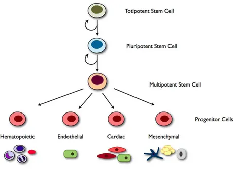
for stem cells-based translational approach to explore the possibility to efficiently regenerate the heart (Gojo et al., 1997; Davani et al., 2005; Smits et al., 2005). Recent reports from different laboratories have shown that the heart of human adult as well as that of other species contains a population of resident stem cells that may differentiate into myoblasts and eventually into adult myocytes (Hierlihy et al., 2002; Abbott and Giordano, 2003; Tateishi et al., 2007). Today, various types of stem cells obtained from different tissues are commonly used for myocardial regeneration (Chiu et al., 1995; Taylor et al., 1998; Ooi et al., 2006; Wu et al., 2007; Piao et al., 2007. For reviews see Marín-García and Goldenthal, 2006; Segers and Lee, 2008). Stem cells are classified in totipotent, pluripotent and multipotent, according to their ability to generate other cell types (Figure 1). More precisely, totipotency refers to the ability of a cell to form cells of all lineages including the extra-embryonic tissue. In mammals, the only cells with this capacity are the zygote and early blastomeres. Pluripotency is the term applied to cells that have the ability to differentiate into all the cell types of the body, except the extra-embryonic tissue (placenta) (Pera, 2001). Multipotent stem cells are those that have the ability to differentiate into a limited number of different cell types (Reyes and Verfaillie, 2001). The stem cells reside in specific regions of tissues to ensure the organ development throughout embryonic, fetal and adult life and divide in case of tissue loss and injury (Nichols, 2001; Thisse and Zon, 2002). A stem cell is clonogenic, capable of unlimited self-renewal by symmetric division, while maintaining a stable diploid karyotype (Linke et al., 2005). Under the control of growth factors and the tissue environment, it is also capable of asymmetric division, one daughter resembling its mother, and one daughter giving rise to multiple types of differentiated cells (Fijnvandraat et al., 2003). In contrast, the progenitor cells that have been identified in adult organs thus far do not meet all of these criteria (Case et al., 2008).
Figure 1 Stem cell hierarchy. Illustration of the stem cell hierarchy. Totipotency refers to the ability of a stem cell to form all cell types including extra embryonic tissue. Pluripotent stem cells are capable of unlimited self-renewal and can differentiate into any adult cell type (e.g. embryonic stem cells (ESCs) and induced pluripotent cells (iPS) cells). Multipotent stem cells have the ability to differentiate into a limited number of different cell types (e.g. hematopoietic stem cells (HSCs)). These multipotent stem cells can give rise to progenitor cells which can be multipotent (e.g. cardiac progenitor cells can differentiate into endothelial cells, smooth muscle cells or cardiomyocytes) or monopotent, giving rise to only one cell type (e.g. endothelial progenitor cells (EPCs)). From Sieveking and Martin, (2009).
Several alternatives approaches, objectives of the regenerative medicine, not mutually exclusive, may be employed to induce heart regeneration as preventing cell death or repopulating myocardium with new contractile cells (Foley and Mercola, 2004) (Figure 2). The most investigated strategy is implantation of stem cells into the heart, although this method is invasive, often clinically unsuitable, and can introduce harmful scar tissue, arrhythmia, calcification, or microinfarction in the heart (Mathur and Martin, 2004). Cells can be injected directly through the coronary arteries or using an intraventricular approach (Zhan-quan et al., 2007). Whit the aim of therapeutic neo-vascularization the cells need to be mobilized from their location with factors such as cytokines in response to an external stimulus, such as tissue ischemia (Wu et al, 2006b).
Figure 2 Repair of damaged heart muscle. From Grounds et al., (2002).
Then they need to successfully home to the site of ischemia and participate in neo-vessel formation. Stem cells treated groups shown improved left ventricular (L.V.) ejection fraction, reduced infarct scar size and decrease L.V. end-systolic volume. A recently developed “cell sheet engineering” technology has greatly improved the efficiency and efficacy of cell engraftment (Masuda et al., 2008). Cell-based cardiac repair offers the promise of rebuilding the injured heart from its component parts (Bai et al., 2007). Work began with the transplantation of authologous skeletal muscle satellite cells (commonly referred as myoblasts), progenitors committed cells that normally mediate regeneration of skeletal muscle (Baroffio et al., 1996; Leor et al., 1996), but recently the field has expanded to explore an array of cell types, including bone marrow cells, endothelian progenitors, mesenchymal stem cells, resident cardiac stem cells, and both mouse and human embryonic stem cells (Jia et al., 1997; Sakai et al., 1999; Rangappa et al., 2002; Sachinidis et al., 2003; Kang et al., 2007) (Figure 3). Recent clinical trials injecting bone marrow or skeletal myoblasts into the injured heart have yielded mixed results in term of cardiac regeneration (Murry et al., 1996; Dorfman et al., 1998; Gulbins et al., 2001; Xu et al., 2007b) and are not able to convert into true cardiomyocytes that could replace those irreversibly loss by heart attack (Atkins et al., 1999).
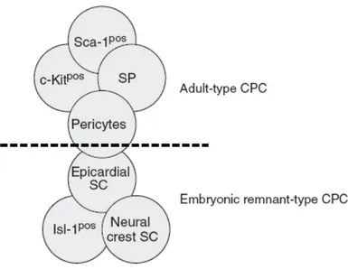
Figure 3 Schematic depiction of the resident cardiac stem cell pool. The majority of Cardiac Progenitors Cells (CPC) represent typical adult stem cells may originate from the bone marrow. They can express c-kit or Sca-1, have SP properties, or be found in the perivascular niche. Other CPC may be remnants from embryologic heart development and express isl-1, display neural crest cell characteristics, or be found within the epicardium. To what extent the two groups and their cell populations overlap is not known. From Stamm et al., (2009).
New insight in experimental cell therapy for heart failure has been given by Messina et al (2004) and Barile et al (2007): in these studies the Authors described the successful achievement of the engraftment and differentiation of the injected cells (adult, autologous, biopsies-derived human cardiac progenitors) in the infracted hearts of SCID mice, with their functional rescue in term of ejection fraction and echocardiogram. An alternative approach is therapeutically stimulating cardiomyocytes to re-enter the cell cycle and progress to mitosis and cytokinesis thought gene-mediated interventions targeting cell cycle regulators, or by injecting genes coding for mitotic cytokines (Bellafiore et al., 2006). In this way, the electromechanical syncytium is more likely to be preserved, since there would be no need for establishing new connections with implanted cells. In addition, the eventuality of rejection of allogenetic cells as well as the ethical problems derived from the use of human embryonic stem cells are avoided (Sugarman, 2007). Although adult mammalian cardiomyocytes show very little or no proliferation when cultured, FGF-1 treatment concomitant with p38 MPA kinase inhibition can stimulate their proliferation in culture. In other studies, the discovery of putative progenitors cells within the hearts of adult mammals has led to the suggestion that the
heart has the potential for homeostatic or regenerative renewal (Nemir et al., 2006). Experimental studies indicate that the delivery or mobilization of stem and progenitor cells may improve tissue perfusion and the contractile performance of the damage heart (Harding et al., 2007; Li et al., 2007b). Directional homing of stem cells is performed via the attraction of cells through the local delivery of various growth factors. Pre-clinical and clinical studies evaluating the therapeutic potential of various cell therapies have reported conflicting results, generating controversy (Min et al., 2002; Shizuru et al., 2005; Sieveking and Martin, 2009). The major barriers to the development of pharmacological agents or cell-based therapies for cardiac repair include obtaining not only an understanding of the identity of the resident cardiac stem/progenitors cells, but also the ability to generate a sufficient number of cells for therapeutic use (Goldenthal and García, 2003). Subsequent challenges involve the optimization of methods for the isolation and delivery of these cells, particularly concerning the timing of cell delivery. Thus, is important to understand how guide the differentiation of stem cells, control their survival and proliferation, identify factors that mediate their homing and modulate the heart’s innate inflammatory and fibrotic responses (Hescheler et al., 1997). The discovery of several types of cardiovascular progenitor cells within the heart provides a unique opportunity to establish pharmacological agents capable of stimulating an increase in the low level of cardiac repair in mammalian hearts, reducing the scar formation and increasing cardiac function (Li et al., 1996; Solloway and Harvey, 2003).
MAMMALIAN HEART: OUTLINE OF STRUCTURE AND REGENERATIVE ABILITY The mammalian heart is a vascular tube construct consisting of more than 20 cell types forming connective, contractile and vascular structures. Specific progenitor cells contribute to formation of the heart during fetal growth. The progenitor cells at the anterior plate mesoderm give rise to the heart. During fetal life, heart is formed from two different committed progenitor cell groups determined according to the expression of specific transcription molecules, and can be classified into primary and secondary heart fields. The primary heart field rises from the anterior splanchnic mesoderm and it gives rises to the heart crescent and later contributes to left ventricle and atrium formation. The secondary cardiac field is also termed the anterior heart field and it originates from the pharyngeal mesoderm located medially at the cardiac crescent. Subsequently it contributes to right ventricle and outflow tract formation (Dyer & Kirby, 2009). The development of a four-chambered heart is a multistep process and depends on unique genetic programs that are highly diverse and unique
for each species. In mice and rats, cardiomyocytes replicate actively during fetal life, but in the perinatal period proliferation ceases and myocytes undergo an additional round of DNA synthesis with nuclear division (karyokinesis), without subsequent cell division (cytokinesis). This leads to binucleation, a form of endoreduplication known as acytokinetic mitosis. The process starts about day four after birth and ends after three weeks, when over 85% of cardiomyocytes are binucleated. With the exception of humans and pigs, in the mammalian species studies so far most cardiomyocytes are binucleated (Klug et al., 1995). Pigs have a majority of tetranucleated myocytes, and cells containing up to 32 nuclei have been reported, but in the human heart most myocytes are mononucleated, and no myocytes containing more than two nuclei have been reported. Mammalian species have little or no ability to replace lost cardiac muscle. This poor regenerative capacity is due in part to the failure of adult cardiomyocytes, the beating cells in the heart, to undergo proliferation (Quaini et al., 2004). In fact, the normal response of mammalian hearts to injury or hypoxia is hypertrophy—the growth of cardiomyocytes without cell division. Although there is evidence that a stem cell-like population may exist in the adult mammalian heart, cell division in this organ is rare. For full-sized adult mammals, homeostasis maintains the status quo, calibrating organ size and function in response to changing physiological conditions, replacing damaged or senescent cells through direct structural cell proliferation, progenitor cell activity or hypertrophy of surrounding cells. Cardiac hypertrophy consists of hypertrophy of cardiomyocytes and hyperplasia of other cell types in the heart, such as fibroblasts (Poss et al., 2002). Indeed, although there is evidence that a stem cell–like population may exist in the adult mammalian heart, cell division in this organ is rare. Prolonged cardiac hypertrophy in humans can lead to dilation, poor contractility, and eventually heart failure and death. Hypertrophy initially results in response to load demands on the heart, and how it progress to cardiac failure is not understood at this time (Nicol et al., 2001). More recently, the discovery of cardiac stem cells and the finding that cardiomyocytes have certain capacities to proliferate challenged that concept and raised significant interest in investigating the molecular mechanisms of myocyte hyperplasia. According to a classical dogmas of mammalian heart regeneration, cardiomyocytes are terminally differentiated and become post-mitotic before or soon after birth and are generally considered to irreversibly withdraw from the cell cycle (Ahuja et al., 2007). Analysis of cardiac myocytes growth during early mammalian development indicates that cardiac myocyte DNA synthesis occurs primarily in uterus, with proliferating cells decreasing from 33% at midgestation to 2% at birth. Throughout life a mixture of young and
old cells is present in the normal myocardium. It was believed that human cardiac myocytes could not self-regenerate after an injury such as acute MI and that the main adaptive response to myocytes loss was hypertrophy, an increase in cardiomyocytes diameter without undergoing cell division. However, this dogma is now being replaced by the idea that there is some in-vivo proliferation of cardiomyocytes after damage. Using a mouse genetic fate-mapping strategy, Hsieh and colleagues (2007) showed strong evidence that stem or progenitor cells refreshed murine cardiomyocytes after heart injury, but not during up to one year of normal aging. Additionally, taking advantages of the incorporation of carbon-14 produced during the Cold War into DNA, Bergmann and colleagues (2009) established a way to determine the age of cardiomyocytes. Using this method, they demonstrated that human cardiomyocytes renew themselves, although at a low frequency. In mammals hearts, after an acute MI, the remaining viable myocardium undergoes a series of structural changes termed cardiac remodeling, consisting in not only in myocardial hypertrophy, but also in myocytes cell loss via apoptosis, coagulative necrosis of the myocardium (myofibrillar hypereosinophilia and loss of nuclei), nuclear ploidy, defective regeneration, infiltration of inflammatory cells such as neutrophils, progressive expansion of the initial infarct area, dilation of the left ventricular lumen and progressive replacement of contractile myocytes by a fibrin deposition at the injury site thought a process that begins around the second week and reaches its maximum about three months after infarction. Then, fibrin is replaced by scar tissue, thought to be permanent, altering left ventricular function. The development, progression and pathogenesis of heart failure is complex and multifactorial. Importantly, remodeling begins with hours of the MI and is initiated by migration of inflammatory cells, mainly macrophages and neutrophilis as well as fibroblasts, which produce tumor necrosis factor (TNF) alpha and transforming growth factor (TGF) (Janczewski et al., 2002). These cytokines stimulates mast cells and cardiac fibroblast proliferation. At the organ level, MI results in thinning of the injured wall and dilatation of the ventricular cavity, a process called ventricular remodeling. These structural changes markedly increase mechanical stress on the ventricular wall and promote progressive contractile dysfunctions. The extent of heart failure after MI is directly related to the amount of myocardium lost. When the heart is submitted to an increased workload, for example after acute MI, the cardiomyocytes enlarge and increase their ploidy status. Since polyploidization implies DNA replication, it can be assumed that adult cardiomyocytes retain the ability to enter into the cell cycle. There are evidences that under certain circumstances adult cardiomyocytes re-enter the cell cycle and advances to
mitosis but the knowledge of the triggering phenomena and the cascade of events leading to cardiomyocyte mitosis is still poor. The four phases of the mammalian cell cycle are tightly regulated at several checkpoints, ensuring that all activities are completed before initiation of the next phase, providing in this way a mechanism for the identification of defective cells. Positive cell cycle regulators (as for example, cyclins, the CDKs, and proto-oncogenes) are highly expressed in embryonic and newborn hearts, and are down-regulated in the adult heart. Various studies have used the approach of genetically targeting the cell cycle regulators to encourage the cardiomyocyte to re-enter the cell cycle and progress into mitosis and cytokinesis (Maltsev et al., 1994). However, whether these cells are derived from a resident pool of cardiomyocyte stem cells or from a renewable source of circulating bone marrow derived stem cells that home to the damaged myocardium is at present not known. Myocytes regeneration is evidenced by a ~2-fold increase in expression of cell cycle markers (Ki67 and phosphohistone H3) and ~13% reduction in mean myocytes diameter. Increased circulating levels of hepatocytes growth factor (HGF), leukemia inhibitory factor (LIF), and macrophage colony-stimulating factor (M-CSF) were associated with mobilization of c-kit+, CD31+, and CD133+ progenitors cells and subsequent increase in myocardial c-kit+ cells. It has been shown that for newt myotubes in tissues culture, the presence of thrombin and serum stimulates myonuclei to re-enter s-phase via phosporylation of the retinoblastoma (mass of undifferentiated/de-differentiated proliferating cells in the growth zone under the wound epidermis formed after damage) of amputated newt limbs (Tanaka et al., 1999). Another integral component of the remodeling process appears to be the development of neo-angiogenesis within the myocardial infarct scar, a process requiring activation of a latent collagenase and other proteinases. Loss of oxygenation to ventricular muscle, usually, because of occlusion of a coronary artery, will result in necrosis and stimulating the new angiogenesis. Moreover, recent observations suggest that a small number of human cardiomyocytes (15-25%) retain the capacity to proliferate and regenerate in response to ischemic injury (Anversa et al., 2006).
ABILITY OF ORGAN REGENERATION IN AMPHIBIAN AND ZEBRAFISH AS COMPARISON WITH MAMMALS
Regeneration is a complex biological process by which animals can restore the shape, structure and function of body parts lost after injury, or after experimental amputation. This process is an evolutionary conserved feature of vertebrate species and requires the concerted action of mechanisms inducing and regulating dedifferentiation, pattern generation, and, in certain instances, trans-differentiation events (Ausoni and Sartore, 2009). Natural scientists have actively pursued the problem of regeneration since the 17th century, largely by utilizing invertebrate and lower vertebrate species possessing exceptional regenerative capacities (Dinsmore, 1991). Newts are the primary experimental model used to study vertebrate regeneration, as they can re-grow a striking number of adult structures, including limbs, tail, spinal cord, jaws, tongue, lens and optic nerve (Brockes, 1997; Ferretti and Géraudie, 1998) (Figure 4). It has long been recognized that Urodele amphibians such as the newt, have a remarkable regenerative ability for many tissues. Consubstantial with survival, tissues and structures of vertebrates cope with everyday wear and tear by being continuously renewed with the progeny of resident or circulating cells, which exhibit a varying degree of plasticity (Wagers et al., 2002). The cellular mechanisms underlying the phenomenon of tissue turnover are highly dependent on the specific type of tissues, and differentiation, but not cell dedifferentiation. The restitution of tissues of structures by increased activity of normal turnover mechanisms is called “tissue restoration”. In contrast with tissues restoration, true regeneration in vertebrates is considered as a type of epimorphic regeneration. It involves the deployment of a complex set of de novo mechanisms including dedifferentiation of post-mitotic cells, cell proliferation, pattern generation and, in some cases, trans-differentiation of adult specialized cells to rebuild parts of the body plan after amputation or injury.
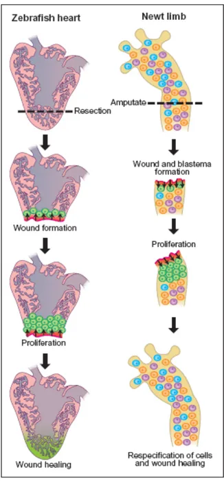
Figure 4. Regenerating hearts and limbs. (Left) Following surgical removal of a portion of the zebrafish ventricle, cardiomyocytes (green cells) adjacent to the wound site (bright red) undergo proliferation, presumably due to signals (arrows) emanating from the wound (Right). In contrast to the heart, the newt limbs is composed of various differentiated cell types (indicated by cells of different colors). Following injury, cells adjacent to the wound epithelium dedifferentiates to form a blastema (green). These cells proliferate and subsequently to re-form a properly patterned limb. From Scott and Stainier, (2002).
Different vertebrate species have different cardiac regeneration rates: high in Teleost fish, moderate in Urodele amphibians, and almost negligible in mammals (Scadding, 1977). Some organs are capable of rapid homeostatic adjustments to tissues loss or gain. Mammals, including humans, exhibit only a few examples of tissues and organs regeneration, most notably the ability of liver hepatocytes to regenerate damaged liver tissue. Progenitor cell populations have been identified within most mammalian organs, including skin, blood, bone, reproductive tissues, skeletal muscle, kidney, lung, liver, intestine, heart and brain. Unfortunately, not all of our organs are equally competent to regenerate, and it is known that heart and central nervous system are particularly resistant to regeneration after injury and that they form scar tissue (Poss et al., 2002). For example, the liver will increase mass through compensatory cell proliferation after partial hepatectomy, or reduce mass through apoptosis after experimentally induced hyperplasia, to maintain the appropriate size. Although robust replacement of structural cells has been discovered in mammalian tissues, such as the liver, blood and skin, most tissues do not share this remarkable ability, like the heart. In animal as salamander, hydra and flatworm, it has been described a huge regenerative capacities indeed, flatworms are able to generate an entirely new animal from a small piece of tissue (Salo and Baguna, 2002). The regeneration on limbs after amputation has been analyzed in detail in these organisms. This process is commonly divided in three sequential steps: (1) wound healing, in which the amputation site is covered by epithelial cells; (2) formation of a mass of undifferentiated progenitor cells, or blastema, by dedifferentiation of cells in the surrounding tissue; and (3) limb redevelopment, in which the correct pattern is generated in the blastema resulting in the regeneration of the amputated portions of the limb (trans-differentiation). The regeneration of limbs and lens in newts, as well as that of fins in zebrafish, relies on the formation, growth and patterning of a blastema. It is well established that, during limb regeneration in newts, multinucleated muscle cells dedifferentiate to adopt muscle or cartilage fates. In addition to limbs, tail, jaw and lens, newts also display the ability to regenerate large portions of their hearts after amputation. Although new heart regeneration does occur to some extent after partial resection of ventricular myocardium, there is only a modest level of tissue replacement that accompanies scarring. Despite the evident significance of understanding this process for the development of potential therapeutic target, heart regeneration in newts has not been addressed from molecular approaches, probably owing to the relative lack of genetic tools in this experimental model. While genetic approaches have been successfully applied to dissect many developmental, physiological and behavioral processes, and could conceivably
be applied to reveal factors required for regeneration, newts and other highly regenerative amphibians are not suitable for this analysis. This is because of their long generation times, enormous genomes, and the difficulty of maintaining large numbers of animals. Nonetheless, selected non-mammalian vertebrates, including Urodele amphibians and Teleost fish, display an elevated regenerative spectrum, with many more tissues capable of impressive regeneration. In particular, because of their amenability to genetic manipulation, zebrafish have proved to be a valuable laboratory model for understanding many aspects of vertebrate embryogenesis (Haffter and Nüsslein-Volhard, 1996). The robust capacity of zebrafish to regenerate a variety of tissues as fins, spinal cord, and retina, position it as an ideal genetic model system for understanding the molecular and cellular events governing regeneration (Wagner and Misof, 1992; Curado et al., 2007).
UTILITY OF USE OF ZEBRAFISH AS MODEL ORGANISM FOR BIOLOGICAL STUDIES AND CARDIAC REGENERATION
Several characteristics have made to use zebrafish as a model organism (Warren et al., 2000), as for instance, the relatively short generation time (3 months), the small size at maturation (3 to 4 cm long as an adult), the simple husbandry requirements, the ease of manipulation and maintenance, and the high fecundity (100-200 eggs per clutch) (Gerhard, 2003). Additionally, external fertilization coupled with optical clarity throughout embryogenesis allow the direct monitoring of developmental processes, and their manipulation using mechanical, chemical, or genetic technique (Nasevicius and Ekker, 2000; Bopp et al., 2006). Moreover, using zebrafish is further supported and facilitated by the sequencing of the zebrafish 1.7-Gb genome (Sanger Institute’s Danio rerio sequencing project), which has facilitated the generation of microarrays for large scale expression profiling (Ton et al., 2002; Lo et al., 2003; Mathavan et al., 2005; Quian et al., 2005). Again, zebrafish can be used as model for pathogenesis study (Neely et al., 2002; Prouty et al., 2003), for hematopoietic and cardiovascular diseases (North and Zon, 2003; Zheng et al., 2008) and for discovering new therapeutic target (Peterson, 2004). Also, zebrafish mutations faithfully phenocopy many human disorders (Goldsmith and Harris, 2003; Meeker and Trede, 2008). Each mutation, once cloned, provides candidate genes and pathways for evaluation in the human (Anderson and Ingham, 2003). The collection of mutations in their entirely potentially provides a medical taxonomy, one based in developmental biology and genetics (Eisen, 1996).
Large-scale genetic screens have been performed in zebrafish after use of ethyl-nitrosourea (ENU) to generate point mutations. More than 7000 mutations in 600 genes have been so generated. Zebrafish orthologos exists for most human genes, and in fact, there are large regions of conserved synteny between chromosomes of fish and mammal (Barbazuk et al., 2000). There is believed to have been a genome duplication at the base of the Teleost radiation 450 million years ago (mya), as well as a second duplication 100 mya, with as many as 20% of genes having been duplicated in the fish as compared to the human. Although such defects are transient and not heritable, morpholinos can be used to assess the effects of diminishing function of orthologs of genes, including those known to be involved in disease, such as dystrophin, presenilin, and apolipoprotein E (For a review see Lieschke and Currie, 2007).
Zebrafish heart share common structures with mammalian hearts, serving as a model for vertebrate animal studies (Wang et al., 1998; Scott and Stainier, 2002; Sehnert and Stainier, 2002). The zebrafish is an ideal organism for the study of heart development because is capable to produce definitive hematopoietic cells and form a simple circulation two-chambered heart (Stainier et al., 1996). Furthermore the detection of perturbations in cardiogenesis and for regeneration because heart formation and function can be assessed visually in the embryo and because the fish is not dependent on blood circulation for survival during embryogenesis (Pelster and Burggren, 1996). Additionally, because the fish is not dependent on blood circulation for survival during embryogenesis (Pelster and Burggren, 1996), and the defects in heart development and/or function are more likely to be detected and recovered. Many zebrafish mutants with defects in cardiac development and function were identified in the large-scale mutagenesis screens and are described in detail elsewhere (Chen et al., 1996). Blood circulation in zebrafish begins by 24hpf and is clearly visible under the microscope. The process of blood development and the morphology of zebrafish blood closely parallels that of mammals (Wingert and Zon, 2003). There are both primitive and definitive waves of differentiation in the zebrafish, which produce primitive erythrocytes and macrophages, followed by definitive erythrocytes, B cells, T cells, monocytes, granulocytes, and thombocytes, respectively (Willet et al., 1999; Bennett et al., 2001; Lawson and Weinstein, 2002; Schoenebeck and Yelon, 2007). Many zebrafish orthologos of blood-specific genes demonstrated to be important in mouse and human development have been isolated, including cmyb, gata1, gata2, globin, hhex, scl, and vegf (Hansen et al., 1997; Gering
et al., 1998; Brownlie et al., 2003). Recently, Holtzinger and Evans (2007) have discovered that gata5 and gata6 in the zebrafish are functionally redundant for specification of cardiomyocytes. In the same year Jia et al., (2007) showed that vertebrate growth is regulated by functional antagonism between Gridlock and Gata5. Gata5 is also required for the development of the heart and the endoderm (Reiter et al., 1999). Although the genetic program appears to be highly conserved during hematopoietic development in the vertebrate embryo, the site of hematopoietic is not as consistent. Whereas primitive hematopoiesis in mice and humans takes place in the yolk sac, in the zebrafish, the primitive wave initiates in a region termed the intermediate cell mass (ICM) (Langeland and Kimmel, 1997; Orkin and Zon, 1997). Definitive hematopoiesis appears to initiate in the dorsal aorta of the zebrafish as it does in all the vertebrate studies to date; however, the site of adult hematopoiesis in the fish is the kidney, not the bone marrow as in mammals (Willet et al., 1997).
In vertebrates, the linear heart tube forms from the migration and fusion of bilateral cardiac progenitors fields at the midline, followed by cardiac looping to form an s-shaped heart. Distinct atrial and ventricular chambers with unique physiological and electrical properties arise, separated by a discrete domain known as the atrio-ventricular canal (AVC) (Stainier, 2001; Beis et al., 2005). The AVC give rise to the valves that ensure unidirectional flow of blood. In mammals, each chamber and valve-forming region becomes septated, resulting in a four-chambered heart. The embryonic heart of the zebrafish has a close anatomic resemblance to that of a human heart at 3 weeks of gestation (Hu et al., 2000; Auman and Yelon, 2004). The zebrafish heart manifests the same pattern of electrical excitation as does the human heart, with impulses generated in the sino-atrial node, propagated through the atrium, pausing in the atrio-ventricular (A-V) node, and thence to the ventricular (Hu et al., 2001; Ho et al, 2002; Beis et al., 2005). Our understanding of heart development has benefited greatly from zebrafish mutants that specifically disrupt cardiovascular form and function (Berdougo et al., 2003; Kinna et al., 2006). Mutations that mimic the most common arrhythmias, including pacemaker problems (slow mo and reggae) (Warren et al., 2001), A-V block (hiphop and breakdance), and atrial fibrillation (island beat, isl) have been described (Baker et al., 1997; Kopp et al., 2005). Indeed, it remains the only laboratory model system that is both amenable to genetic manipulation and capable of carrying out a robust regenerative response after the loss of complex tissue (Ho et al., 2007).
Zebrafish have a population of haemangioblasts, which is absent in chick and mammalian embryos, raising the possibility that these cells represent the evolutionary
ancestror of the second heart field. The genetic programs of these anterior haemangioblasts and the adjacent heart field are co-regulated, by transcriptional factors previously associated with heart but not blood or endothelial development (Chen and Fishman, 2000). The anterior lateral plate mesoderm (ALM) in zebrafish is a source of hematopoietic, endothelial and cardiogenic cells, with the blood and endothelium coming from the most rostral region and cardiac tissue deriving from the adjacent more posterior population. Differences between zebrafish and mouse/chick may in part reflect the different origins of the endoderm: whereas in chick and mouse, yolk sac haematopoiesis and vasculogenesis occur adjacent to the visceral endoderm, in zebrafish the ALM is adjacent to the definitive endoderm.
DEVELOPMENT AND STRUCTURE OF THE ZEBRAFISH HEART
The majority of biological research concerning the zebrafish has focused on its developmental stages because of its transparency of the embryo. The heart is enclosed in the pericardial cavity with paired pericardial muscles running in caudo-cranial direction and is covered by an epicardial membrane. Zebrafish heart develops from a simple tube, which bends and twists (loops) rightward to create the basic plan of the mature heart that consists of only one atrium and one ventricle (1 mm3) composed of two concentric layers: an inner endothelial layer called the endocardium and an outer muscular layer termed the myocardium (Stainier and Fishman, 1992; Stainier et al., 1993; Glickman and Yelon, 2002). Two additional smaller compartments are the sinus venosus (SV) and the bulbus arteriosus (BA) (Forouhar et al., 2006). The embryonic zebrafish heart at 24 hpf is nearly identical to the two-chambered human heart at three weeks of gestation (Fishman and Chien, 1997). It is spontaneously contractile, emptying atrium and then ventricle sequentially to generate unidirectional blood flow (Bendig et al., 2006).
Heart formation involves the specification and differentiation of cardiac precursors, the integration of precursors into a tube, and the remodeling of the embryonic tube to create a fully functional organ (Glickman and Yelon, 2002). Cardiac progenitors can be identified at approximately 11 hours post fertilization (hpf) as marked by nkx2.5 expression, termed the cardiac field (Stainier et al., 1993; Lee et al., 1994; Griffin et al., 2000). Angioblasts in zebrafish are first evident around 14 hpf and the formation of the zebrafish heart begins at approximately 16 hpf when zebrafish cardiac precursors begin to migrate to the midline from bilateral regions of the anterior lateral plate. Each bilateral cardiac sheet contains lateral atrial precursors that express Cardiac myosin light chain 2 (Cmlc2), and medial ventricle precursors
that express both Cmlc2 and Ventricular myosin heavy chain (Vmhc) (Yelon et al., 1999; Yelon, 2001; Huang et al., 2003; Rottbauer et al., 2006). For the embryonic heart development, it is also important the expression of NXT2, member of NXT proteins, involved in exporting nuclear RNA in eukaryotes, as demonstrated by Huang et al., (2005b). The timing of zebrafish myogenesis is regulated by smacd3 (Ochi et al., 2008). For normal myocyte proliferation during early cardiac development is necessary the expression of ndrg4 (Qu et al., 2008). Early cardiac connexin, cx36,7/Ecx, regulates myofibril orientation and heart morphogenesis by establishing Nkx 2.5 expression (Sultana et al., 2008). Hrt is required for cardiovascular development in zebrafish (Szeto et al., 2002). The aorta begins to form in the posterior trunk at 17.5 hpf and complete its formation after the blood circulation starts (Thisse and Zon, 2002). The cone tilts and extends to produce the heart tube, which begins to beat as a peristaltic wave at 22 to 24 hpf (Stainier et al., 1993). Heart formation is completed through a series of remodeling steps that properly position the ventricle and atrium, and produce cardiac valves to ensure unidirectional blood flow through the heart (Stainier et al., 1992; Glickman and Yelon, 2002). By 36 hpf cardiac looping is completed and coordinated contractions of atrium and ventricle provide circulation to the head and trunk (Glickman and Yelon, 2002; Cha and Weinstein, 2007). Growth of a muscular septum late in human embryonic development, which divides the primitive atrium and ventricle into left and right parts, differentiates the adult human from the adult zebrafish heart, the latter retaining a single atrium and single ventricle. By 5 dpf, the larvae heart is essentially formed as noted in the adult Teleost configuration. The four compartments are now separated from another by three sets of fully functional valves (Weinstein and Fishman, 1996). In each compartment, the myocardium is about one cell layer thick except for the ventricle, which consists of two to three cell layers.
More than 35 mutations in the zebrafish prevent the normal acquisition of contractile function, without disturbing the generation of normal chamber cell fate or formation, and therefore functionally mimic dilated cardiomyopathies (Sehnert and Stainier, 2002; Basset and Currie, 2003; Huang et al., 2005a). Pickwick, a mutation in titin, the myofibrillar element around wich the actin-myosin arrays assemble and contributor of most of the elastic force to the muscle, has been cloned. It is interesting to note that a human dilated cardiomyopaty family has also recently been documented to have a titin mutation (Xu et al., 2002). These “heart failure” mutations in zebrafish will reveal not only candidate genes for disease
propensity in humans, but also potential factors and pathways that could be pharmacologically manipulated to improve contractile function (Chun and Chen, 2007).
Concern the contractile function, it is well known that vertebrates have evolved a nerve system to modulate cardiac activity in a sophisticated fashion (Incardona et al., 2004). These activities include heart rates, contractile force, action potential and conduction velocity that can be exposed to several anomalies (Ernest et al., 2000). Congenital anomalies of the heart affect 8 of 1000 live births. Three percent of these are due to inadequate chamber growth. Although there is a high sibling recurrence, suggesting a genetic component, these diseases in general are not transmitted in a Mendelian fashion. Several mutations in zebrafish disrupt generation of early heart form. For example, heart and soul causes the ventricle to form inside of the atrium and is due to a mutation in PKCλ. Handsoff and Pandora have diminutive ventricles. The former due to mutation in the bHLH transcription factor dHAND. Jekyll is due to a defect in the enzyme UDP-glucose dehydrogenase and lacks an A-V valve. Casanova is a sox-related gene that lacks endoderm and causes cardia bifida. The zebrafish homologue of caldesmon (CaD, an actin-binding protein implicated in the cytoskeletral organization and various signaling pathways) is similar to the mammalian low molecular weight caldesmon (l-CaD, associated with the development of tumor vasculature). It is essential for normal vertebrate cardiac looping, chamber formation, muscularization and proper cardiac function. The obvious contribution of CaD to cardiac muscularization offers clues for future therapeutic strategies in regeneration of cardiomyocytes.
Despite a two-chambered heart and lack of pulmonary vasculature the zebrafish heart parallels that of humans in terms of QT interval and heart rate (Arnaout et al., 2007). The atrium is medially dorsal and posterior to the ventricle. Similar to humans, the zebrafish heart is enchased by a pericardial sac in the thoracic cavity, and is situated below the pectoral bone of the pectoral fins. The bulbous arteriosus is analogous to the human aortic arch with thick contractile smooth muscle. Blood returns into the sinus venosus, which is analogous to the vena cava. The fundamental electrical properties are remarkably similar to those of humans, and the critical pathways in cardiovascular development parallel higher vertebrates (Kopp et al., 2007). In 2004, Forouhard at al., reported an electrocardiogram (ECG) of an embryonic zebrafish, revealing similar atrial and ventricular electrical signals as found in a human ECG. It has been demonstrated that zebrafish embryos are sensitive toward a range of QT-prolonging drugs inducing severe arrhythmia (Langheinrich et al., 2003). Ventricular trabeculae are more prominent in the zebrafish heart than in those of humans (Grimes et al.,
2006). Histological studies show that the ventricle of an adult zebrafish is composed of trabecular and compact myocardium, and surrounded by epicardium and endocardium. Using zebrafish mutant tr265/tr265, whose Band 3 mutation disrupts erythrocyte formation and result in anemia, Sun and colleagues (2009a) showed that cardiac hypertrophy involves both myocyte hypertrophy and hyperplasia in anemic zebrafish.
ZEBRAFISH HEART REGENERATION
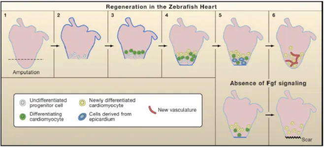
The zebrafish has emerged as an excellent model for cardiovascular research (Curado and Stainier, 2006). Recently, it has been shown that adult zebrafish possess a unique yet poorly understood capacity for cardiac regeneration, restoring in 1-2 months, the ventricular muscle removed by surgical resection, offering new possibilities for experimentally approaching this fascinating biological phenomenon (Lepilina et al., 2006) (Figure 5). The regenerative ability of an adult zebrafish heart was uncovered by surgical resection of a large portion (20-30%) of the ventricular chamber including the apex (Poss et al, 2002). This was easily accomplished in anesthetized adult fish, whose hearts were readily accessible after incision of the skin, muscle, and pericardial sac. The ventricle was then gently pulled at the apex and cut at the desired position with iridectomy scissors. This surgical procedure was highly reproducible and relatively safe, with greater than 80% survival.
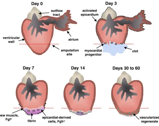
Figure 5. Model for Zebrafish Heart Regeneration Resection of the ventricular apex stimulates rapid expansion of the entire epicardium by 3 dpa (black dots), at which time myocardial progenitor cells first originate in the wound, express pre-cardiac markers, and associate with existing muscle. By 7 dpa, raldh2/tbx18-positive epicardial cells begin to surround and invade the wound. Meanwhile, there is continued seeding, maturation, and proliferation of myocardial progenitor cells, contributing the first layers of new muscle (stage 1). To coordinate these epicardial and myocardial events, regenerating myocardium synthesizes Fgf17b and possibly other factors with the potential to recruit Fgfr2/Fgfr4-presenting epicardial cells. Epicardial-derived cells undergo EMT in response and vascularize the regenerate (green dots). Presence of new coronary vasculature by 14 dpa extends progenitor cell activity and facilitates restoration and expansion of the ventricular wall (stage 2). From Lepilina et al., (2006).
In response to amputation, thrombosis immediately develops to achieve homeostasis in the ventricle. After a short initial phase of intense bleeding that stops within 1 minute, a blood clot formed that sealed the ventricle and kept fish from exsanguinations.The formation of the blood clot in the low-pressure fish circulatory system is remarkably efficient. Probably because of this, amputated fish did not, in contrast to newts, exhibit intense myocardium contraction at the site of resection, or circulatory stasis. First, the clot that seals the apex
matures within several days (1-4 day post-amputation -dpa), into fibrin, a complex milieu containing serum factors and degenerated erythrocytes. Functional cardiomyocytes and new contractile muscle infiltrated the injured area and sealed off the wound. Interestingly, the regenerated myocardium displayed a transiently hypertrophied compact zone, from 21-30 dpa, most likely reflecting a compensatory reaction to the hemodynamic overload subsequent to myocardial loss. Scarring and collagen deposition, characteristic of damaged mammalian hearts, did not occur. Remarkably, 60 days after surgery, the zebrafish heart appeared roughly normal both histologically and based on examination of heartbeat. In contrast, mouse hearts subjected to similar damage induced by freezing do not regenerate, but instead form scar tissue (Wills et al., 2008). In the infarcted mammalian ventricle, fibrin deposition attracts fibroblasts and inflammatory cells, and is a precursor to scarring. The normal response of mammalian hearts to injury or hypoxia is hypertrophy, the growth of cardiomyocytes without cell division. Cardiac hypertrophy refers to the cardiac remodeling process in response to a variety of intrinsic and extrinsic stimuli that stress the heart (Baohua et al., 2006). Initially, the heart compensates for the stress though increasing cardiac mass to normalize wall tension. However, if the underlying stress is untreated, cardiac hypertrophy can lead to sudden death or heart failure. The hallmarks of pathological hypertrophy include enlargement of individual cardiomyocytes, disarray or myofibrils, fibrosis in the extracellular matrix, reactivation of fetal transcriptional programs, and decreased cardiac function (Kitzmann et al., 2006).
The ventricular myocardium displays histological characteristics of hypertrophy at 21 and 31 dpa, most likely reflecting compensatory reaction to the hemodynamic overload subsequent to myocardial loss. Milan et al., (2006) obtained zebrafish ECG by inserting two needle electrodes through the ventral epidermis. They saw that the mean rate of the adult male zebrafish is 151±30 beats/min. Sun and colleagues (2009b) applied a minimally invasive methodology to monitor zebrafish heart function, electrical activities, and regeneration in real-time. They performed a micro-electrocardiograms to study post-ventricular amputation of the zebrafish heart and, unlike the human counterpart, they didn’t observed ventricular tachycardia or fibrillation in response to ventricular amputation 2 and 4 dpa. Atrial arrhythmia was recorder after prolonged sedation. Ventricular amputation led to a shortened QTc interval without affecting the PR and QRS intervals from non-anesthetized and non-paralyzed adult zebrafish. It has been demonstrated that agents known to induce QT prolongation in humans led to QT prolongation in zebrafish embryos and drugs that induce repolarization abnomalies cause bradicardia in zebrafish (Milan et al., 2003). Understanding the cellular and molecular
mechanisms of this regenerative process can have exciting implications for human cardiac biology and diseases.
CELLULAR BASIS OF ZEBRAFISH HEART REGENERATION
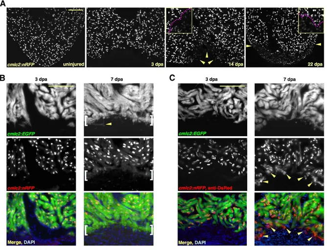
Regeneration of the zebrafish heart appears to differ from that described in other tissues, including the newt limb and zebrafish fin, where differentiate cells (muscle, cartilage, and skin) adjacent to the wound site, first dedifferentiate to form a blastema or mass of pluripotent cells, which then give rise to a fully formed and patterned limb or fin (Biga and Goetz, 2006). The formation of the blastema that regenerates these tissues requires successive process of dedifferentiation, transdifferentiation, and pattern formation. The zebrafish myocardium is a relatively simple structure, composed of one major cell type, so the initial step of dedifferentiation observed during limb regeneration may not be required for cardiac regeneration. Using double transgenic animals with both nuclear-localized DsRed2 fluorescent reporter for cmlc2 (cmlc2:nRFP, where RFP is red fluorescent protein) and enhanced green fluorescent protein (EGFP), that can reveal temporal and spatial characteristics of promoter activation and inactivation, Lepilina and colleagues (2006) demonstrated that new myocardium arises from undifferentiated progenitors cells and maintained by a dynamic epicardial injury response (Figure 6).
Figure 6. New Myocardium Arises from Undifferentiated Progenitor Cells. (A) Sections through uninjured and injured cmlc2:nRFP ventricles. A subpopulation of RFPlo nuclei manifests by 14 dpa (arrowheads), representing ostensibly the entire regenerate by 22 dpa. Magenta line in high-magnification insets delineates RFPhi CMs (above line) from RFPlo CMs. (B) cmlc2:nRFP; cmlc2:EGFP ventricles. EGFP and RFP expression from the cmlc2 promoter is reported at the same basoapical level at 3 dpa. By 7 dpa, an RFPneg front of newly differentiated muscle reporting the faster-fluorescing EGFP (brackets) appears apical to the EGFPposRFPpos portion. Arrowhead indicates an EGFPpos cell process extending into the clot. (C) cmlc2:nRFP; cmlc2:EGFP ventricles stained for DsRed immunoreactivity with an anti-DsRed antibody. At 3 dpa, there is no difference in appearance from the unstained 3 dpa ventricle in (B). By 7 dpa, a front of RFPcyto muscle (arrowheads), representing the most recently differentiated CMs, colabels EGFPpos tissue apical to natural RFPnuc fluorescence. Scale bar = 100 mm. From Lepilina et al., (2006).
To directly investigate the nature of cells that contribute to the regenerating tissue in the zebrafish heart, Poss and colleagues (2002) undertook an exhaustive series of bromodeoxyuridine (BrdU) pulse-chase analyses. Bromodeuxiridine (BrdU), a chemical that is only incorporated into cells undergoing de novo DNA synthesis (S phase), was injected into fish at various time point following the surgery. Cardiomyocytes from uninjured hearts rarely incorporate BrdU, even after prolonged exposure. However, there was a marked increase in BrdU incorporation following injury, which peaked at 14 days after surgery and was primarily localized to the outermost layer of the myocardium adjacent to the wound area. The regeneration resulted from proliferation of cardiomyocytes adjacent to the area of injury. Surprisingly, uninjured zebrafish hearts displayed a significant number of cardiomyocytes labeled with BrdU (Raya et al., 2003), indicating that normal cardiac cell turnover in this animal is much higher than in other organisms. Remarkably, a ten-fold increase in BrdU-labeled cardiomyocytes was detected in the area surrounding the lesion 2 weeks after amputation. These results were further confirmed by staining with anti-phosphorylated histone 3, a marker of condensed chromatin (Poss et al., 2002). The Authors then analyzed the source and fate of regenerating cardiomyocytes by administering BrdU at 7–14 dpa and analyzing labeled cells at 14, 30, and 60 dpa. These studies identified a leading edge of proliferation in the new layer of epicardial cardiomyocytes, which were continuously displaced inward to replenish the amputated region (Poss et al., 2002). The identification of a population of differentiated cardiomyocytes that re-enter the cell cycle and proliferate in response to heart amputation (Poss et al., 2002), together with the absence of molecular markers of de novo cardiomyocyte differentiation in the injured area (Raya et al., 2003), provide strong support for a scenario in which heart regeneration in zebrafish is carried out by resident cardiomyocytes. It has been recently reported that resident stem cells exist in the adult mammalian heart, which can participate in a regenerative response (Beltrami et al., 2003). Whether such cells exist in the zebrafish heart, and indeed whether they contribute to its regeneration after amputation, was not directly addressed in the studies by Poss and colleagues (2002) or by Raya and colleagues (2003). The ventricular resection induces developmental gene expression and proliferation within the epicardial layer, activated epicardial cells soon surround the wound with a portion of them penetrating several cell layers deep into the wound and regenerating muscle. Concomitantly, the new myocardium is substantially vascularized. Thus, it is assumed that the epicardial cells have similar roles as in the embryonic heart; that is, as a progenitor tissue that contributes smooth muscle and/or