Research Article
Vitamin D Effects on Osteoblastic Differentiation of
Mesenchymal Stem Cells from Dental Tissues
Francesca Posa,
1Adriana Di Benedetto,
1Graziana Colaianni,
2Elisabetta A. Cavalcanti-Adam,
3,4Giacomina Brunetti,
2Chiara Porro,
1Teresa Trotta,
1Maria Grano,
2and Giorgio Mori
11Department of Clinical and Experimental Medicine, Medical School, University of Foggia, Foggia, Italy
2Section of Human Anatomy and Histology, Department of Basic and Medical Sciences, Neurosciences and Sense Organs,
University of Bari, Bari, Italy
3Institute of Physical Chemistry, Department of Biophysical Chemistry, University of Heidelberg, Heidelberg, Germany
4Max Planck Institute for Intelligent Systems, Stuttgart, Germany
Correspondence should be addressed to Francesca Posa; [email protected] Received 12 July 2016; Revised 24 August 2016; Accepted 25 September 2016 Academic Editor: Dario Coletti
Copyright © 2016 Francesca Posa et al. This is an open access article distributed under the Creative Commons Attribution License, which permits unrestricted use, distribution, and reproduction in any medium, provided the original work is properly cited. 1𝛼,25-Dihydroxyvitamin D3(1,25(OH)2D3), the active metabolite of vitamin D (Vit D), increases intestinal absorption of calcium and phosphate, maintaining a correct balance of bone remodeling. Vit D has an anabolic effect on the skeletal system and is key in promoting osteoblastic differentiation of human Mesenchymal Stem Cells (hMSCs) from bone marrow. MSCs can be also isolated from the immature form of the tooth, the dental bud: Dental Bud Stem Cells (DBSCs) are adult stem cells that can effectively undergo osteoblastic differentiation. In this work we investigated the effect of Vit D on DBSCs differentiation into osteoblasts. Our data demonstrate that DBSCs, cultured in an opportune osteogenic medium, differentiate into osteoblast-like cells; Vit D treatment stimulates their osteoblastic features, increasing the expression of typical markers of osteoblastogenesis like RUNX2 and Collagen I (Coll I) and, in a more important way, determining a higher production of mineralized matrix nodules.
1. Introduction
Vitamin D (Vit D) is crucial for many biological pro-cesses, that is, the bone mineralization of vertebrates, the maintenance of calcium homeostasis, cell proliferation, and differentiation.
The actions of Vit D are carried out by its active
metabolite, 1,25-dihydroxycholecalciferol [1,25(OH)2D3 or
calcitriol], that is produced through a series of enzymatic
steps starting from cholecalciferol or vitamin D3.
The main portion of vitamin D3 is derived from the
conversion of 7-dehydrocholesterol (Provitamin D) after the exposure of the skin to ultraviolet radiation. Age, skin surface exposed to the sun, thickness, and irradiation time are all factors that control Vit D synthesis [1].
Foods provide only few units of Vit D compared to the amount produced by the skin in response to sunlight.
Vit D is fat-soluble and is absorbed in the duodenum and jejunum and subsequently distributed through the lymphatic circulation [1].
1,25(OH)2D3 exerts its action by binding to a specific
nuclear receptor, that is, the vitamin D receptor (VDR). It is a member of the class II steroid hormones [2].
Mice with targeted ablation of the nuclear VDR (conventional-VDR knockout mice) represent an important
model to study the actions of the system 1,25(OH)2D3/VDR.
The KO mice showed, after weaning, alopecia, an early development of hypocalcemia, which in turn induces a state of secondary hyperparathyroidism, infertility, and severely impaired bone formation: these are typical features recurrent in humans with vitamin D-dependent rickets type II [3–6].
Osteoblasts are among the cells expressing VDR;
there-fore they represent a functional target of 1,25(OH)2D3action.
Volume 2016, Article ID 9150819, 9 pages http://dx.doi.org/10.1155/2016/9150819
1,25(OH)2D3can affect human osteoblast growth and dif-ferentiation stimulating bone formation and mineralization
[7]. Moreover 1,25(OH)2D3modulates bone deposition
pro-cess preventing expro-cessive and pathological mineralization [8].
The effects of 1,25(OH)2D3 on bone matrix protein
expression have been also studied in cultures of calvaria cells, comparing young cells with more differentiated ones for
dif-ferent time periods. Following treatment with 1,25(OH)2D3,
mature osteoblasts were more inhibited, favoring those which were in an earlier state of differentiation [9].
Vit D effects on osteoblast differentiation may be dissim-ilar according to the animal species considered; a discrepant responsiveness has been shown between two categories: human/rat osteoblasts and murine osteoblasts. Thus, in con-trast to the stimulatory effect of Vit D on human and rat osteoblasts, Vit D has been demonstrated to determine an inhibitory effect on murine osteoblasts [10].
Vit D has been reported to induce alkaline phosphatase activity and to increase Collagen Type I expression in the course of proliferation and differentiation of human osteoblasts; this effect is less evident when lower concentra-tions are used [11].
Kveiborg et al. have shown that 1,25(OH)2D3 is able
to offset, in aging osteoblasts in vitro, the reduction of gene expression which is normally required for osteoblast functionality [12].
Osteoblasts precursors are present on periosteal surface and in bone marrow; moreover they can differentiate from Mesenchymal Stem Cells (MSCs).
Adult stem cells are pluripotent cells capable of regenerat-ing different tissues. Bone marrow contains cells with a high clonogenic capacity, the Colony-Forming Units Fibroblasts (CFU-Fs), which give rise to the cells of bone tissue, cartilage tissue, adipose, and fibrous tissues [13]. These CFU-Fs cells are called MSCs.
MSCs can be induced to differentiate into osteocytes, adipocytes, or chondrocytes [14] and also neuronal cells [15] or hepatocytes [16].
It is estimated that about 15% have a “stem” power, and among them there are those who can give origin to osteoblasts [13]. The commitment of MSCs towards differentiated cell lines is regulated by transcriptional mechanisms called mas-ter switches.
RUNX2 is a transcription factor with a key role in the control of osteoblast differentiation and function.
1,25(OH)2D3 is an important regulator of the RUNX2,
with which it cooperates in inducing the expression of osteocalcin, that is, the key protein regulating bone matrix mineralization [17].
There are a lot of data in literature about the effect of Vit D on MSCs isolated from bone marrow, in particular concerning their osteogenic differentiation.
1,25(OH)2D3influences human MSCs (hMSCs)
inhibit-ing cell proliferation and stimulatinhibit-ing their differentiation into osteoblasts [18, 19]. It has also been demonstrated, in hMSCs preimplantation cultures, that Vit D is able to determine the formation of mature osteoblasts, resulting in an increase of mineralized matrix formation, but after implantation of these cells the same Vit D is not sufficient in enhancing bone
formation [20]. That leads to assuming a different in vivo effect of Vit D.
In order to overcome some negative aspects connected with the use of MSCs from bone marrow, that is, morbidity, pain, and low yield of cells, other sources of MSCs have been examined.
MSCs can be isolated from tissues different from the bone marrow such as adipose tissue, brain, skin, liver, and several fetal tissues; little is known about Vit D influence in osteogenic differentiation of MSCs from these alternative sources. Anyway, with MSCs from adipose tissue, the capacity of Vit D in osteoblast formation has been confirmed [21].
According to the evidence based research dental tissues represent an alternative and promising source of postnatal MSCs [22–26].
Deciduous teeth, or the wisdom tooth, that is a tooth with a limited chewing function and that often creates problems of overcrowding, can be used to isolate MSCs. In the case of immature teeth, a productive source of MSC is the dental bud (DB) that consists in noncalcified tissues; in the case of the mature tooth the sources are the periodontal ligament [27], the Dental Pulp (DP) [27–30], and the apical papilla [31].
Dental Pulp Stem Cells (DPSCs) are obtained from wis-dom teeth pulp of adult donors, while Dental Bud Stem Cells (DBSCs) come from the DB. The DB, which is the immature form and therefore not yet fully calcified of the tooth, is an excellent source of stem cells; since it is considerably larger, it is comprised of cells more undifferentiated than the ones composing the pulp and can be removed in children of age between 8 and 12 in case of expected overcrowding with a safe technique called piezosurgery.
The advantage of the tooth bud, compared to the pulp, is first of all dimensional, it contains a greater number of cells, and moreover almost all the tissue is made from stem cells that have a high proliferative capacity and an excellent degree of stemness [24, 32, 33].
The tissues constituting the mature tooth, cement and periodontal ligament, enamel, dentin, pulp, and the central part of the bud, corresponding to the dental papilla, are all derived from DBSCs, obtained from the DB, thus containing MSCs which can successfully differentiate into osteoblast-like cells.
DBSCs are more undifferentiated MSCs, if compared to the ones isolated from the bone marrow; for this reason they can be considered an ideal model for studying the early stages of the osteoblastic differentiation process.
The differentiation into osteogenic lineage has already been demonstrated in DPSCs [27–29, 34] and a considerable mineral matrix deposition has been observed using inno-vative scaffolds for their culturing [35, 36]; moreover these
cells, if cultured on 𝛽-tricalcium phosphate/poly (l-lactic
acid/caprolactone) three-dimensional scaffolds, showed an increased osteogenesis in the presence of Vit D compared to dexamethasone [37].
The aim of this study is to analyze the effect of the active
metabolite of Vit D, 1,25(OH)2D3, on DBSCs which represent
a useful model to understand the anabolic activity of the bone cells, starting from very undifferentiated osteoblasts precursors.
2. Patients, Materials, and Methods
2.1. Materials. 1𝛼,25-Dihydroxyvitamin D3, ascorbic acid, 𝛽-glycerophosphate, dexamethasone, Alizarin Red powder, and alkaline phosphatase kit were from Sigma Aldrich, Milan, Italy.
Anti-RUNX2 antibody was from Abnova, anti-Coll I, anti-OPN, and anti-BSP II were from Abcam, Cambridge, UK.
2.2. Patients and Cell Cultures. Normal human third molar
buds were collected from tooth buds of 10 healthy pediatric patients, 8–12 years of age, that underwent extractions for orthodontic reasons, mainly overcrowding, after informed consent from both patient parents.
The central part of DBs, corresponding to the dental papilla, was cut in small pieces and digested with agitation
for 1 hour at 37∘C in a solution of 3 mg/mL type I collagenase
plus 4 mg/mL dispase (Gibco Ltd., Uxbridge, UK). Single-cell suspension was obtained by passing the Single-cells through
a 70𝜇m BD Falcon strainer (Falcon) (Becton & Dickinson,
Sunnyvale, CA).
After filtration, single-cell suspension was centrifuged at 1300 rpm for 5 min; the pellet was resuspended and cultured in Mesenchymal Stem Cell Culture medium supplemented with 5% fetal bovine serum (FBS), 100 U/mL penicillin-G,
and 100𝜇g/mL streptomycin (Gibco Limited, Uxbridge, UK).
Cells were seeded at a density of5 × 103cells/cm2.
Flasks were incubated at 37∘C and 5% CO2 and the
medium was changed every 3 days.
In order to induce osteoblastic differentiation, 1500
cells/cm2 were seeded and cultured in osteogenic medium
consisting of 𝛼-MEM supplemented with 5% FBS, 10−8M
dexamethasone, and 50𝜇g/mL ascorbic acid (Sigma Aldrich,
Milan, Italy).
For the evaluation of DBSCs ability to form mineralized matrix nodules in vitro, cells were cultured in the osteogenic
medium supplemented with 10 mM 𝛽-glycerophosphate
(Sigma Aldrich, Milan, Italy).
1,25(OH)2D3(Sigma Aldrich, Milan, Italy) was
reconsti-tuted at 10−4M in 95% ethanol and stored at−20∘C.
For all cell cultures, a 95% ethanol (vehicle) control was included at a concentration equivalent to that of Vit D. Thus
cells were cultured in replicates with 1,25(OH)2D3 and an
equivalent dilution of 95% ethanol.
2.3. Alizarin Red Staining (ARS). The ability of DBSCs to
gen-erate mineralized matrix nodules in vitro, a feature normally attributed to osteoblasts, was assessed by performing Alizarin Red Staining on cells cultured for 21 days in osteogenic medium.
Thus, cells were gently rinsed with PBS and fixed with 10% formaldehyde at room temperature for 10 min.
Then they were washed twice with deionized water and incubated with 1% ARS solution for 10 min at room temperature. Cells were afterwards washed twice with deion-ized water, to remove excess staining, and air dried. The monolayer appeared to be red stained.
To quantify the ARS, cells were incubated with 10% acetic acid at room temperature for 30 min with shaking. The cell layer was scraped and vortexed and then the solution
was incubated for 10 min at 85∘C. After 5 min on wet ice,
the suspension was centrifuged at 20,000×g for 15 min and
500𝜇L of the supernatant was treated with 10% ammonium
hydroxide to neutralize the acid. The Optical Density (OD) was read in triplicate at 405 nm.
2.4. Alkaline Phosphatase (ALP). The expression of alkaline
phosphatase, which is a marker of osteoblast differentiation, was assessed with the Leukocyte Alkaline Phosphatase kit (Sigma Aldrich).
Briefly, cell media were removed and cells were fixed in 0.5 mL of a fixative solution for 5 min at room temperature, according to manufacturer’s instructions.
Subsequently, the wells were washed with deionized water and cells were stained with 0.3 mL ALP solution (a mixture of FRV-Alkaline Solution, Naphthol AS-BI Alkaline Solution, Sodium Nitrite Solution) in each well. Following a 15 min incubation in the dark, the wells were washed again with deionized water and air dried and cells were then inspected under the microscope.
2.5. Western Blot. Detection of osteoblastic markers as
pro-tein levels was performed by SDS-PAGE gel electrophoresis and western blot analysis. DBSCs were lysed after 7, 14, and 21 days of osteogenic differentiation; the cell lysates were cleared
with a centrifugation at 13000 rpm for 15 min at 4∘C. The total
protein concentration of the supernatant was determined using a protein assay (BIORAD).
Equal amounts of protein for each sample were separated by SDS-PAGE and transferred to nitrocellulose membranes (Amersham, UK) with a Trans-Blot (Biorad, USA). The
mem-branes were probed with primary antibodies overnight at 4∘C
and then samples were incubated for 90 min with secondary antibodies conjugated to horseradish peroxidase at room temperature. The reaction was analyzed with the Odyssey Infrared Imaging System of LI-COR (LI-COR Biotechnology Lincoln, Nebraska, USA).
2.6. Statistical Analyses. Statistical analyses were performed
by Student’s𝑡-test with the Statistical Package for the Social
Sciences (spssx/pc) software (SPSS, Chicago, IL, USA). The
results were considered statistically significant for𝑃 < 0.05.
3. Results
3.1. ALP Positivity and Calcium-Rich Deposits in DBSCs. In
order to investigate Vit D ability to induce the differentiation of DBSCs into osteoblasts, cells were cultured in osteogenic
medium and stimulated with 1,25(OH)2D310−8M. This
con-centration has been observed as the most efficacious among Vit D physiological concentrations analyzed in previous experiments.
DBSCs cultures were stopped at 7, 14, and 21 days after continuous Vit D treatment in differentiating conditions.
We used histochemical assay to evaluate the expression of alkaline phosphatase, a marker of osteoblastic differentiation, a key enzyme in the process of osteodeposition.
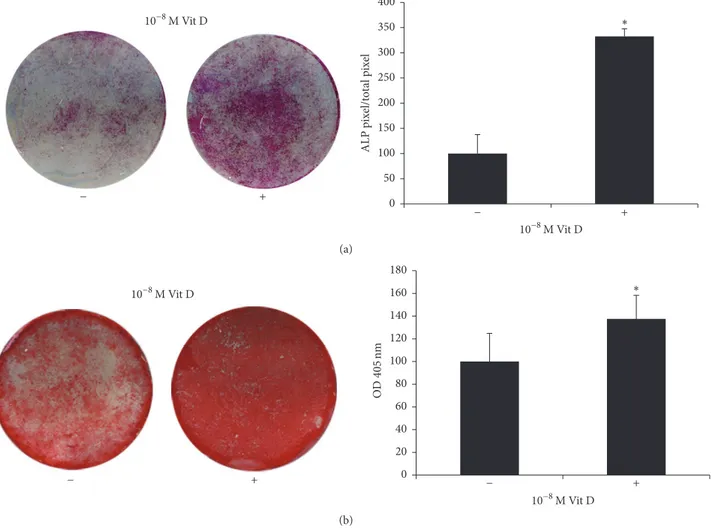
+ − 10−8M Vit D 10−8M Vit D ∗ + − 0 50 100 150 200 250 300 350 400 ALP p ix el/t ot al p ix el (a) + − 10−8M Vit D ∗ 10−8M Vit D + − 0 20 40 60 80 100 120 140 160 180 OD 40 5nm (b)
Figure 1: ALP positivity and mineralized nodules formation. (a) ALP histochemical assay (purple staining) performed on DBSCs seeded and differentiated for 7 days with vehicle (−) and 1,25(OH)2D3(+). The graph shows quantification of positive staining as percentage compared to
untreated cells (−) (∗𝑃 < 0.05) and is representative for 3 independent donors. Data are presented as mean ± SEM. Student’s 𝑡-test was used
for single comparisons. (b) Alizarin Red (red staining) shows mineralized nodules formation by DBSCs incubated for 21 days in osteogenic medium with vehicle (−) and 1,25(OH)2D3(+). The graph shows quantification of ARS with the Optical Density (OD) at 405 nm (b);∗indicates
statistical significance (𝑃 < 0.001). Data are representative for 3 independent donors and are presented as mean ± SEM. Student’s 𝑡-test was used for single comparisons.
We found that DBSCs cultured in an osteogenic medium
and treated with 1,25(OH)2D3expressed higher levels of ALP
after 7 days, compared to those of the control (Figure 1(a)). There was a considerable increase of ALP levels after 14 days, with slight differences between vehicle and treatment (data not shown).
Moreover, long-term cultures of DBSCs demonstrated the capacity to form calcium-rich deposits; this activity, representing the osteoblast final step matrix secretion, was assessed with ARS quantification.
Interestingly DBSCs deposition of mineral matrix nod-ules was significantly higher in cells cultured with Vit D, compared to the control (Figure 1(b)).
3.2. Osteoblast Markers Expression in DBSCs. The main
osteoblastic markers, such as Collagen I (Coll I), its precursor Pro-Collagen I (Pro-Coll I), RUNX2, Bone Sialoprotein
(BSP), and Osteopontin (OPN), were analyzed in DBSCs during the different steps of their osteogenic differentiation.
RUNX2 is the master gene of osteogenic differentiation; it directs MSCs to an osteoblastic lineage and inhibits their differentiation into other lineages such as adipocytes and chondrocytes [38].
As shown in Figure 2(a), RUNX2 expression raised in a quite constant way during DBSCs osteogenic differentia-tion; however the addiction of Vit D greatly increased its expression level after 7 and 14 days of treatment. At 21 days of culture, no difference between vehicle and Vit D was observed.
A trend similar to the one already described for RUNX2 was observed for Coll I (Figure 2(b)): Vit D upregulated its expression in the first 7 days of differentiation; the effect was still present, although attenuated, after 14 and 21 days of treatment.
Runx2 Differentiation (days) Und 7 14 21 56 kD 50 kD 𝛽-Tubulin + − + − + − − 10−8M Vit D Differentiation (days) Und 7 14 21 ∗ ∗ ∗ ∗ ∗ ∗ + − + − + − − 10−8M Vit D # # 0 100 200 300 400 500 600 O D ra tio 700 800 R unx 2/ 𝛽-t ub ulin (a) Coll I 40 kD 𝛽-Actin Differentiation (days) Und 7 14 21 + − + − + − − 10−8M Vit D 70–90 kD Differentiation (days) Und 7 14 21 + − + − + − − 10−8M Vit D ∗ ∗ ∗ ∗ ∗ ∗ # # 0 50 100 150 200 250 300 350 O D ra tio C oll I/ 𝛽-ac tin 400 450 (b) 40 kD Pro-Coll I 𝛽-Actin Differentiation (days) Und 7 14 21 + − + − + − − 10−8M Vit D + − + − + − − − 10−8M Vit D 130–140 kD O D ra tio P ro-C ol l I/ 𝛽-ac tin 0 20 40 60 80 100 120 140 Differentiation (days) Und 7 14 21 ∗ ∗ ∗ ∗ (c) BSP 𝛽-Tubulin 35 kD 50 kD O D ra tio BS P/ 𝛽-t ub ulin Differentiation (days) Und 7 14 21 + − + − + − − 10−8M Vit D 0 20 40 60 80 100 120 140 160 180 200 + − + − + − − − 10−8M Vit D Differentiation (days) Und 7 14 21 ∗ ∗ ∗ ∗ ∗ # ∗# (d) Figure 2: Continued.
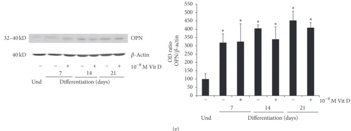
OPN 𝛽-Actin ∗ ∗ ∗ ∗ ∗ ∗ O D ra tio OPN /𝛽 -ac tin 32–40 kD Differentiation (days) Und 7 14 21 + − + − + − − 10−8M Vit D 0 50 100 150 200 250 300 350 400 450 500 550 + − + − + − − − 10−8M Vit D Differentiation (days) Und 7 14 21 40 kD (e)
Figure 2: Protein expression. Immunoblots showing the trend expression of RUNX2 (a), Coll I (b), Pro-Coll I (c), BSP (d), and OPN (e) during the osteogenic differentiation process (0–21 days) of DBSCs cultured in osteogenic medium with vehicle (−) and 1,25(OH)2D3(+).
Each graph represents means± SEM of 3 independent experiments.∗𝑃 < 0.01 compared to undifferentiated samples (und) and#𝑃 < 0.05
compared to untreated samples (−). Student’s 𝑡-test was used for single comparison.
Interestingly Pro-Coll I, the immature precursor form of Coll I, showed a reverse trend (Figure 2(c)): Vit D reduced its expression at 7, 14, and 21 days of culture suggesting that this treatment promotes the conversion of Pro-Coll I in the mature Coll I.
It is remarkable how these data confirmed the typical trends of Coll I and its precursor during osteoblastogenesis, indicating that DBSCs are a reliable model of osteoblastic differentiation: moreover Vit D can sustain and increase this process.
Then we evaluated the expression of OPN and BSP which are considered the most important noncollagenous proteins produced by bone cells during the formation of bone matrix
in vitro [39–41].
The expression of BSP was slightly upregulated after 7 days of Vit D treatment, while at 14 days there was a remark-able reduction; at 21 days BSP did not exhibit significant expression changes (Figure 2(d)).
No significant effect was attributed to the Vit D treatment on the expression of OPN (Figure 2(e)).
4. Discussion
The main actions of Vit D are those concerning mineral and skeletal homeostasis. The protracted deficiency of Vit D has different skeletal consequences in humans deter-mining decreased bone mass and mineralization, with the manifestation of diseases known as Rickets in children and osteomalacia in adults [42, 43]. Low levels of Vit D also result in osteopenia or osteoporosis mainly attributed to an increased osteoclast bone resorption. These effects depend
mostly on the indirect actions of 1,25(OH)2D3. Vit D may
affect the bone indirectly, stimulating the intestinal calcium absorption, or directly, that is, by acting on bone cells; which of the two actions is prevailing is still debated [44] and the
mechanisms whereby Vit D affects osteoblasts are mostly unknown [45].
Some studies have described 1,25(OH)2D3as being able
to increase human osteoblasts mineralization, leading to earlier and higher rate of mineral deposition [46], and able to accelerate hMSCs commitment with the subsequent osteoblast maturation [47, 48].
The bone marrow has been considered the major source of MSCs; however these cells can be obtained from other tissues and organs, which in some cases could represent an easier harvesting site being perceived as less invasive for donors. This is the case of stem cells isolated from the dental tissues.
Contrary to other sources used in adults to obtain MSCs, the dental tissues are formed at an older age, this is due to the late completion of odontogenesis and tooth eruption; consequently these tissues contain a higher amount of stem cells that have been discovered to be multipotent cells [22, 49]. DPSCs and DBSCs can be isolated from the third molar, in adult donors or in children, respectively. The dental bud is an immature organ, which originates several years after birth and is composed of undifferentiated cells with a higher proliferation rate than that observed for MSCs from bone marrow.
DBSCs fully satisfy the requirements to be considered MSCs; in fact it has been shown that they express more than 95% of mesenchymal stem markers (CD44, CD73, CD90, CD105, CD146, and HLA-I) and express the typical mesenchymal adhesion molecules [33]. Therefore all these mesenchymal features of DBSCs, together with their easy accessibility, make MSCs from dental tissues an excellent substitute to the bone marrow cells.
We have already demonstrated that osteoblastogenesis can successfully, and in a very productive way, take place from dental follicle, which is the peripheral part of the dental
bud [32] and from dental bud [33]. These cells differentiate toward an osteoblastic phenotype; they express RUNX2, Coll I, and ALP, characteristic osteoblast markers, and produce mineralized matrix nodules.
In this work we analyzed how Vit D could influence the osteogenic differentiation of DBSCs. To this purpose we studied the expression of typical osteoblastic markers and mineral matrix deposition during DBSCs osteogenic
differentiation in the presence, or not, of 1,25(OH)2D3. Our
results confirmed the functional osteogenic differentiation of DBSCs [33]; in addition in this work we demonstrated that Vit D treatment enhances the commitment of DBSCs toward osteoblastic lineage. Indeed we observed that DBSCs treated
with 1,25(OH)2D3 expressed increased levels of the main
osteoblastic markers, RUNX2, Coll I, and ALP. Furthermore our results showed that enhanced commitment of DBSCs in presence of Vit D was also accompanied with an augmented production of mineralized matrix.
The enhanced expression of RUNX2, which is an early marker of osteoblastic differentiation, indicated that Vit D acts on uncommitted cells, prompting them to differentiate toward osteogenic lineage and then to express the typical osteoblastic markers ALP and Coll I. These proosteogenic
effects exerted by 1,25(OH)2D3 on undifferentiated
mes-enchymal progenitor cells result subsequently in an acceler-ated formation of mineralized matrix nodules in vitro after 21 days of differentiation.
Our results also showed that the expression of RUNX2 and Coll I was accentuated in cells treated with Vit D during the early phases of osteogenic differentiation (7–14 days), while their expression turned to the control levels around 21 days of culture, indicating that the effects of Vit D are predominant at the beginning of the culture.
These results suggest that Vit D probably acts on the first steps of MSCs differentiation toward the osteoblastic phenotype, becoming less efficacious on differentiated cells. Our data are in line with previous data on human osteoblasts that the effect of Vit D depends on the time and the cells differentiation phase seeming to vanish in ongoing mineralization [46].
Our observations demonstrated that also stem cells from dental tissues respond to Vit D signal and are consistent with the data in literature which attribute to Vit D a key role in inducing osteogenic differentiation of MSCs [18, 47, 50] from human bone marrow. These results confirm that MSCs from dental tissues share similar features with MSCs from bone marrow and suggest that cultures of DBSCs in presence of Vit D could be taken in consideration for bone regenerative therapies.
Thus these data reflect the mesenchymal origin of DBSCs and their osteogenic capacity; moreover this study shows that osteoblastic differentiation of DBSCs was stimulated
by 1,25(OH)2D3; our observations suggest that Vit D acts
directly on these cells that can be considered osteoblast precursors, directing them to an increased bone matrix deposition.
The point of force of this work is that DBSCs are postnatal stem cells more undifferentiated than those isolated from bone marrow, that is, adult stem cells comparable to
embryonic stem cells. The limit of our data is that all these conclusions have been already reported for MSCs from bone marrow; they are anyway a confirmation.
5. Conclusion
It is known that the efficacy of calcium intake on osteoporosis and fracture prevention is conditioned by the concomitant assumption of Vit D. Without adequate Vit D assumption only the 10–15% of the calcium can be utilized for building new bone [51].
Our finding that Vit D stimulates osteoblastic differentia-tion of DBSCs with the subsequent increase of bone mineral matrix deposition suggests in addition a possible use of Vit D as food aid in reconstructive therapies of bone with MSCs.
Widening the 1,25(OH)2D3 intake in the population
could be recommend for both preventive and therapeutic purposes in bone diseases and trauma.
Abbreviations
MSCs: Mesenchymal Stem Cells
DB: Dental bud
DBSCs: Dental Bud Stem Cells
Vit D, 1,25(OH)2D3: Vitamin D
VDR: Vitamin D receptor Coll I: Collagen I Pro-Coll I: Pro-Collagen I BSP: Bone Sialoprotein OPN: Osteopontin DP: Dental Pulp
DPSCs: Dental Pulp Stem Cells
DFSCs: Dental Follicle Stem Cells
ALP: Alkaline phosphatase
ARS: Alizarin Red Staining.
Competing Interests
The authors have declared that no competing interests exist.
Acknowledgments
Funding for this work was awarded from Ministero dell’Is-truzione, dell’Universit`a e della Ricerca—PRIN 20098KM9RN Mori (Progetto di Ricerca d’Interesse Nazionale—Grant 2009); EACA is grateful for the support from the DFG (SFB TRR 79 TPB5); the author A. Di Benedetto is funded by Fondo di Sviluppo e Coesione 2007-2013, APQ Ricerca Regione Puglia “Programma regionale a sostegno della specializzazione intel-ligente e della sostenibilit`a sociale ed ambientale-FutureInRe-search”.
References
[1] M. F. Holick, “Resurrection of vitamin D deficiency and rickets,”
Journal of Clinical Investigation, vol. 116, no. 8, pp. 2062–2072,
2006.
[2] H. F. DeLuca, “Overview of general physiologic features and functions of vitamin D,” The American Journal of Clinical
[3] Y. C. Li, A. E. Pirro, M. Amling et al., “Targeted ablution of the vitamin D receptor: an animal model of vitamin D-dependent rickets type II with alopecia,” Proceedings of the
National Academy of Sciences of the United States of America,
vol. 94, no. 18, pp. 9831–9835, 1997.
[4] K.-I. Takeyama, Y. Yamamoto, and S. Kato, “VDR knockout mice and bone mineralization disorders,” Clinical Calcium, vol. 17, no. 10, pp. 1560–1566, 2007.
[5] T. Yoshizawa, Y. Handa, Y. Uematsu et al., “Mice lacking the vitamin D receptor exhibit impaired bone formation, uterine hypoplasia and growth retardation after weaning,” Nature
Genetics, vol. 16, no. 4, pp. 391–396, 1997.
[6] R. G. Erben, D. W. Soegiarto, K. Weber et al., “Deletion of deoxyribonucleic acid binding domain of the vitamin D recep-tor abrogates genomic and nongenomic functions of vitamin D,”
Molecular Endocrinology, vol. 16, no. 7, pp. 1524–1537, 2002.
[7] J. van de Peppel and J. P. T. M. van Leeuwen, “Vitamin D and gene networks in human osteoblasts,” Frontiers in Physiology, vol. 5, article 137, 2014.
[8] V. J. Woeckel, B. C. J. Van der Eerden, M. Schreuders-Koedam, M. Eijken, and J. P. T. M. Van Leeuwen, “1𝛼,25-dihydroxyvitamin D3 stimulates activin A production to fine-tune osteoblast-induced mineralization,” Journal of Cellular
Physiology, vol. 228, no. 11, pp. 2167–2174, 2013.
[9] C. G. Bellows, S. M. Reimers, and J. N. M. Heersche, “Expression of mRNAs for type-I collagen, bone sialoprotein, osteocalcin, and osteopontin at different stages of osteoblastic differentiation and their regulation by 1,25 dihydroxyvitamin D3,” Cell and
Tissue Research, vol. 297, no. 2, pp. 249–259, 1999.
[10] M. van Driel and J. P. T. M. van Leeuwen, “Vitamin D endocrine system and osteoblasts,” BoneKEy Reports, vol. 3, article 493, 2014.
[11] M. van Driel, H. A. P. Pols, and J. P. T. M. van Leeuwen, “Osteoblast differentiation and control by vitamin D and vita-min D metabolites,” Current Pharmaceutical Design, vol. 10, no. 21, pp. 2535–2555, 2004.
[12] M. Kveiborg, S. I. S. Rattan, B. F. C. Clark, E. F. Eriksen, and M. Kassem, “Treatment with 1,25-dihydroxyvitamin D3 reduces impairment of human osteoblast functions during cellular aging in culture,” Journal of Cellular Physiology, vol. 186, no. 2, pp. 298–306, 2001.
[13] A. Friedenstein, “Osteogenic stem cells in bone marrow,” in
Bone and Mineral Research, J. N. M. Heersche and J. A.
Kanis, Eds., pp. 243–272, Elsevier Science, Amsterdam, The Netherlands, 1990.
[14] M. F. Pittenger, A. M. Mackay, S. C. Beck et al., “Multilineage potential of adult human mesenchymal stem cells,” Science, vol. 284, no. 5411, pp. 143–147, 1999.
[15] M. Dezawa, H. Kanno, M. Hoshino et al., “Specific induction of neuronal cells from bone marrow stromal cells and application for autologous transplantation,” The Journal of Clinical
Investi-gation, vol. 113, no. 12, pp. 1701–1710, 2004.
[16] J. M. Luk, P. P. Wang, C. K. Lee, J. H. Wang, and S. T. Fan, “Hepatic potential of bone marrow stromal cells: development of in vitro co-culture and intra-portal transplantation models,”
Journal of Immunological Methods, vol. 305, no. 1, pp. 39–47,
2005.
[17] Y.-L. Li and Z.-S. Xiao, “Advances in Runx2 regulation and its isoforms,” Medical Hypotheses, vol. 68, no. 1, pp. 169–175, 2007. [18] P. Lui, B. O. Oyajobi, R. G. G. Russell, and A. Scutt, “Regulation
of osteogenic differentiation of human bone marrow stromal
cells: interaction between transforming growth factor-𝛽 and 1,25(OH)2 Vitamin D3in vitro,” Calcified Tissue International, vol. 65, no. 2, pp. 173–180, 1999.
[19] S. Geng, S. Zhou, and J. Glowacki, “Effects of 25-hydroxyvita-min D3 on proliferation and osteoblast differentiation of human marrow stromal cells require CYP27B1/1𝛼- hydroxylase,”
Jour-nal of Bone and Mineral Research, vol. 26, no. 5, pp. 1145–1153,
2011.
[20] I. J. De Kok, K. C. Hicok, R. J. Padilla, R. G. Young, and L. F. Cooper, “Effect of vitamin D pretreatment of human mesenchy-mal stem cells on ectopic bone formation,” The Journal of Oral
Implantology, vol. 32, no. 3, pp. 103–109, 2006.
[21] L. V. Logovskaya, T. B. Bukharova, A. V. Volkov, E. B. Vikhrova, O. V. Makhnach, and D. V. Goldshtein, “Induction of osteogenic differentiation of multipotent mesenchymal stromal cells from human adipose tissue,” Bulletin of Experimental Biology and
Medicine, vol. 155, no. 1, pp. 145–150, 2013.
[22] S. Gronthos, M. Mankani, J. Brahim, P. G. Robey, and S. Shi, “Postnatal human dental pulp stem cells (DPSCs) in vitro and
in vivo,” Proceedings of the National Academy of Sciences of the United States of America, vol. 97, no. 25, pp. 13625–13630, 2000.
[23] G. Mori, G. Brunetti, A. Ballini et al., “Biological characteristics of dental stem cells for tissue engineering,” Key Engineering
Materials, vol. 541, pp. 51–59, 2013.
[24] A. Di Benedetto, C. Carbone, and G. Mori, “Dental pulp stem cells isolation and osteogenic differentiation: a good promise for tissue engineering,” Methods in Molecular Biology, vol. 1210, pp. 117–130, 2014.
[25] M. Miura, S. Gronthos, M. Zhao et al., “SHED: stem cells from human exfoliated deciduous teeth,” Proceedings of the National
Academy of Sciences of the United States of America, vol. 100, no.
10, pp. 5807–5812, 2003.
[26] A. Ballini, S. Capodiferro, M. Toia et al., “Evidence-based den-tistry: what’s new?” International Journal of Medical Sciences, vol. 4, no. 3, pp. 174–178, 2007.
[27] A. Ballini, G. De Frenza, S. Cantore et al., “In vitro stem cell cul-tures from human dental pulp and periodontal ligament: new prospects in dentistry,” International Journal of
Immunopathol-ogy and PharmacolImmunopathol-ogy, vol. 20, no. 1, pp. 9–16, 2007.
[28] G. Mori, G. Brunetti, A. Oranger et al., “Dental pulp stem cells: osteogenic differentiation and gene expression,” Annals of the
New York Academy of Sciences, vol. 1237, no. 1, pp. 47–52, 2011.
[29] G. Mori, M. Centonze, G. Brunetti et al., “Osteogenic properties of human dental pulp stem cells,” Journal of Biological Regulators
and Homeostatic Agents, vol. 24, no. 2, pp. 167–175, 2010.
[30] E. Giorgini, C. Conti, P. Ferraris et al., “FT-IR microscopic anal-ysis on human dental pulp stem cells,” Vibrational Spectroscopy, vol. 57, no. 1, pp. 30–34, 2011.
[31] W. Sonoyama, Y. Liu, T. Yamaza et al., “Characterization of the apical papilla and its residing stem cells from human immature permanent teeth: A Pilot Study,” Journal of Endodontics, vol. 34, no. 2, pp. 166–171, 2008.
[32] G. Mori, A. Ballini, C. Carbone et al., “Osteogenic differentia-tion of dental follicle stem cells,” Internadifferentia-tional Journal of Medical
Sciences, vol. 9, no. 6, pp. 480–487, 2012.
[33] A. Di Benedetto, G. Brunetti, F. Posa et al., “Osteogenic differentiation of mesenchymal stem cells from dental bud: role of integrins and cadherins,” Stem Cell Research, vol. 15, no. 3, pp. 618–628, 2015.
[34] A. Ballini, F. Mastrangelo, G. Gastaldi et al., “Osteogenic differentiation and gene expression of dental pulp stem cells
under low-level laser irradiation: a good promise for tissue engineering,” Journal of Biological Regulators and Homeostatic
Agents, vol. 29, no. 4, pp. 813–822, 2015.
[35] F. Paduano, M. Marrelli, L. J. White, K. M. Shakesheff, M. Tatullo, and X. Liu, “Odontogenic differentiation of human dental pulp stem cells on hydrogel scaffolds derived from decellularized bone extracellular matrix and collagen type i,”
PLoS ONE, vol. 11, no. 2, Article ID e0148225, 2016.
[36] M. Marrelli, G. Falisi, A. Apicella et al., “Behaviour of dental pulp stem cells on different types of innovative mesoporous and nanoporous silicon scaffolds with different functionalizations of the surfaces,” Journal of Biological Regulators and Homeostatic
Agents, vol. 29, no. 4, pp. 991–997, 2015.
[37] R. Khanna-Jain, B. Mannerstr¨om, A. Vuorinen, G. K. B. S´andor, R. Suuronen, and S. Miettinen, “Osteogenic differentiation of human dental pulp stem cells on𝛽-tricalcium phosphate/poly (l-lactic acid/caprolactone) three-dimensional scaffolds,”
Jour-nal of Tissue Engineering, vol. 3, no. 1, pp. 1–11, 2012.
[38] T. Komori, “Regulation of osteoblast differentiation by tran-scription factors,” Journal of Cellular Biochemistry, vol. 99, no. 5, pp. 1233–1239, 2006.
[39] T. Nagata, C. G. Bellows, S. Kasugai, W. T. Butler, and J. Sodek, “Biosynthesis of bone proteins [SPP-1 (secreted phosphoprotein-1, osteopontin), BSP (bone sialoprotein) and SPARC (osteonectin)] in association with mineralized-tissue formation by fetal-rat calvarial cells in culture,” Biochemical
Journal, vol. 274, no. 2, pp. 513–520, 1991.
[40] T. Nagata, H. A. Goldberg, Q. Zhang, C. Domenicucci, and J. Sodek, “Biosynthesis of bone proteins by fetal porcine calvariae in vitro. Rapid association of sulfated sialoproteins (secreted phosphoprotein-1 and bone sialoprotein) and chondroitin sul-fate proteoglycan (CS-PGIII) with bone mineral,” Matrix, vol. 11, no. 2, pp. 86–100, 1991.
[41] S. Kasugai, R. Todescan Jr., T. Nagata, K.-L. Yao, W. T. Butler, and J. Sodek, “Expression of bone matrix proteins associated with mineralized tissue formation by adult rat bone marrow cells in vitro: inductive effects of dexamethasone on the osteoblastic phenotype,” Journal of Cellular Physiology, vol. 147, no. 1, pp. 111– 120, 1991.
[42] M. F. Holick, “High prevalence of vitamin D inadequacy and implications for health,” Mayo Clinic Proceedings, vol. 81, no. 3, pp. 353–373, 2006.
[43] M. F. Holick, “Vitamin D deficiency: what a pain it is,” Mayo
Clinic Proceedings, vol. 78, no. 12, pp. 1457–1459, 2003.
[44] D. D. Bikle, “Vitamin D and bone,” Current Osteoporosis Reports, vol. 10, no. 2, pp. 151–159, 2012.
[45] R. St-Arnaud, “The direct role of vitamin D on bone homeosta-sis,” Archives of Biochemistry and Biophysics, vol. 473, no. 2, pp. 225–230, 2008.
[46] V. J. Woeckel, R. D. A. M. Alves, S. M. A. Swagemakers et al., “1𝛼,25-(OH)2D3acts in the early phase of osteoblast differenti-ation to enhance mineralizdifferenti-ation via accelerated production of mature matrix vesicles,” Journal of Cellular Physiology, vol. 225, no. 2, pp. 593–600, 2010.
[47] E. Piek, L. S. Sleumer, E. P. van Someren et al., “Osteo-transcriptomics of human mesenchymal stem cells: acceler-ated gene expression and osteoblast differentiation induced by vitamin D reveals c-MYC as an enhancer of BMP2-induced osteogenesis,” Bone, vol. 46, no. 3, pp. 613–627, 2010.
[48] S. Zhou, J. Glowacki, S. W. Kim et al., “Clinical characteristics influence in vitro action of 1,25-dihydroxyvitamin D3in human
marrow stromal cells,” Journal of Bone and Mineral Research, vol. 27, no. 9, pp. 1992–2000, 2012.
[49] G. T.-J. Huang, S. Gronthos, and S. Shi, “Mesenchymal stem cells derived from dental tissues vs. those from other sources: their biology and role in regenerative medicine,” Journal of Dental
Research, vol. 88, no. 9, pp. 792–806, 2009.
[50] S. Zhou, M. S. LeBoff, and J. Glowacki, “Vitamin D metabolism and action in human bone marrow stromal cells,”
Endocrinol-ogy, vol. 151, no. 1, pp. 14–22, 2010.
[51] M. F. Holick and M. Garabedian, “Vitamin D: photobiology, metabolism, mechanism of action, and clinical applications,” in Primer on the Metabolic Bone Diseases and Disorders of
Mineral Metabolism, pp. 106–114, American Society for Bone
and Mineral Research, Washington, DC, USA, 6th edition, 2006.
Submit your manuscripts at
http://www.hindawi.com
Hindawi Publishing Corporation
http://www.hindawi.com Volume 2014
Anatomy
Research International
Peptides
Hindawi Publishing Corporation
http://www.hindawi.com Volume 2014
Hindawi Publishing Corporation http://www.hindawi.com
International Journal of
Volume 2014
Zoology
Hindawi Publishing Corporation
http://www.hindawi.com Volume 2014
Molecular Biology International
Genomics
International Journal of Hindawi Publishing Corporation
http://www.hindawi.com Volume 2014
The Scientific
World Journal
Hindawi Publishing Corporationhttp://www.hindawi.com Volume 2014
Hindawi Publishing Corporation
http://www.hindawi.com Volume 2014
Bioinformatics
Advances inMarine Biology
Journal ofHindawi Publishing Corporation
http://www.hindawi.com Volume 2014 Hindawi Publishing Corporation
http://www.hindawi.com Volume 2014
Signal Transduction
Journal ofHindawi Publishing Corporation
http://www.hindawi.com Volume 2014
BioMed
Research International
Evolutionary Biology International Journal of
Hindawi Publishing Corporation
http://www.hindawi.com Volume 2014
Hindawi Publishing Corporation
http://www.hindawi.com Volume 2014
Biochemistry Research International
Archaea
Hindawi Publishing Corporation
http://www.hindawi.com Volume 2014
Hindawi Publishing Corporation
http://www.hindawi.com Volume 2014 Genetics
Research International
Hindawi Publishing Corporation
http://www.hindawi.com Volume 2014
Advances in
Virology
Hindawi Publishing Corporation http://www.hindawi.com
Nucleic Acids
Journal ofVolume 2014
Stem Cells
International
Hindawi Publishing Corporation
http://www.hindawi.com Volume 2014
Hindawi Publishing Corporation
http://www.hindawi.com Volume 2014
Enzyme
Research
Hindawi Publishing Corporation
http://www.hindawi.com Volume 2014
International Journal of