Feeding behaviour and digestive physiology in larval fish:
current knowledge, and gaps and bottlenecks in research
Ivar Rønnestad1, Manuel Yu´fera2, Bernd Ueberscha¨r3, Laura Ribeiro4, Øystein Sæle5andClara Boglione6
1 Department of Biology, University of Bergen, Bergen, Norway
2 Instituto de Ciencias Marinas de Andalucı´a (ICMAN-CSIC), Puerto Real, Ca´diz, Spain 3 Helmholtz Centre for Ocean Research Kiel– GEOMAR, Kiel, Germany
4 Aquaculture Research Center of Portuguese Institute of Sea and Atmosphere, Olha˜o, Portugal 5 National Institute of Nutrition and Seafood Research (NIFES), Bergen, Norway
6 Department of Biology, University of Rome ‘Tor Vergata’, Rome, Italy
Correspondence
Ivar Rønnestad, Department of Biology, University of Bergen, Pb 7803, 5020 Bergen, Norway. Email: ivar.ronnestad@bio.uib.no Received 15 April 2012; accepted 23 August 2012.
Re-use of this article is permitted in accordance with the Terms and Conditions set out at http://wileyonlinelibrary.com/ onlineopen#OnlineOpen_Terms
Abstract
Food uptake follows rules defined by feeding behaviour that determines the kind and quantity of food ingested by fish larvae as well as how live prey and food par-ticles are detected, captured and ingested. Feeding success depends on the pro-gressive development of anatomical characteristics and physiological functions and on the availability of suitable food items throughout larval development. The fish larval stages present eco-morpho-physiological features very different from adults and differ from one species to another. The organoleptic properties, dimensions, detectability, movements characteristics and buoyancy of food items are all crucial features that should be considered, but is often ignored, in feeding regimes. Ontogenetic changes in digestive function lead to limitations in the abil-ity to process certain feedstuffs. There is still a lack of knowledge about the diges-tion and absorpdiges-tion of various nutrients and about the ontogeny of basic physiological mechanisms in fish larvae, including how they are affected by genetic, dietary and environmental factors. The neural and hormonal regulation of the digestive process and of appetite is critical for optimizing digestion. These processes are still poorly described in fish larvae and attempts to develop optimal feeding regimes are often still on a ‘trial and error’ basis. A holistic understanding of feeding ecology and digestive functions is important for designing diets for fish larvae and the adaptation of rearing conditions to meet requirements for the best presentation of prey and microdiets, and their optimal ingestion, digestion and absorption. More research that targets gaps in our knowledge should advance larval rearing.
Key words: absorption, digestion, gut, ingestion.
Introduction
The principal objectives of this review are (i) to analyse current knowledge, research trends and efforts in basic and applied aspects of the feeding behaviour and digestive phys-iology of fish larvae focusing primarily on marine species and (ii) to identify the gaps and bottlenecks that need to be tackled in future research on advanced and more efficient production of fish larvae.
Several recent reviews have covered various aspects of larval development, with emphasis on feed acquisition and digestive processes (i.e. Morais et al. 2007;
Rønnes-tad et al. 2007b; Yu´fera & Darias 2007a; RønnesRønnes-tad & Morais 2008; Finn & Kapoor 2008; Gisbert et al. 2008; Zambonino-Infante et al. 2008; Holt 2011). These reviews illustrate the advances made in knowledge from different perspectives, highlighting the essential role of feeding and digestive organs, as well as behaviour and the digestive machinery that enables rapid growth (larvae multiply their body mass to adult size by a factor of 105–107; Finn et al. 2002), in addition to the anatomical status and transformations exhibited by fish during the first weeks of life. Although there is a substantial litera-ture regarding the direct effect of some abiotic factors
such as temperature on exogenous feeding activity, less attention has been paid to the impact of temperature, illumination and feeding protocol in a rhythmic or cir-cadian perspective (Yoseda et al. 2008; Villamizar et al. 2009). The potential importance of environmental and endogenous rhythms and transient conditions on food uptake and feeding physiology is recognized but not well understood, as are even less so their implications for nutritional composition, growth rates and survival. Another important aspect that should be taken into account is the variety in ontogeny, feeding physiology and behaviour even within the same family. Thus, spe-cies-specific findings for a process or function in a model species cannot be extrapolated directly to other teleosts and specific validation studies are essential.
It is obvious that better knowledge of larval feeding behaviour and digestive physiology will contribute to the optimization of diets and feeding protocols and will eventually improve growth rates and survival, even in many common aquaculture species. An integrated under-standing of the various factors and events interacting in food acquisition and digestion is necessary for the design of diets that meet the requirements for optimal inges-tion, digestion and absorption. Not least, better knowl-edge in this area would mean reductions in the costs of fry production.
In comparison with earlier reviews of this field, this review focuses more tightly on identifying and describing the most significant gaps in our understanding of larval rearing that relate to feeding and digestion and on recom-mending future research strategies in the area.
Feeding behaviour and appetite
Good knowledge of the complexity of feeding behaviour and of the factors that modulate food detection, acqui-sition and processing is essential if we are to improve feeding protocols with live prey and to improve the design of microdiets to replace these. Successful food consumption depends, on the one hand, on a series of anatomical characteristics and physiological functions that should be ready to work at the appropriate time, and on the other, on the availability of appropriate food items. Given that the main activities of fish larvae are to eat and avoid being eaten, the differentiation of sen-sory organs, mouth and digestive elements and the capacity for locomotion is of primary importance (Osse & van den Boogaart 1999; Boglione et al. 2003; Yu´fera & Darias 2007a).
Feeding behaviour comprises several activities that take place before and after consumption of a food item. In a hatchery scenario they can be summarized in terms of detection, capture and ingestion, and digestion and
assimi-lation. This review discusses them according to this sequence.
Detection
Fish larvae receive information from their environment via their sensory organs. The type and intensity of their responses depend on the nature of the stimulus, the stage of development and the species. Food is detected via a wide range of chemical (olfaction and taste buds), visual (eyes) and mechanical (free neuromasts and lateral line) stimuli. Olfaction detects the most distant stimuli while touch and gustation detect the closest ones (Pavlov & Kasumyan 1990). Vision plays the most prominent role in prey detec-tion in the larvae of most fish species (Hunter 1981; Blaxter 1986). However, some species are capable of eating and growing independent of light, at low light intensities, in turbid waters or in the dark (Chesney 1989; Downing & Litvak 2001; Mukai et al. 2008). These species can detect food with the help of the olfactory epithelium and taste buds (chemical stimuli), neuromasts and lateral line (mechanical) and/or electroreceptive ampullary (electrical) organs. Furthermore, scotopic (low light) vision varies among species and developmental stages and changes in the light spectrum may affect activity, feeding behaviour and larval growth (Villamizar et al. 2009; Blanco-Vives et al. 2010).
The timing and sequence of development of sensory tis-sues and organs differs among species. In most fish, the rudimentary sensory organs develop rapidly in the course of the first few days after hatching and permit prey to be detected at first feeding (O’Connell 1981; Fukuhara 1985; Evans & Browman 2004; Pankhurst 2008). These organs continue to develop and new tissue appears in order to optimize prey detection in accordance with the body’s new capabilities (O’Connell 1981; Fukuhara 1985; Osse & van den Boogaart 1999).
The fish visual system is capable of wavelength discrimi-nation, high spatial acuity, motion detection and both sco-topic (low light intensity) and phosco-topic (high light intensity) vision, but each species has its peculiar attributes. There is also significant inter- and intra-specific variation in the developmental trajectory of fish visual systems (Evans & Browman 2004) and what we report here cannot be extended to all fish larvae. The fish larval eye is usually functional at the onset of feeding and in Atlantic halibut (Hippoglossus hippoglossus) (Helvik et al. 2001) at least, the retina is equipped with green-sensitive single cones as the only type of photoreceptor. This structure provides rela-tively good vision and object detection under bright light. Double and mosaic cone structures and rod photoreceptors generally appear later in development. With the structure of the retina complete the larvae can see at low light
intensities and are able to detect movement (Kawamura et al. 1984; Higgs & Fuiman 1996; Shand et al. 2002; Evans & Browman 2004). As development progresses (according to species), the diameter of the eye increases, providing a wider angle of vision and longer-distance perception (Hunt von Herbing & Gallager 2000; Chesney 2008).
Visual disorders and pathologies related to nutritional deficiencies have been described in several fish species (Hughes 1985): the importance of retinoic acid for retinal development in larval fish was demonstrated by Marsh-Armstrong et al. (1994) and Rønnestad et al. (1999). A lack of docosahexanoic acid affects rod formation (Bell & Dick 1993; Shields et al. 1999) and reduced vision under this dietary deficiency has been reported in several species such as herring (Clupea harengus) (Bell et al. 1995), European sea bass (Dicentrarchus labrax) (Bell et al. 1996), Atlantic halibut (Shields et al. 1999) and gilthead seabream (Sparus aurata) (Benı´tez-Santana et al. 2007). Some recent trends in aquaculture are based on the use of 24 h artificial light-ing or differently coloured tank walls in order to enhance the ingestion rates and growth of larvae and juveniles; if these practices are employed in the absence or ignoring of knowledge of larval ecology (i.e. larvae that develop in deep, dark waters or in surface waters), or the ocular struc-ture and capacity of the species concerned, there is a possi-bility of increasing stress in the larvae and undermining the intention of the measure.
The first olfactory sensory cells generally differentiate at hatching, and olfaction plays an active role in prey detec-tion in most larvae from first feeding (Dempsey 1978; Knutsen 1992; Kolkovski et al. 1997a). The olfactory organ comprises a number of different types of sensory cells (Hara & Zielinski 1989; Hara 1993, 2005; Devitsina & Kazhlayev 1995; Boglione et al. 2003). Olfaction, in combi-nation with the detection of current direction by the lateral line system, enables the odour source to be located: olfac-tory stimuli trigger a feeding excitaolfac-tory state that starts and maintains klinotactic swimming activity until the food is located (Valentincˇicˇ 2005). This is particularly relevant when live prey characteristics are to be mimicked in micro-diets. Leaching hydrosoluble compounds such as amino acids from inert food particles after rehydration can improve their acceptability to larvae (Kolkovski et al. 2000). Therefore, although excessive nutrient leakage should be avoided, particles should not be completely hermetic.
Olfactory sensory cells differentiate very early. Thus, cili-ated and/or microvillous receptors develop during the embryonic stages, and aggregate in two placodes or pits at hatching. However, the olfactory bulbs are believed to dif-ferentiate later in larval development, and only when they are connected with olfactory receptors do larvae acquire the ability to detect chemical stimuli. At present, data are
lacking for most species. As larvae grow, the primordial placodes or pits continue to develop, becoming the nares and olfactory epithelium (Noakes & Godin 1988; Tanaka et al. 1991; Boglione et al. 2003; Kawamura et al. 2003). As olfaction takes place in water, fish primarily perceive small water-soluble molecules. Most of the substances that stimu-late olfactory and gustatory sensory cells are characterized by low molecular weight (<1000 Da) non-volatility; they tend to be nitrogenous and amphoteric (Hara 1993). All these characteristics apply to amino acids, betaines, nucleo-tides and other substances similar to amino acids. The amino acids that stimulate the chemical senses of fish, even in mixture, are species-specific and exist in a large number of kinds and concentrations, without any identifiable pat-tern. Fish may be grouped according to the sensitivity of their olfactory cells to different amino acids: (i) wide response range species, which respond to a wide range of amino acids (e.g. channel catfish (Ictalurus punctatus), red sea bream (Pagrus major), black mullet (Mugil cephalus); (ii) limited response range species, sensitive to a limited number of amino acids (Salmonidae, Cyprinidae and other freshwater species) (Marui & Caprio 1992; Hara 1993). Any diet administered to fish larvae should therefore take into account the species-specific sensitivity to and attractiveness of chemical substances in the diet, particularly if new candi-date species for aquaculture are involved. Unfortunately, such data are currently only available for a few species.
The other chemoreceptors involved in feeding behaviour, the taste bud system, are extremely sensitive to a smaller number of chemical stimuli. These trigger reflex responses such as turning and biting/snapping (Valentincˇicˇ 2005). While the olfactory chemo-detection of food is involved in long-range detection, taste buds intervene in determining palatability; the external taste buds play a role in the preli-minary determination of the suitability of food, intervening in the decision whether to seize or reject an item. The taste receptors located in the mouth are more sensitive (Kasum-yan & Kazhlayev 1993) and detect a wider range of sub-stances than the inner buds, which are involved in the final verification of the quality of the prey seized (swallow or reject) (Pavlov & Kasumyan 1990). Although taste buds may be present at the opening of the mouth in some spe-cies, they usually proliferate days or weeks after the onset of feeding (Boglione et al. 2003; Cobcroft & Pankhurst 2006; Sa´nchez-Amaya et al. 2007); their implication in the food acceptation is thus more relevant at later stages, e.g. in Sparidae. In sharpsnout seabream (Diplodus puntazzo), the external and internal taste buds differentiate only during the post-larval phase (5–28.6 mm total length) (Boglione et al. 2003). The rainbow trout taste system is narrowly tuned, as it detects only a small number of amino acid stimuli and alone does not release feeding excitation, since either a visual or an olfactory input is also necessary
(Valentincˇicˇ 2005). Very few data exist on the ontogenesis of taste buds in reared fish, and the individual ontogeneti-cal stages of different types of taste bud still need to be defined.
Mechanical stimuli such as touching or water move-ments are detected by neuromasts, which may be either free-standing or sunk within the lateral line system. In lar-val fish, a few free neuromasts are already present at hatch-ing, and these proliferate during growth and development (Higgs & Fuiman 1996; Kawamura et al. 2003). Originating from the neural crest, they early differentiate near the audi-tory capsules in the lateral cephalic region soon after hatch-ing, and then develop and migrate to the trunk and the head. Both the ratio of superficial neuromasts to those sunk in canals or in single pores and the pattern of the canal sys-tem differ according to species and during development. At first, they differentiate as free superficial neuromasts with a mucopolysaccharidic cupula that covers the stereo- (non sensory) and cnido- (sensory) cilia. They are involved not only in schooling, in the timing of responses to a predator stimulus, in detecting currents, in obstacle avoidance and in controlling swimming movements (Blaxter et al. 1983), but also in larval food detection (Mukai et al. 1994; Myr-berg & Fuiman 2002). In fact, neuromasts are able to detect a low range of frequencies (about 70 Hz vs 50–200 Hz) and small velocities (0.03 mm s 1 vs 0.3–20 mm s 1) by organisms in the surrounding water: swimming and feeding movements of various types of zooplankton produce fre-quencies in the range of 10–50 Hz (Myrberg & Fuiman 2002); motile particles can stimulate neuromasts from a relatively short distance, 0.5–3 mm (Appelbaum & Riehl 1997; Harvey et al. 1992; Jones & Janssen 1992; Poling & Fuiman 1998; Webb 1989). Furthermore, the location of the free neuromasts on the head and trunk reveals the range within which the larvae can perceive prey movements; Iwai (1980) considered the symmetrical head arrangement of free neuromasts as typical of larvae that pursue their prey through the water column.
As the larvae develop, most of the free neuromasts move deeper into the derma, at first forming a groove and then becoming enclosed in the intradermal canal, opening to the exterior via canal pores. The canalization extends the sensi-tivity of this system to a wider range of frequencies and velocities. Unlike vision, the potential effects of nutritional deficiencies on olfactory epithelium, taste bud and neuro-mast development have yet to be explored.
Capture and ingestion
Both the mouth apparatus and swimming ability are important for the capture and ingestion of food. In most cultured species, teeth and the ability to bite appear during the later stages of the larval phase (Kohno et al. 1983;
Koumoundouros et al. 2000; Boglione et al. 2003) and therefore, larval fish ingest food primarily by swallowing directly the particle identified as food by the sensory organs. The selection of prey is mainly done by choosing sizes that can be swallowed as well as prey organisms that display inadequate escape behaviour. Mouth gape and oesophagus diameter are therefore defining factors in food-particle preferences during development (Busch 1996; Yu´fera & Darias 2007a; Russo et al. 2009). More specifi-cally, the prey size/mouth gape ratio determines the accessi-bility of a given prey. Thus, although fish larvae may ingest preys that are similar in size to the mouth gape (Russo et al. 2009), ratios ranging between 25% and 60% have been reported as positively selected in several species (Shi-rota 1970; Ferna´ndez-Dı´az et al. 1994; Østergaard et al. 2005). When food items are considered as 3-D objects, it is found that width and depth tend to differ, and that depth is the usually smallest dimension. In this way, preys are ingested whose depth corresponds to the width of the mouth. This hypothesis is consistent with those of Ham-bright (1991) regarding largemouth bass (Micropterus sal-moides), Blaxter (1965) for herring, Qin and Hillier (2000) for snapper (Pagrus auratus) and Economou (1991) for five gadoid species.
The range of accessible particle sizes increases as the larva grows. However, the ingested prey also needs to supply mass and energy. The growing larvae presumably try to ingest the maximum quantity of nutrients required for stable growth, while maintaining a favourable ratio between the energy gained by the ingestion of prey and that spent in capturing it (Hunt von Herbing et al. 2001; Puvanendran et al. 2004). Thus, the developing larvae gradually move to larger and more energy-rich prey. Yu´fera et al. (1995), who tested a widely accepted experimental microdiet, found that gilthead seabream larvae ingested a greater mass of inert food than live prey, probably to compensate for the lower nutritional value of the microdiet.
In cultured larvae, suitable prey size is obtained by sup-plying progressively larger strains of rotifers, followed by Artemia nauplii and metanauplii (Polo et al. 1992; Olsen et al. 2000). In some species with very small mouths, other zooplankton species have been tested at first feeding, although not at industrial scale (Strøttrup & Norsker 1997; Russo et al. 2009; Wullur et al. 2009). This is not a prob-lem with microdiets, since a wide range of particle diame-ters can easily be produced.
Locomotory capacity during the early larval stages depends largely on the length and development of anatomi-cal features such as the notochord (which becomes the ver-tebral column), fins and muscular system of the trunk. These structures gradually develop; total length increases, notochord flexion occurs, then the primordial finfold pres-ent at hatching is replaced within a few weeks by the lateral,
median and caudal fins, differentiated vertebrae replace the notochord in sustaining the body, the swim-bladder is inflated, and the size and number of muscle white fibres also increases (Fukuhara 1985; Blaxter 1986; Webb & Weihs 1986; Vegetti et al. 1990). Swimming capacity is also highly dependent on hydrodynamic conditions and larval body shape. The relationship between inertial and viscous forces in the water, characterized by the Reynolds number, depends on the size and shape of the growing larvae (Webb & Weihs 1986). During the first days of feeding, when the total length of marine fish larvae ranges between 2 and 4 mm, frictional forces dominate (Webb & Weihs 1986; Batty & Blaxter 1992) and the speed and duration of swim-ming episodes are limited (Hunt von Herbing & Gallager 2000). The inertial forces become more relevant after noto-chord flexion and caudal fin development, usually occurring between 5 and 6 mm of total length. After these develop-mental events swimming resistance notably increases. As fish larvae grow, they develop better swimming perfor-mance (Webb & Weihs 1986; Fuiman et al. 1999).
During the first days of feeding with limited locomotory capacity, the detection and capture of prey is highly depen-dent on encounter opportunities. As swimming capacity improves, the larvae can actively search for food and pursue prey, the maximum reaction distance increases as does the distance covered in burst pursuit of the prey (Blaxter 1986; Hunt von Herbing & Gallager 2000; Chesney 2008). In other words, attack and feeding success rates rise as larvae grow.
Swimming work involves the rhythmic contractions of the segmentally arranged myotomes helped by fins (John-ston 2006). Restricted feeding and dietary imbalances nega-tively affect several of these structures, reducing locomotion and foraging capacity. The negative effects on ossification lead to abnormal bone development. Malfor-mations in the spinal column and fin rays have been observed in several species fed on inadequate amounts of certain classes of lipids (Cahu et al. 2003a), vitamins A and D (Haga et al. 2002; Villeneuve et al. 2006; Ferna´ndez et al. 2008, 2009; Darias et al. 2010) or unbalanced amino acid composition (Akiyama et al. 1986; Saavedra et al. 2009). Nutritional effects on skeletal anomalies have been covered in recent reviews by Boglione (2012) and Hamre et al. (2012). Likewise, myotome structure is immediately affected by feeding restrictions: stunting can easily be observed in larvae kept without food after the opening of the mouth or poorly fed during growth (Margulies 1993; Yu´fera et al. 1993; Gisbert et al. 2004). In some species, deficient lipid nutrition also appears to induce functional disorders in the swim-bladder (Kitajima et al. 1981; Katavic´ 1986). Irregular bladder inflation affects the larva’s capacity to remain at the desired level in the water column, leading to higher energy expenditure and seriously interfering with feeding.
Feeding rhythms
As mentioned above, most fish larvae are visual feeders and thus depend on a minimum illumination level to detect and ingest prey. Day/night cycles thus mark feeding rhythmicity in larval fish. Some studies of wild popula-tions have indicated that larval fish feed preferentially at sunrise and sunset (Kane 1984; McLaren & Avendano 1995) and also show the highest digestive enzyme capacity at night (Ueberscha¨r 1995). This may be attributed to the particular light intensity of these crepuscular hours and to nycthemeral migration of zooplankton. An adequate com-bination of the appropriate irradiance for the visual search and prey encounter rate has been suggested as a determi-nant of feeding success in larval Atlantic cod (Gadus mor-hua) (Fiksen & Jørgensen 2011). Laboratory studies have also revealed a daily feeding pattern in spite of the continu-ous presence of prey (Dou et al. 2000; Ma et al. 2006; Ko-tani & Fushimi 2011). These daily rhythms differ according to species. During the first week of life, most fish species feed during the day, while feeding ceases during the hours of darkness. Ingestion can be greater at sunrise and during the early hours of morning, at sunset or at both of these times of day (Shoji et al. 2001; Østergaard et al. 2005; Wang et al. 2008). This rhythmic feeding activity may be maintained even under continuous 24 h illumination (Yo-seda et al. 2008), or may disappear (Dou et al. 2000). The daily feeding pattern may also change throughout develop-ment as observed in tongue sole (Cynoglossus semilaevis) (Ma et al. 2006) and loach (Misgurnus anguillicaudatus) (Wang et al. 2008). Feeding may thus be continuous, diur-nal, nocturnal or preferentially crepuscular. The feeding pattern may change in the course of development and some species exhibit different pre-metamorphosis and post-metamorphosis patterns. Finally, the larvae may have endo-genous rhythms that are strongly influenced by exoendo-genous factors such as light level or the time at which food is more accessible.
Neuroendocrine control of appetite and ingestion
The physiological regulation of appetite and ingestion involves a complex integration of peripheral and central signals by the brain. As in mammals, the centre that regulates ingestion and energy homeostasis in fish seems to be located in the hypothalamus (Demski & Northcutt 1983). The appetite-regulating hormones described in fish so far have been reviewed in several papers (Lin et al. 2000; Volkoff et al. 2005, 2009, 2010; Volkoff 2006). Many of the mammalian neuropeptides and hor-mones involved in the central regulation of appetite have homologues in fish, and several neuropeptides in goldfish have a regulatory effect on appetite similar to
that described in mammals (e.g. Volkoff et al. 2009, 2010).
Satiety signals from the gastrointestinal tract (GI tract), including the stomach if present, have a major impact on appetite and meal size in both juvenile and adult stages. Studies of rainbow trout (Oncorhynchus mykiss), for instance, have shown that appetite returned when 80–90% of the stomach content from the previous meal had been transferred downstream into the proximal gut (Grove et al. 1978). Several GI tract hormones seem to be involved together with neural pathways in this satiety feedback mechanism. Furthermore, the central regulator of appetite also receives afferent signals regarding the energy stored in the body. It has been suggested that leptin produced in the liver and fat plays an important role in this signalling func-tion in fish (Murashita et al. 2011) (Table 1).
There are almost no studies of how neuropeptides and endocrine factors affect appetite and ingestion in larvae. In Atlantic cod, one study describes gene expression at the whole-larva level during ontogeny as well as the effects of the dietary regime (Kortner et al. 2011a,b), although analy-sis of whole larvae has limitations (see below). In adult fish, the list of appetite-regulating endocrine factors cur-rently includes orexigenic factors such as neuropeptide Y (NPY), apelin and galanin, and a long list of anorexigenic factors such as cocaine-amphetamine-regulated transcript (CART), cholecystokinin (CCK) and amylin (Volkoff et al. 2010). The precise physiological role of each factor is not yet properly understood, and has been explored in only a few species. What we know of mammalian systems suggests that the list of endocrine factors that affect appetite and energy homeostasis in fish will be significantly extended and that it will include both central and peripheral signal-ling pathways. Adding to the complexity are the integration and interactions of the various factors involved. However, one of the key questions in larval stages is when these regulatory loops become functional. The fact that larvae continue to feed in spite of having a full gut suggests that satiety factors may play a relatively minor role, at least in the first feeding stages. Under natural feeding conditions, these larvae may never encounter prey densities that are high enough to permit ingestion rates equal to those in intensive culture. It is not yet known whether altricial fish larvae possess a system that regulates appetite and to what extent it depends on feedback from the digestive tract (for satiety) and/or from the body (nutrients/energy stored/ adiposity).
Adaptation of feeding protocols to the feeding behaviour Larval feeding activity and digestion capacity define food preferences, daily ration and daily feeding rhythm. Good knowledge of all these processes, which are changing during
the transition to the juvenile stage, is needed for the design of appropriate feeding protocols for each species. Identify-ing the optimum temporal sequence for the different prey types (usually rotifers and Artemia nauplii and metanau-plii) during development has been the aspect of feeding behaviour most often examined in all reared species. Simi-larly, the questions of how much food should be offered and how often it should be supplied to the rearing tanks have been studied since the start of larvi culture. Providing sufficient food has been always a priority. Feeding protocols tend to maintain a permanent, or at least a better than min-imum, prey density by supplying food twice or more often per day. However, ‘when and how often’ are important issues that have not been sufficiently studied. Protocols for supplying live planktonic prey tend to be adapted to work-ing hours, or diurnal period. This protocol works for most species although obviously it can be optimized. Besides, as mentioned above, some species develop nocturnal habits. An inappropriate feeding schedule may limit ingestion, either because the larvae do not eat or do not detect the prey at feeding time or because they are already satiated. In both cases, the prey will spend hours in the tanks before being eaten, with a consequent drop in nutritional quality (Romero-Romero & Yu´fera 2012). On the other hand, in many species that tend to eat continuously during the early stages of life, an excess of food during the feeding period may shorten the gut passage time, while the prey are less efficiently digested (for details see section Gut transit rate vs dietary protein utilization).
Digestive physiology
Once larvae have captured and ingested their food, the role of the digestive system is to reduce it to very simple mole-cules (absorbable units) that are transported across the intestinal epithelium into the blood. Understanding the functions and limitations in processing capacity of the digestive system during early life stages continues to be a key area for aquaculture-related research on fish larvae. Ontogeny and plasticity of the digestive system
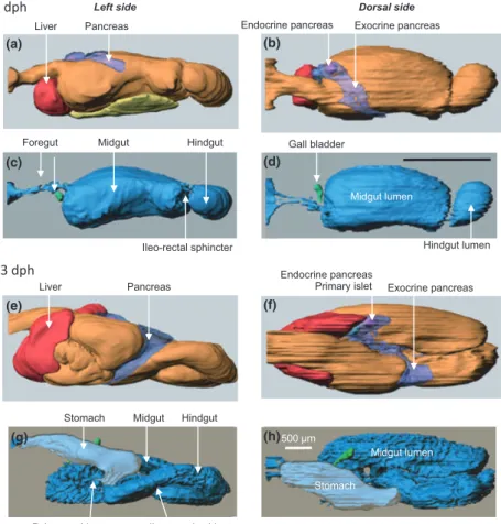
An ample literature describes the gross morphological ontogeny of the fish digestive tract. In recent years, these descriptions have gradually gone into more detail as new tools become available to describe the embryogenesis of the GI tract and its associated organs. Recently, 3D models of the digestive tract at various developmental stages have started to become available (Fig. 1). Fish larvae are often categorized on the basis of the presence of a stomach: agas-tric fish will not develop a stomach, even in the adult stage, precocial fish have a functional stomach at the onset of first feeding, while altricial larvae develop a functional stomach
during metamorphosis. Most research on feeding-related physiology has targeted altricial larvae, among those most of the marine species common in aquaculture, since they present the largest challenge to successful rearing.
In altricial fish, the digestive tract gradually develops from a short and straight tube, often closed at both the mouth and anal ends in yolk-sac larvae, into a segmented and histologically differentiated tract in juvenile fish. At the onset of exogenous feeding the digestive tract has histologi-cally and functionally distinct regions, the bucco-pharynx, oesophagus, stomach anlage, intestine and anus. Some authors use a different terminology and group these differ-ent regions into the foregut, midgut and hindgut. At the onset of exogenous feeding, the gut length is often less than half the body length. The midgut is individually separated from the foregut and hindgut by muscular sphincters. With few exceptions, the gut is coiled into a loop before exoge-nous feeding begins. In the subsequent phase of develop-ment, the digestive tract increases its absorptive capacity through elongation and mucosal folding. The wall of the tract also increases in thickness due to a thicker gut epithe-lium and maturation of enterocytes that includes develop-ment of the intestinal brush border with larger microvilli.
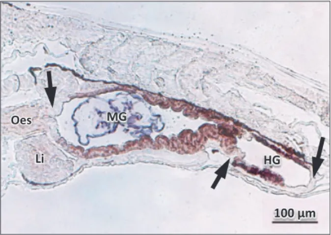
The shape of the midgut may differ from a narrow tube. A 3D model of the GI tract of the Atlantic cod shows the rela-tively large volume of the anterior midgut, resulting in a bulb-like appearance of this compartment (Kamisaka & Rønnestad 2011; Fig. 1c,d). This feature probably increases the residence time of food and enables good mixing with secretions from the pancreas and bile that enter close to the oesophagus. Systematic studies linking morphology with functional studies of the digestion could provide valuable insight into the process, particularly at a general level.
During the transition period from larva to juvenile, the stomach epithelium becomes structurally differentiated, glands develop and by the end of this period gastric diges-tion has been established. However, the pyloric sphincter in Japanese flounder (Paralichthys olivaceus) for instance, seems to be functional before the onset of metamorphosis (Rønnestad et al. 2000b), which suggests that the stomach may start to function as a short-term reservoir before assuming its role as an acid proteolytic chamber. It has been suggested that the appearance of gastric glands acts as a marker to indicate the juvenile stage (Tanaka 1973). However, gastric glands may develop some days before HCl- and pepsinogen-secretion mechanisms are established
Dorsal side Left side
Liver Pancreas
Midgut lumen Endocrine pancreas Exocrine pancreas
Ileo-rectal sphincter
Pylorus sphincter Ileo-rectal sphincter
Foregut Hindgut Midgut
500 m
Midgut lumen Stomach
Stomach Hindgut Midgut
Liver Pancreas
Endocrine pancreas
Primary islet Exocrine pancreas Gall bladder Hindgut lumen 53 dph 4 dph (a) (b) (c) (d) (e) (f) (g) (h)
Figure 1 Three-dimensional model of the digestive tract with associated organs in Atlantic cod, an altricial fish that does not possess a stomach at first feeding. 4 dph, first feeding larvae; 53 dph, later stages of metamorphosis (modified from Kamisaka & Rønnestad 2011).
(Douglas et al. 1999; Darias et al. 2005, 2007a), leading to a gap between gastric gland appearance and its secretory function.
Pyloric appendices (caeca) develop during metamorpho-sis, usually by the time that gastric glands develop. The caeca increase the volume of the anterior midgut and also the absorptive area of the digestive tract and may be impor-tant for protein and fat absorption. In some species, changes in enterocytes during ontogeny include enhanced brush border digestive capacity, due to increased activity of membrane-associated enzymes that contribute to a change in digestive abilities of the organism. In European sea bass, this shift occurs 3–4 weeks after hatching (Zambonino-Infante et al. 2008) and is believed to represent a shift to the adult mode of digestion. There is an elongation of the digestive tract during the larval stages, and this seems to continue for some species after metamorphosis. There are small changes in gut morphology after metamorphosis and these may include further increases in its relative length and, possibly, adaptation to changes in diet.
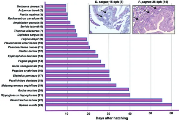
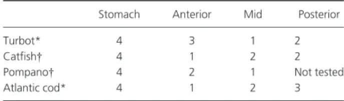
The anatomical and functional ontogeny of the digestive tract is similar in most teleosts once the above-mentioned differences between precocious and altricial, and gastric and agastric species are taken into account. However, there are important inter-species variations in the sequence of appearance of some tissues and enzymatic activities. The most relevant difference is probably the timing of stomach development. The point at which the first gastric glands appear ranges from a few days to several months after first feeding, even within the same family (Fig. 2). This event marks the beginning of the end of the larval mode of diges-tion. After the appearance of gastric glands and pepsin activity, the completion of the stomach and full acidic digestion capacity takes some weeks or months, depending on the species. However, in the Senegalese sole (Solea senegalensis) in spite of the presence of gastric glands, the acidification process has not been detected in either juve-niles or adults (Yu´fera & Darias 2007b). The species differ-ences are presumably evolutive adaptations to the diets they encounter in their natural environment and the appar-ently ‘abnormal’ functionality in sole might be explained by their diet of invertebrates that continues into the adult stage.
In conclusion, the ontogeny of the digestive tract has been described in a number of species by means of methods that have usually targeted gross morphology, histology and to a certain extent ultrastructure, often in combination with biochemical, immunochemistry or recently, molecular techniques. However, cellular changes, the differentiation and the turnover of intestinal cells as well as the underlying signalling pathways that control the genetically pro-grammed development of different sections of the digestive tract during the early stages remain to be described. More
attention also needs to be focused on understanding how, and to what extent, environmental and dietary factors induce effects (plasticity) on the digestive tract and diges-tive function.
Accessory digestive organs
The liver, pancreas and gallbladder with their outlets are differentiated as early as the embryo and yolk-sac stages, and are functional at the onset of exogenous feeding, which normally occurs some time before the yolk-sac has been absorbed completely (Sarasquete et al. 1995; Guyot et al. 1995, 1998; Ribeiro et al. 1999; Hoehne-Reitan & Kjørsvik 2004; Micale et al. 2008). Taking Atlantic cod as an exam-ple, the pancreas is normally a distinct organ at the onset of first feeding (Kamisaka & Rønnestad 2011; Fig. 1a,b). Dur-ing ontogeny, the pancreas develops from a compact organ to an elongated and branched (but still not diffuse) organ along the posterior midgut (Fig. 1e,f). Most of the pancreas tissue is exocrine, but endocrine tissue can also be identi-fied. The first islet of Langerhans can usually be observed from first feeding (Tanaka 1969; Sarasquete et al. 1993; Guyot et al. 1998). Additional islets appear later, although with a high interspecific variability. For instance, in red banded seabream (Pagrus auriga) the start of proliferation was detected at 21 days post-hatch (dph) (Sa´nchez-Amaya et al. 2007) while in Atlantic cod it becomes evident at 53 dph (Kamisaka & Rønnestad 2011).
As in adult fish, the liver plays a role as an energy reservoir in fish larvae. Mature hepatocytes with accu-mulation of glycogen and protein granules in the cyto-plasm have been observed in the liver of several species from first feeding (Tanaka 1969; Guyot et al. 1995; Micale et al. 2008). As development progresses and body size increases, the hepatic sinusoids and vacuolization proliferate. Glycogen is progressively stored in the liver with a species-specific time sequence (Hoehne-Reitan & Kjørsvik 2004). In Senegalese sole liver glycogen deposi-tion was observed at 7 dph (19 °C; Ribeiro et al. 1999) and in Atlantic cod the visible storage of glycogen increases after yolk resorption (Kjørsvik et al. 1991). A key role for liver in digestion is to produce the bile that is required for proper lipid digestion. The synthesis of biliary products has been observed from the beginning of the trophic life of larval fish in European sea bass, gilthead seabream and pike-perch (Stizostedion luciop-erca) (Diaz et al. 2002), but generally speaking, little is known about either the qualitative and quantitative aspects of this process in larvae, nor to what extent bile is recovered from the hindgut in the entero-hepatic pathway in larvae.
Most work related to the accessory digestive organs has focused on the production and the activity of pancreatic enzymes, particularly trypsin and how enzyme activities
respond to diets. There is little in-depth knowledge of the digestive process and mechanisms by which the produc-tion, release and activity of enzymes are regulated by nutri-ents, either directly or via neuronal and hormonal factors. Likewise, we possess very little knowledge of the endocrine pancreas in early life or of the role of pancreatic hormones on post absorptive metabolism.
Intestinal modelling and remodelling
The intestinal epithelium is capable of continuous self-renewal, providing the intestine with the plasticity it needs to adapt to changing feeding conditions and playing an essential role in organ homeostasis, tissue repair and regeneration (Henning 1979; Buddington et al. 1997; Shi et al. 2011). Intestinal development is mediated by pre-programmed intrinsic factors and external cues. However, inadequate dietary and environmental conditions may interfere with the complex regulation mechanisms involved in epithelial development, disrupting epithelial integrity and affecting its capacity for self-renewal (Buddington 1994). Weaning onto inert diets with inadequate dietary composition when gastrointestinal motility, digestion and absorption are not yet fully developed will usually result in epithelial disorders that depress growth and survival rates,
as observed for fish larvae (Zambonino-Infante et al. 2008; Kamisaka et al. 2010) and other vertebrates, including rats, pigs and chickens (Henning 1979; Buddington 1994; Buddington et al. 1997; Zabielski et al. 2008). On the other hand, reduced proliferative capacity of the intestinal epithe-lium has been observed in Vimba bream larvae (Vimba vimba) fed diets based on free amino acids (FAA) and dipeptides than in larvae fed live food and a commercial microdiet (Ostaszewska et al. 2008). The identification of factors affecting the epithelium proliferation and intestinal maturation is therefore a task of primary importance for the prevention of developmental delays and future malfor-mations and improvement of the quality of the produced juvenile.
In other vertebrates, such as mammals and especially anurans, this is a research area of intense activity, due to its importance for the treatment of human diseases and mal-formations (van der Flier & Clevers 2009; Heimeier et al. 2010), but it is also extremely relevant to improvements in animal production (Rawdon 2001; Zabielski et al. 2008). Remodelling demands a continuous coordination of prolif-eration, differentiation and apoptosis. Mammalian intesti-nal stem cells are able to differentiate into the four types of cells present in the intestinal epithelium: enterocytes,
Figure 2 Age of detection of first gastric glands in various species of interest in aquaculture. Inserts show two examples of first developing gastric glands (arrows). (1) Zaiss et al. 2006; (2) Gisbert et al. 1999; (3) Cousin & Baudin-Laurencin 1985; (4) Faulk et al. 2007; (5) O¨nal et al. 2008; (6) Chen et al. 2006; (7) Kaji et al. 1999; (8) Ortiz-Delgado et al. 2003; (9) Tanaka 1971; (10) Douglas et al. 1999; (11) Mai et al. 2005; (12) Santamarı´a et al. 2004; (13) Kato et al. 2004; (14) Darias et al. 2005; (15) Ribeiro et al. 1999; (16) Micale et al. 2006; (17) Micale et al. 2008; (18) Bisbal & Bengtson 1995; (19) Hamlin et al. 2000; (20) Kamisaka & Rønnestad 2011; (21) Garcı´a-Herna´ndez et al. 2001; (22) Elbal et al. 2004; (23) Luizi et al. 1999.
entero-endocrine cells, mucous (goblet) cells and Paneth cells. Proliferative cells are located in the crypts of Lieberku¨hn, which are epithelial invasions into the underly-ing connective tissue (van der Flier & Clevers 2009). The definitive intestine in mammals and adult amphibians is normally attained after intense, or even drastic, morphoge-netic transformations that are associated with a drastic change in diet from suckling to solid food in mammals, or from herbivorous to carnivorous in amphibian (Henning 1979; Buddington 1994; Ramalho-Santos et al. 2000; Shi & Ishizuya-Oka 2001; Zabielski et al. 2008; Schreiber et al. 2009; Heimeier et al. 2010). Complex molecular pathways coordinate and regulate intestinal remodelling in mammals and amphibians. In mammals, stem cell proliferation seems to be via the Wnt and Notch molecular pathways, respec-tively responsible for the secretory and absorptive lineages (van der Flier & Clevers 2009). Furthermore, the self-renewal system appears to be dependent on endogenous thyroid hormone (triiodothyronine– T3) levels, which are high during amphibian metamorphosis and around birth in mammals (Shi et al. 2011).
During development, the intestine is lined by a mono-layer of rapidly renewing cells, in which cellular prolifera-tion and apoptosis are coordinated and regulated by cell-signalling events between the epithelial and mesenchymal layers (Theise 2005; Shi & Ishizuya-Oka 2001; Ramalho-Santos et al. 2000; van der Flier & Clevers 2009). Adult epi-thelial cells have been identified as small islets of undiffer-entiated cells in the transition layer between epithelium and connective tissue (Shi & Ishizuya-Oka 2001). In mammals, the number of proliferative cells falls during embryonic development and rises again after birth in the crypts of Lieberku¨hn (Henning 1979; van der Flier & Clevers 2009). In anurans, it is still not clear whether the larval epithelium as a whole (Schreiber et al. 2005) or a subpopulation of adult stem cells (Ishizuya-Oka & Shi 2005) are the progeni-tors of the adult epithelium. Studies in larval stages of the rosy barb, a cyprinid (Barbus conchonius) and zebrafish (Danio rerio) demonstrated that these species exhibited a comparable pattern of gastrointestinal development, since proliferative cells along the intestine are observed. How-ever, these cells tend to be restricted to small bundles at the base of the epithelial folds (Rombout et al. 1984; Ng et al. 2005; Wallace et al. 2005), resembling the Lieberku¨hn crypts of mammals, although some proliferative cells can also be identified in other fold levels (Stroband & Debets 1978). No such cell agglomerations were observed in the intestine of larval lampreys (Petromyzon marinus) during the transition from larval to adult epithelium (Youson & Horbert 1982), suggesting that other mechanisms are in operation.
The use of zebrafish as a model organism for several lines of biomedical research has revealed similarities in several
aspects of development and physiology between mammals and this small teleost (Ho¨ltta¨-Vuori et al. 2010). The infor-mation available on the zebrafish digestive tract shows some differences in the sequence of events of digestive tract morphogenesis, but that it is similar to that of mammals in terms of its development, organization and function, indi-cating conserved cellular mechanisms (Ng et al. 2005; Wal-lace & Pack 2003; WalWal-lace et al. 2005). The first signs of digestive tract morphogenesis in the zebrafish were observed during gastrulation, and 21 h post-fertilization (hpf), and until hatching (48–72 hpf) organization and polarization of the columnar epithelium occurred (Wallace & Pack 2003). Between hatching and mouth opening (74 hpf) the formation of epithelial junctions, the elabora-tion of dense brush border microvilli, and the differentia-tion of the three cell lineages – absorptive enterocytes, secretory goblet and enteroendocrine cells– were observed, while the anus opened later at 96 hpf (Ng et al. 2005; Wal-lace et al. 2005). The gut was fully functional by day 5 post-fertilization (dpf), a regular pattern of spontaneous motility is clearly visible, and exogenous feeding commences (Holmberg et al. 2004). This pattern of gastrointestinal development is identical to what is described for marine fish larvae (this review).
The expression of the intestinal fatty acid-binder protein (I-FABP) mRNA is regarded as a useful marker of intestinal development (Shi & Ishizuya-Oka 2001). However, in zebrafish I-FABP mRNA expression has been demonstrated in tissues other than intestine during embryogenesis. This indicates the existence of physiological differences between species (Sharma et al. 2004). In zebrafish, I-FABP was detected after hatching (3 dpf) (Andre´ et al. 2000). Sharma et al. (2004) detected the expression of this gene as early 11 hpf (beginning of somatogenesis) in other tissues, such as the syncytial layer. Some hours later these authors observed expression in the intestinal bulb (36 hpf) and in the liver and the pancreas primordium cells (48 hpf). I-FABP belongs to the intracellular lipid-binding family, whose functions have been suggested to be involved in dietary fatty acid uptake and intracellular lipid transport (Baier et al. 1996; Levy et al. 2001). These authors showed that genes involved in mammal gut morphogenesis, such as faust/gata-5 (functional orthologue of gata-4) and sonic hedgehog, are also essential for the formation of the teleost digestive system.
An understanding of clues to epithelium proliferation and remodelling in the gut of developing fish is important for the design of appropriate feeds and feeding protocols. During development, feeding habit changes from strictly zooplanktivorous to carnivorous, herbivorous and mostly to omnivorous, with more or less of a preference for flesh or plant material. In fact, adaptation to a changing food spectrum offered by the habitat is a characteristic of many
fish species. Studies that use model species such zebrafish are contributing to our knowledge in this still poorly understood process. Nevertheless, it is important to take into account that the zebrafish is an agastric fresh water species and have other feeding habits and preferences than target teleost marine species. It is therefore necessary to study this process in other species in order to build up a more complete picture of the factors and mechanisms involved in gut maturation and in the capacity to adapt to different foods, particularly to inert microdiets.
Digestion: an overview
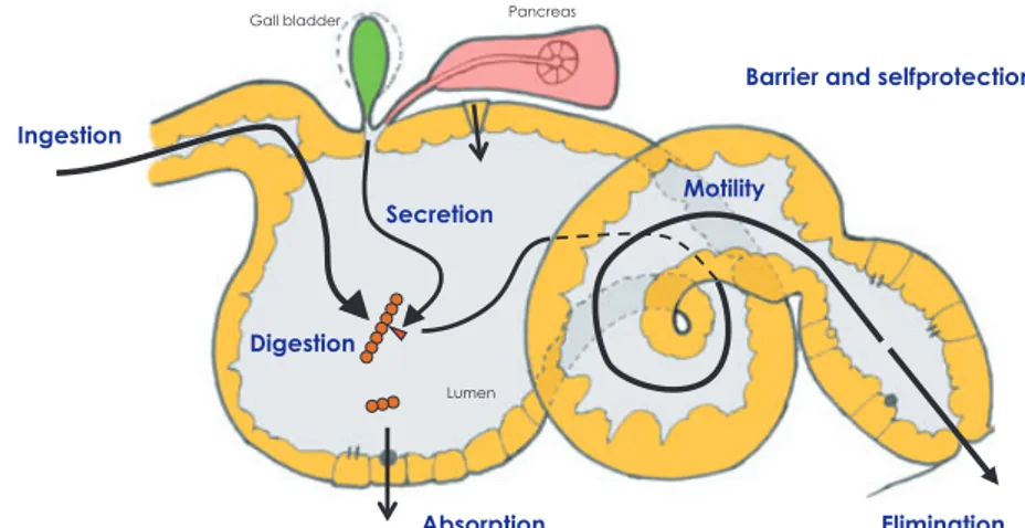
The digestive process involves a series of events that require the coordination of a range of basic processes in the diges-tive tract (Fig. 3), starting with ingestion of the food, and including the secretion of enzymatic and fluid secretions, digestion via mechanical and enzymatic processes, absorp-tion, motility (including evacuation) and finally regulation of the different processes. The efficiency of the digestive processes determines the delivery of nutrients to the rapidly growing larval tissues. As mentioned above, the gut also acts as an important primary barrier to prevent the entry of unwanted agents into the body tissues. The last topic is out-side the scope of this review, but has been dealt with in a recent review by Vadstein et al. 2012 (accepted for Reviews in Aquaculture).
Digestion of proteins and peptides Pancreatic enzymes
Proteolytic enzymes from the pancreas are regarded as being particularly significant in the early life stages of pre-cocious and altricial fish because of the absence of a func-tional stomach with its acid protease, pepsin. There is a wealth of literature describing the activities of the major pancreatic enzymes during ontogeny and/or their response
to various nutritional conditions in a variety of teleosts (e.g. Hjelmeland et al. 1988; Ueberscha¨r et al. 1992; Cahu & Zambonino-Infante 1995; Moyano et al. 1996; Martı´nez et al. 1999; Ribeiro et al. 1999, 2002; Srivastava et al. 2002; Ma et al. 2005; Rathore et al. 2005; Ueberscha¨r 2006; Cara et al. 2007; Fujii et al. 2007; Lamarre et al. 2007; Lazo et al. 2007; Kamaci et al. 2010; Buentello et al. 2011; Galaviz et al. 2011; Kortner et al. 2011a). Such studies have dem-onstrated that proteolytic enzymes are produced and secreted from the pancreas at the onset of first feeding, although the enzyme activities vary between species. For instance, in yellowfin tuna (Thunnus albacares), the activity levels of several enzymes were much greater than in simi-larly aged/sized marine fish of other species (Buentello et al. 2011). This has been explained as an evolutionary adaptation of scombrid fish larvae (Tanaka et al. 1996; Kaji et al. 1999) that exhibit early piscivory habits and remark-ably higher growth rates than most other marine species. Further comparative studies that take the life history of each species into account should be performed, in order to provide a better understanding of differences in the proteolytic capacity of each species, as this is believed to be important for further optimization of the nutrient supply in the early larval stages.
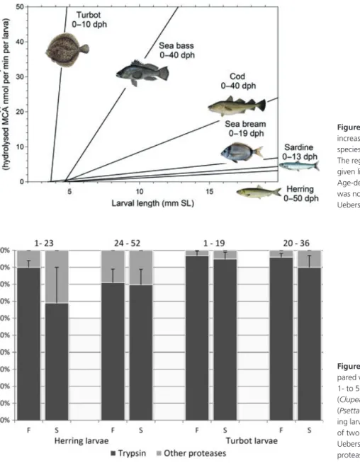
Figure 4 depicts the development of tryptic enzyme capacity in six species in order of increasing size. In order to accommodate the comparison within a single graph, dif-ferent age ranges had to be employed. There is some coinci-dence of species with rapid development of trypsin capacity (turbot, European sea bass, Atlantic cod) with their success in aquaculture (except for gilthead seabream in this exam-ple).
It is a generally accepted that trypsin is the most impor-tant proteolytic enzyme in the early stage of marine fish lar-vae (Fig. 5). Trypsin is a serine protease found in the digestive system of many vertebrates, where it hydrolyses proteins and also plays a key role in activating other
Ingestion Secretion Digestion Absorption Motility Elimination Lumen Gall bladder Pancreas
Barrier and selfprotection
Figure 3 The digestive tract is a multifunc-tional organ. Digestion includes a range of closely orchestrated processes that are integrated in ways that is believed to optimize efficiency and maximize absorption in feeding larvae under natural conditions. See text for more information.
pancreatic enzymes in the gut lumen. Trypsin is produced in the pancreas as the inactive precursor trypsinogen and cleaves peptide chains, mainly on the carboxyl side of the amino acids lysine or arginine.
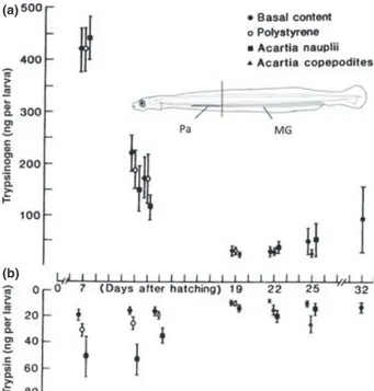
Considering the development of trypsin-like enzyme capacity in early larval stages, a ’four-phase model’ of the ontogenetic development of tryptic activity in marine fish larvae from hatching to metamorphosis has been suggested (Ueberscha¨r 1993). In Phase I, at the yolk-sac stage and beyond at commencement of exogenous feeding, the tryp-sin level increases; in Phase II, which is regarded as a ‘criti-cal’ stage with decreasing tryptic activity, poor growth and high mortality rates may occur; in Phase III, sufficient pro-duction of trypsin (at optimal food supply) and high growth rates can be observed; in Phase IV, at the beginning
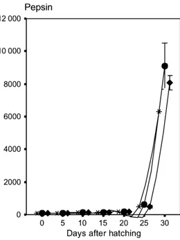
of metamorphosis, tryptic activity decreases again since it is in the course of being partly replaced by the activity of pep-sin in the developing stomach. Although this is a rather schematic approach, this pattern has been observed in a number of species (Ueberscha¨r 1993, 1995, 2006). Figure 6 depicts a typical example that demonstrates the postulated four different ontogenetic stages related to the development of tryptic enzyme capacity in larval common pandora (Pagellus erythrinus). The related pepsin activity (Fig. 7) demonstrates that tryptic activity is decreasing in the late larval stages, when the stomach is becoming functional and peptic activity is gradually increasing.
The PCR analyses have shown that mRNA transcripts of several proteolytic enzymes including trypsin are present already from hatching, which for most species takes
Figure 4 Increase in tryptic activity with increasing larval size. Comparison of six species reared under laboratory conditions. The regression line was calculated within the given lifespan in days after hatching. Age-dependent variability of tryptic activity was not considered here (re-drawn from Ueberscha¨r 2006).
Figure 5 Significance of tryptic activity com-pared with total alkaline proteolytic activity in 1- to 52-day-old laboratory-reared herring (Clupea harengus) and l–36 day old turbot (Psetta maxima) larvae (F, fed larvae; S, starv-ing larvae). Mean values with error bars (±SD) of two time intervals are shown (adapted from Ueberscha¨r 1993). ( ) Trypsin; ( ) Other proteases.
place several days before the onset of exogenous feeding (Srivastava et al. 2002; Lo & Weng 2006; Perez-Casanova et al. 2006; Drossou et al. 2006; Darias et al. 2007b). As far as trypsin is concerned, there appear to be temporal differences between the expressions of the various enzy-matic paralogues. In winter flounder (Pseudopleuronectes americanus), for instance trypsin 2 expression was detected from 5 dph, while trypsin 1 levels of expression only became significant in the late larval stages and during metamorphosis and trypsin 3 showed expression only after 20 dph (Murray et al. 2004). Whether these ontogenetic differences in mRNA expression of individual paralogues have functional implication for protein digestion is not known.
It is still not clear why tryptic enzyme activity decreases shortly following the onset of first feeding before increas-ing again. This phenomenon warrants further in-depth study.
Chymotrypsin is another pancreatic proteolytic enzyme of significance in the larval intestine. Like trypsin, chymo-trypsin is a serine alkaline protease produced in the pan-creas and secreted as its precursor chymotrypsinogen into the lumen of the intestine. The activation is catalysed by cleavage of a small peptide chain. Chymotrypsin displays a substrate-specific pattern of activity like trypsin and hydrolyses peptide bonds on the carboxyl side of the aromatic side-chains of tyrosine, tryptophan and
phenylal-anine, and of large hydrophobic residues such as methio-nine. Its activity is complementary to that of trypsin, which cleaves peptide chains mainly on the carboxyl side of lysine or arginine. Aktulun et al. (2008) observed that chymotrypsin was present immediately after hatching in sharpsnout seabream larvae and continuously increased during the larval period. In red drum (Sciaenops ocellatus) larvae too, chymotrypsin activity was detected before the onset of exogenous feeding (Applebaum et al. 2001). Total chymotrypsin activity increased with age and standard length. Specific activity was greatest on day 10 post-hatch. These results confirm that chymotrypsin contributes to protein digestion also in the early stages of larval develop-ment. However, considering the quantitative significance of trypsin and chymotrypsin in early stages of marine lar-vae, it was shown that trypsin is the most important alka-line protease in the early life stages (Fig. 5). Compared with trypsin, our current knowledge about chymotrypsin and its dynamics in the early larval stages is still limited, as is its significance in the digestive processes during these stages.
Chymotrypsin has also been regarded as an appropriate indicator of the nutritional condition of marine larvae (Applebaum & Holt 2003). The approach to using the tryp-sin/chymotrypsin ratio as an indicator was suggested by e.g. Cara et al. (2007) for European sea bass larvae close to and beyond the weaning stage. The idea here is that the
Figure 6 Development of tryptic activity in experiments on larvae from common pandora (Pagellus erythrinus). Newly hatched larvae were fed rotifers from day 3. Tryptic activity was measured in individual larvae and shown with standard deviations. Re-used from Suzer et al. (2006). (*) Group A; (●) Group B; (♦) Group C.
Figure 7 Development of pepsin activity in experiments using larvae from common pandora (Pagellus erythrinus). Pepsin was detected on day 25 in connection with stomach formation and a sharp increase until 30 dph. Re-used from Suzer et al. (2006). (*) Group A; (●) Group B; (♦) Group C.
ratio might indicate the extent to which chymotrypsin is activated by trypsin and that this in turn might indicate the growth potential of the fish. The higher the trypsin/chymo-trypsin ratio, the higher is the absorption rate of essential amino acids for protein synthesis and growth potential. However, this approach has its limitations, since trypsin usually decreases significantly at the onset of a functional stomach with gastric digestion established, while chymo-trypsin apparently continues to increase (compare the four-phase model mentioned above). Further, trypsin and chymotrypsin activation is also catalysed by the enzyme enterokinase, which is produced in the intestinal entero-cytes. This mechanism was not considered in the above-mentioned experiments. Obviously, more research is needed on the dynamic and the role of chymotrypsin.
Pepsin is found in the functional stomach and is active in an acidic environment with pH as low as 1.5–2.0. In fish that lack a functional stomach in the early stages, pepsin-like enzyme activities and acidification capacity develop slowly during metamorphosis (Yu´fera et al. 2004; Darias et al. 2007a; Yu´fera et al. 2012b). For example, the first record of significant pepsin-like activity in larval barra-mundi started at 17 dph, coinciding with the acidification of the developing stomach (Walford & Lam 1993), while in turbot larvae, peptic activity not was observed before day 22 post-hatch (Ueberscha¨r 1993). Moreover, in this experi-ment it was observed that the peptic level reacts to food deprivation by a decrease in activity. In common pandora, peptic activity began to rise between 20 and 24 days post hatch (Suzer et al. 2006; compare Fig. 7). Nevertheless, the measurements of pepsin activity in fish have been done at pH 2 instead of buffering at the actual gastric pH with the consequent overestimation of this activity in developing larvae (Ma´rquez et al. 2012; Yu´fera et al. 2012b). Unlike chymotrypsin and trypsin, which have specific sites of action (Keil 1971; Blow et al. 1972; Blow 1976), pepsin has a broad range of active sites (Fruton 1970). Pepsin diges-tion yields polypeptides of very diverse sizes, but few FAA (Lied & Solbakken 1984; Espeland 2006). The combined effects of HCl and pepsin make the stomach a highly efficient organ for degrading complex proteins such as col-lagen-rich connective tissues (Gildberg 2004). As a conse-quence, tryptic activity, as described above, decreases when the stomach becomes functional (Fig. 6). The digestive processes that combine pepsin and pancreatic enzyme activity are thus likely to hydrolyse proteins to a greater extent than when pepsin is missing. The acquisition of functional proteolytic digestion in the stomach thereby allows a wider range of ingredients to be incorporated in fish diets. At the same time the absence of a functional stomach and its associated acid-pepsin-mediated digestion, could be a limiting influence on the digestibility of dietary proteins in the youngest stages of marine fish larvae (e.g.
Tonheim et al. 2005; Rønnestad et al. 1999, 2007b). Based on the aforementioned points, considerable efforts are being put into assessing protein digestibility by both in vivo and in vitro methods and also into establishing protocols for efficient treatments and handling of dietary protein ingredients to optimize amino acid (AA) bioavailability at different life stages (Tonheim et al. 2007; Fox & Lawrence 2009; Hansen et al. 2009; Johnson et al. 2009; Nordgreen et al. 2009; Saenz de Rodrigan˜ez et al. 2011). This impor-tant work needs to be kept in focus.
In conclusion, the weak ability of early larval stages effi-ciently to digest currently available microdiets (compared with live food) may be attributed to the lack of denaturiz-ing and proteolytic cleavage of food proteins by combined peptic and acid activity. This is one of the main challenges that remain to be tackled when formulated feeds are used as first feed for the young stages of marine fish larvae (see General protein-processing capacity).
Enzymes in the mucosal layer
Enzymes associated with the enterocytes also contribute to the final digestion of proteins and peptides. The brush-border membrane (BBM) enzymes are located in the api-cal cell membrane facing the gut lumen. These enzymes are aminopeptidases that hydrolyse small peptides that are the result of cleaving by pancreatic proteases, into FAA and small peptides that are small enough to be absorbed. The activities of BBM enzymes such as the leucine amino-peptidase (LAP) of the enterocytes in Atlantic cod and Atlantic halibut are at a low level at first feeding, but increase during larval development (Kva˚le et al. 2007). The activity of cytosolic (intracellular) peptidases such as leucine-alanine peptidase which, it has been suggested, participate in protein degradation after pinocytosis, are high in some species around first-feeding but tend to decrease as larvae develop, concurrent with rising levels of alkaline phosphatase (Cahu & Zambonino-Infante 2001; Kolkovski 2001). This picture is less clear in cod than in halibut, which have a stable level of LAP activity through-out the start-feeding period, apart from a peak in activity around day 35 after the start of feeding (Kva˚le et al. 2007).
It has been suggested that the ontogenetic pattern of trypsin, membranous and cytosolic peptidase activity dur-ing larval development reflect changes in the mode of larval digestion, which is believed to become increasingly depen-dent on luminal digestion and less so on intracellular diges-tion (Cahu & Zambonino-Infante 2001).
Exogenous enzymes
In the past, it was argued that first-feeding marine fish lar-vae had insufficient endogenous digestive enzyme capacity and therefore rely on exogenous enzymes originating from
their prey. Lauff and Hofer (1984) concluded for freshwater coregonid species that external enzymes obtained from zooplankton might contribute 70–80% of the total enzyme activity in the fish digestive tract, supporting the earlier results of Dabrowski and Glogowski (1977). Consequently, it was assumed that compound diets result in poor growth and survival in the earliest larval stages, since no exogenous enzymes were supplied with the artificial diets. This led to the supply of exogenous enzymes in compound diets (Kol-kovski et al. 1997c) but this approach has never been intro-duced in routine feeding protocols in fish culture (Kolkovski 2001).
Related research in the course of the past two decades has disclosed that the marine species studied to date possess sufficient levels of endogenic digestive enzymes to allow the digestion of suitable live prey and well-designed artificial diets from first feeding (for trypsin, e.g. Ueberscha¨r 1993; Cahu & Zambonino-Infante 1994). Results regarding com-parisons of the quantity of endogenous and exogenous enzymes specifically in herring larvae allow the conclusion to be drawn that the exogenous enzymes that are ingested with live food (e.g. rotifers, Artemia nauplii, copepod nau-plii), are of minor significance for the overall digestive capacity of marine fish larva (Hjelmeland et al. 1988; Pedersen & Hjelmeland 1988; Pedersen & Andersen 1992; Ueberscha¨r 1995). In recent experiments, it was demon-strated that exogenous enzymes from live food (Brachionus sp., Acartia tonsa, Temora sp., newly hatched Artemia nau-plii) contributes only 1–2% of the total tryptic enzyme activity measured in young herring larvae (Ueberscha¨r, unpublished).
Absorption Free amino acids
It has been suggested that dietary proteins, peptides and free amino acids (FAA) in larval fish are absorbed primarily by the midgut (Rønnestad & Conceic¸a˜o 2005). However, our understanding of the mechanisms and ontogenetic changes involved in protein, peptide and FAA absorption in larvae is still limited. Amino acid absorption involves a number of transport processes, some of them with overlap-ping functions. The in vivo transport rates reported for free amino acids (Rønnestad et al. 2000a,b; Applebaum & Rønnestad 2004) will depend on several elements, includ-ing the luminal concentration of AA, the transport affinity and capacity of each amino acid transporter (AAT), and the amount of each AAT in the brush border membrane. It is also important to emphasize that these data represent transintestinal absorption rates and reflect the net flux of free amino acids from the midgut lumen, across the diges-tive tract cells, and into the body. In their studies Apple-baum and Rønnestad (2004) failed to find a saturable
component for free amino acid absorption for luminal con-centrations up to 20 mM for the four amino acids tested; dispensable (alanine, glutamate) and indispensable (argi-nine, lysine). Further studies need to be performed to improve our understanding of the absorption kinetics of individual amino acids and of interactions between various amino acids.
In mammals, there is a range of functional systems that mediate amino acid transport in the apical and basolateral membranes of epithelial cells. Each system accepts groups of amino acids rather than individual amino acids, and most of them have several genetic representatives. For instance, in mammals more than four separate systems are involved in transepithelial transport of the cationic amino acids lysine and arginine in the GI tract. Some AAT are heterodimeric and thus consist of two subunits and genes. Molecular studies of the AAT that were described on the basis of their functional characteristics have identified the genes involved that are now classified into solute carrier families (slc). While many of the genes coding for AATs have been cloned in mammals, such efforts have only started in fish (Narawane 2011; Pinto et al. 2012). In zebra-fish, several of the transporters for cationic amino acids have been identified (Narawane 2011). This list includes the presence of system b0,+and y+. Zebrafish system b0,+, which is a heterodimeric transporter that comprises mem-bers of the solute carrier family memmem-bers slc3a1 and slc7a9 and displays conservation of the essential features that determine its functions and interactions. Analysis of the zebrafish system y+transporter slc7a1 also revealed conser-vation of essential functional features and a high affinity for cationic amino acids. Both slc3a1 and slc7a9 were expressed in the developing intestine as well as in the proximal tubule segments of the nephrons (Narawane 2011). Due to the ubiquitous distribution in tissue of some of these AAT, functional studies in larval stages will be a challenge. Narawane (2011) demonstrated that slc7a1 is widely expressed in the developing eyes, somites, distal nephrons and branchial arches of zebrafish. Gene-specific morpholi-no-mediated knockdown that induces loss of gene function had major effects on early zebrafish embryos and these AAT are essential for normal embryonic development and organ-ogenesis during endogenous feeding. However, due to the temporary effect of the morpholino technique used (which ceases to work around the time of first feeding), combined with the abundant expression of these AAT, these studies have yet to provide any specific information regarding the role of these transporters in the digestive tract.
A recent study by Pinto et al. (2012) has described the cloning and ontogenetic expression of the taurine trans-porter (TauT; slc6a6) in Senegalese sole. The results showed a high degree of similarity between TauT in Senegalese sole and other vertebrates, but some differences in TauT amino