DOTTORATO DI RICERCA IN
"BIOCHIMICA, BIOLOGIA MOLECOLARE E
BIOTECNOLOGIE"
CICLO XXIV
COORDINATORE Prof. Francesco Bernardi
Cell encapsulation systems based on hybrid
hydrogels
Settore Scientifico Disciplinare BIO/10
Dottorando Tutore
Dott. Mazzitelli Stefania Prof. Gambari Roberto
Cotutore
Prof. Claudio Nastruzzi
TABLE OF CONTENTS
ABSTRACT
4PREFACE
5ACKNOWLEDGMENTS
7CHAPTER 1
8Bioencapsulation for cell therapy: general considerations
1.1. Introduction 8 1.2. Hydrogels 11 1.2.1. Collagen 12 1.2.2. Gelatin 14 1.2.3. Hyaluronic acid 15 1.2.4. Agarose 16 1.2.5. Alginate 17
1.3. Scaffold design and characteristics 21
1.3.1. Dimensional, permeability and morphological properties 24
1.3.2. Mechanical properties 25
1.3.3. Biocompatibility properties 26
CHAPTER 2
32 Bioencapsulation for cell therapy: experimental procedures forthe production and characterization of microcapsules
2.1. Cell encapsualtion in microparticles: methods of preparation 33
2.2. Cell encapsulation by coaxial bead generator 35 2.2.1. Application of a coaxial bead generator to IB3-1 cells 37
2.2.1.1. Materials and methods 38
2.2.1.2. Results 41
2.2.1.3. Discussion 50
2.3. Cell encapsulation by a vibrating-nozzle procedure 54 2.3.1. Application of a vibrating-nozzle procedure to 55 Mesenchymal stem cells
2.3.1.1. Materials and methods 57
2.3.1.2 Results 62
2.3.1.3. Discussion 76
2.4. Cell encapsulation by a microfluidic based approach 78 2.4.1. Application of a microfluidic based approach to Sertoli Cells 79
2.4.1.1. Materials and methods 81
2.4.1.2. Results 85
2.4.1.3. Discussion 94
CHAPTER 3
102 Bioencapsulation for cell therapy: engineered microparticlesand fibrous multifunctional scaffolds
3.1. Engineered microparticles 103
3.1.1. Materials and methods 105
3.1.2. Results 108
3.1.3. Discussion 121
3.2. Fibrous multifunctional scaffolds 123
3.2.1. Materials and methods 126
3.2.2. Results 129
3.2.3. Discussion 148
3.3. References 150
CHAPTER 4
155PREFACE
This PhD thesis was focused on the production and characterization of encapsulation systems for cell therapy and tissue engineering applications. The structure of the work is organized in four main chapters: (i) the general characteristics of hydrogel based devices for cell encapsulation, (ii) the description of the experimental encapsulation procedures for microcapsular devices for the embedding of different cell types, (iii) the design of new strategies for engineered and or fibrous scaffolds and finally (iv) the main conclusion gathered by the entire experimental results of the research project.
In detail, chapter 1 presents an overview of commonly applied biomaterials, as well as the characteristics of optimal encapsulation systems for in vivo application. In particular hydrogels performances are discussed analysing advantages and disadvantages for cell therapy applications. Chapter 2 and 3 are devoted to the description of different fabrication techniques to process hydrogels containing different cell types into scaffolds.
Each chapter refers to one or more journal articles dealing with the experiments performed to produce the encapsulation devices. Particularly, chapter 2 is related to papers on various procedures for microcapsule production, while chapter 3 is relative to contributions about new scaffolds in form of engineered microcapsules or multifunctional microfibres.
The scientific work presented in the current PhD thesis program has beneficed of the skilled collaboration of researchers and scientific Institutions:
1. Prof. S.F. Badylak, MacGowan Institue for Regenerative Medicine, University of Pittsburgh, Pittsburgh, PA, USA
2. Prof. X. Zhang, Engineering and the Environment, University of Southampton, Southampton, UK
3. Biomaterials Group, King‟s College, London, UK
Studi di Ferrara, Ferrara, Italy
5. Prof. R. Piva, Dipartimento di Biochimica e Biologia Molecolare, Università di Ferrara, Ferrara, Italy
6. Prof. E. Becchetti, Dr. G. Luca, Dr. M. Calvitti, Dr. F. Mancuso, Dipartimento di Medicina Sperimentale e Scienze Biochimiche, Università degli Studi di Perugia, Perugia, Italy.
ACKNOWLEDGMENTS
The important thing is not stop questioning. AlbertEinstein Foremost, I would like to express my sincere gratitude to Prof. Claudio Nastruzzi. During more than seven years of knowing him, he showed me science in its full depth and taught me how to appreciate the good scientific work. His knowledge with his originality has triggered and nourished my intellectual maturity that I will benefit from, for a long time to come. I am deeply grateful to my supervisor, Professor Roberto Gambari for his important support throughout this work.
I owe my gratitude to Professor S.F. Badylak of the McGowan Institute of Regenerative Medicine who gave me the opportunity to work with him and to join his group in Pittsburgh.
All the work would not have been possible without the guidance of several colleagues who in one way or another contributed and extended their valuable assistance in the preparation and completion of this study. First of all, the researchers from the University of Perugia (G. Luca, F. Mancuso, M. Calvitti) that introduced me to cell work and gave me extraordinary experiences through out the work. Moreover all the colleagues from the groups of Prof. Gambari and Prof. Piva, University of Ferrara, who helped me with my experimental project.
During the thesis I had the opportunity to work at the Biomaterials Group of the King’s college in London and at the School of Engineering Sciences of the University of Southampton, where I met and collaborated with people for whom I have great regards (Prof. S. Deb, Prof. L. Di Silvio, Prof. X. Zhang, Obi, Cristian, Daniel, Emanuele, Dario).
Special thanks to my colleagues and most of all friends Lorenzo and Stefano, with whom not only I had many productive scientific discussions but also chats in front many beers.
From the non-scientific side, individual acknowledgments are also owed to my childhood friends in Tropea (Lucia, Gemma, Rosaria, Francesca), my friends in Perugia (Mino, Cinzia, Pasquale, Kasia, Nicoletta) and the new ones in Ferrara (Emilia, Elena, Francesca). They provided me unflinching friendship, making me always smile and enjoy the life.
My parents, brother and my sister-in-law deserve special mention for their inseparable support and encouragement. My Father, Pino, in the first place is the person who put the fundament of my learning character and my Mother, Luciana, is the one who raised me with care and love.
And last but not least I wish to extend my thanks to Giusa and Federico. The first one for always letting me know that I can count on her even when far away form each other and for the good advice, even if never follow them. The second one for the special person he is and for having come along at the right time, supporting me with an incredible amount of patience.
CHAPTER 1
Bioencapsulation for cell therapy:
general considerations
1.1. Introduction
Millions of surgical procedures are performed each year to replace or reconstruct damaged tissue resulting from chronic/degenerative diseases, injury, congenital malformations and cancer. Unfortunately, there is a wide gap between the demand for organs and replacement tissue and patients actually receiving transplants. The cellular therapy and tissue engineering based protocols offer a potential solution to the shortage of organ donors and the problems relating with the repair of damaged tissues (i.e. risk of graft failure) [1]. Cellular therapy is indeed a novel technology based on the use of cells to treat a wide range of human diseases by replacing damaged cells or tissues.
These approaches are based on the administration/transplant of living cells to specific body sites, where cells can exert therapeutic effects through: (a) the repair/replacement of damaged tissue/organs or (b) the production/release of specific bioactive molecules (e.g. enzymes, growth factors and antibodies) [2].
Recently, cells have been increasingly exploited as alternative controlled drug delivery vehicles. Cells can indeed act as drug depots enabling the delivery of “de novo” produced therapeutic products with site specificity over an extended time period, providing the possibility to treat various diseases that cannot be cured with currently available therapeutic protocols [3].
In this respect, different cell types (primary, stem and bioengineered cells) have been considered as potential therapeutic tools and tested in preclinical and clinical studies for the treatment of many pathologies, such as diabetes, anemia, hemophilia, bone defects and cancer [4-6].
For instance, biomolecules secreted by cells may be delivered continuously such as an angiogenesis inhibitor to a tumor [7], or in response to a physiologic signal, such as insulin to glucose for the treatment of diabetes [8]. Erythropoietin cell production in response to oxygen delivery can found application for the treatment of anemia [9]. A selected protein may also be expressed and produced by the cell implant after genetic modification, such as the coagulation factors VIII and IX for the treatment of hemophilia A and B. Moreover, stem cells can secrete a diverse array of growth factors, including vascular endothelial growth factor (VEGF) and nerve growth factor, which are used to treat ischemia and neuronal damage, respectively [10,11].
Over the years, two conceptually different strategies to deliver cells into diseased tissue have been developed: the first approach consists of the injection of free autologous, allogenic or xenogenic cells, the second one is based on cells seeded or embedded in scaffold; notably, the latter approach finds applications both in vitro (basic and applied research) and in vivo (reconstructive medicine) [12].
Unfortunately, the use of free cells, in spite of the superior handling, holds the major drawback to elicit the host immune rejection. Furthermore, transplanted cells not protected from external mechanical loadings or not surrounded by the proper microenvironment may rapidly lose their viability and functionality. Therefore, an increasing number of natural and synthetic structures have been developed as potential immunoisolating scaffold for cell embedding [13].
Cell immunoisolation into scaffolds represents the major advance in cell based therapy since it avoids constraints associated with cell sources, making allogenic and xenogenic cells a good alternative to the limited autologous donors.
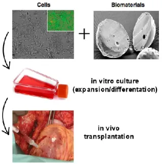
A cellular based therapy requires the combined use of suitable scaffolds and selected cell populations to create biocompatible devices that once transplanted, can immunoisolate the cells from the host's immune system,
possibly eliminating or reducing the requirement for immunosuppressant systemic drug administration (Fig. 1). Non-specific suppression of the immune system may lead to a variety of unwanted complications in patients, including infections and failure of tumor surveillance [14].
A key role in preserving cell functions is played by the biomaterial used to produce the cellular scaffolds suitable for implanting cells into the host in order to regenerate a tissue or to obtain a long-term systemic delivery of biomolecules.
Fig. 1. Schematic representation of a system for cell therapy protocols based on cells and biomaterials.
Cells can be adequately combined with biomaterials following two main approaches. On the one hand, cells are isolated from the host‟s body by a microcapsular structure allowing exchange of nutrients; on the other one, cells can be seeded onto a preformed scaffold, that is generally implanted into the host after a given cultivation time.
The concept of cell immunoisolation was initially described by Chang in 1960s and since then, various biomaterials have been proposed to embed cells in immunoisolating microenvironments, manly in form of microcapsules, creating a semi-permeable membrane/barriers that control the outward and inward diffusion of gases (oxygen and carbon dioxide), metabolic and therapeutic molecules.
Irrespectively of the strategy employed, the success of any bioencapsulation approach is mostly related to the properties of the biomaterial, that should be biocompatible, easy to sterilize and biodegradable over an appropriate length of time into products metabolized or excreted by the recipient without inducing adverse inflammatory responses.
Ideally, the selected biomaterials should be reproducibly processed into a desired shape and structure (i.e. spherical or tubular configurations), maintained even after the in vivo implantation.
Moreover, an immunoisolation biomaterial must provide a structured protective environment with tissue-specific mechanical properties and porous niche for cells capable of delivering potentially therapeutic factors.
Finally, the biomaterial should offer an appropriate regulation of cell behaviour including adhesion, proliferation, migration and differentiation. When in vivo implanted, the biomaterials must provide temporary mechanical support sufficient to withstand forces exerted by the surrounding tissue.
It is important to underline that the different, and in a certain sense opposite biomaterial characteristics, are provided by a very limited number of materials. In this respect, hydrogel forming biomaterials occupy a prominent position, thanks to their special physical properties and feasibility for cell encapsulation procedures. Hydrogels provide a highly controlled, synthetic 3D environment that is structurally and biomechanically similar to native extracellular matrix (ECM) topology and provides a rich ligand landscape to influence cell behaviour [15].
1.2. Hydrogels
Hydrogels represent an important class of biomaterials for biotechnology and biomedical applications, since they exhibit excellent biocompatibility with minimal inflammatory responses and tissue damage. By definition, the hydrogel structure is constituted of a polymeric network with three-dimensional configuration capable of imbibing high amounts of water or
biological fluids. The high water affinity is attributed to the presence of hydrophilic groups such as –OH, –CONH2– and –SO3H, in the polymers
forming hydrogels, that create a hydrated network with different degrees (sometimes, more than 90% of water by weight), depending on the nature of the aqueous environment and the polymer chemical composition. The polymeric network is usually cross-linked by different modalities, including covalent bonds, hydrogen binding, van der Waals interactions, or physical entanglements. Hydrogels are extremely suitable for a variety of applications in the pharmaceutical and medical industry since they hold a number of appealing features [16]. Thanks to the presence of large amounts of water, hydrogels resemble certain native tissues; their framework presents a defined pore size allowing the diffusion of water/metabolites and exchange of nutrients/wastes, but strictly avoiding the overall migration or leakage of individual cells. Moreover, hydrogels can be easily fabricated into different shapes and sizes, from nano-scale particles to centimetre long sheets. In conclusion, the embedding of cells on hydrogels is very suitable for in situ cell delivery and cell therapy as demonstrated by numerous articles having as topic the regeneration of cartilage, cornea, nerve and liver [17].
Classification of hydrogels can be made according to various criteria including: the material used for their preparation, the preparation method, the overall charge and the mechanical characteristics.
With respect to materials, hydrogels can be obtained by natural or synthetic polymers. To the first class belongs polysaccharides (e.g. agarose, alginate) and proteins (e.g. collagen and gelatin); to the other one poly(ethylene glycol) (PEG), poly(vinyl alcohol) (PVA), poly-lactic-co-glycolic acid (PLGA) and poly (hydroxylethyl methacrylate) (PHEMA) [18].
Concerns regarding synthetic polymers inducing non-physiologic cellular responses have caused a shift of interest to using more natural materials, such as stand-alone products, chemically derived, physically derived, or extracellular matrix-derived materials.
The advantage of employing some natural biomaterials, such as collagen, fibrin, hyaluronan (HA), gelatin is their ability to mimic certain features of
native extracellular matrix (especially those of ECM origin), facilitating cell adherence, migration, differentiation. Cells indeed in the body are exposed to a complex milieu regulated by their interactions with other cells, the surrounding cell matrix and soluble factors. A key element of this microenvironment is the three dimensional (3D) architecture of the extracellular matrix. By removing the cells from this microenvironment many cell types quickly loose their function. Therefore, the ability to resemble the in vivo microenvironment of cells outside the body could be a potentially powerful tool.
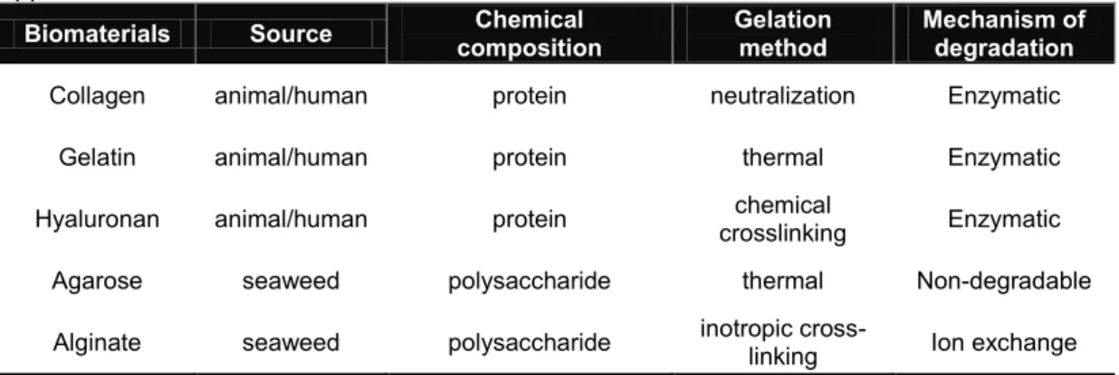
In table 1 and in the following sections are briefly described some example of natural polymer usually employed for the preparation of hydrogels that have found a large number of applications in cell based protocols.
Table 1. Natural biomaterials used for cell based therapy and tissue engineering applications.
Biomaterials Source composition Chemical Gelation method Mechanism of degradation Collagen animal/human protein neutralization Enzymatic
Gelatin animal/human protein thermal Enzymatic Hyaluronan animal/human protein crosslinking chemical Enzymatic
Agarose seaweed polysaccharide thermal Non-degradable Alginate seaweed polysaccharide inotropic cross-linking Ion exchange
1.2.1. Collagen
Collagen is the major structural component of mammalian connective tissue and has been used in cell immobilization due to its biocompatibility, biodegradability, abundance in nature and the ability to bind cells. The main sources of collagen are generally represented by cartilage, tendon, skin, bone, cartilage and ligament, where it is present in high concentrations. Collagen can be readily processed into different shaped scaffold including porous sponges, fibres, films, membranes and injectable cell immobilization carriers, since the gelation process occurs without chemicals modifications [19]. However, to improve the mechanical properties of scaffolded gels
different methods have been proposed such as crosslinking by chemicals, UV, temperature or polymeric agents. Collagen plays an important in regulating essential cellular events, such as proliferation, migration and differentiation via cell–matrix interaction and via integrin binding. Its natural ability to bind cells makes it a promising material for controlling cellular distribution within immunoisolated systems and its enzymatic degradation can provide appropriate degradation kinetics for tissue regeneration in microporous scaffolds. Moreover, collagen contains cell-adhesion domain sequences (e.g. RGD) that exhibit specific cellular interactions that can help to retain the phenotype and activity of many types of cells, including fibroblasts and chondrocytes.
Collagen has been extensively used to produce scaffold for many biomedical/regenerative applications, nevertheless it still has some constrains mainly related to the presence of antigenic components associated to the collagen molecules that can evoke host immunoreactions [20].
1.2.2. Gelatin
Gelatin is a naturally derived protein from collagen via a partial hydrolysis of the native collagen. During the manufacturing of gelatin, raw animal material is treated with dilute acid or alkali, resulting in partial cleavage of the crosslinks. Moreover, gelatin is a product of the meat-processing industry and it is a readily and economically available material, which has been used for decades in the food and pharmaceutical industries [21]. At a temperature of about 40°C, gelatin aqueous solutions are in the sol state and form physical thermoreversible gels after cooling. During the gelation process, the chains undergo a conformational disorder-order transition and tend to recover the original collagen triple-helix structure.
It is widely used for its non-toxicity, non-irritant and biodegradable properties and its mild gelling process that makes it is an attractive candidate as starting material for preparing hydrogels for drug delivery systems (i.e. hard and soft capsules, microspheres) and tissue engineering approaches (i.e. vascular
prostheses and wound dressing). As a biomaterial, gelatin displays several advantages: it is a natural polymer that has not shown antigenicity, it is completely resorbable in vivo and the presence of the large number of functional groups in the side chain offers the possibility to bind and release growth factors or incorporate proteins that can influence cell adhesion and growth in a controlled manner [22].
Compared to collagen, gelatin does not express antigenicity in physiological conditions, due to the denaturing process, and it is much cheaper and easier to obtain in concentrate solutions. Conversely, gelatin based hydrogels exhibits poor mechanical properties that can be overcame only via chemically cross-linking with various bifunctional agents, including glutaraldehyde or water-soluble carbodiimide that unfortunately reduce the initial high gelatin gel biocompatibility [23].
1.2.3. Hyaluronic acid
Hyaluronic acid (HA) is a nonsulphated glycosaminoglycan, composed of alternating units of D-glucuronic acid and D-N-acetylglucosamine, linked together via alternating β-1,4 and β-1,3 glycosidic bonds. HA is one of the major components of the extracellular matrix of skin, cartilage and the vitreous humor.
It plays a vital role in maintaining tissue integrity, as well as in facilitating adhesion and differentiation of cells during inflammation, wound repair, and embryonic development. Covalently cross-linked HA hydrogels can be formed by means of multiple chemical modifications. Hyaluronan is highly non- antigenic and non-immunogenic, owing to its high structural homology across species, and poor interaction with blood components [24].
A variety of commercially available preparations of HA derivatives and cross-linked HA materials have been developed for the production of drug delivery systems (microspheres, liposomes, fibres) or hydrogel-based scaffolds. Application of HA as a cell delivery vehicle has been investigated for cartilage, bone and osteochondral regeneration. Although the main mechanism of HA is unknown, in vivo, in vitro and clinical studies have
demonstrated various physiological effects of exogenous including chondroprotective properties.
Recently, HA has become recognized as an important building block for the creation of new biomaterials for use in cell therapy, three-dimensional (3-D) cell culture and tissue engineering applications. One of the key advantages of using HA gels for tissue engineering is that their degradation can be mediated by hyaluronidase, an enzyme secreted by a various cell types. On the other hand, the obtained HA based hydrogels are characterized by a weak mechanical strength resulting in scaffolds difficult to handle or to maintain the characteristics during the sterilization process.
1.2.4. Agarose
Agarose is a natural polysaccharide extracted from the cellular walls of agarophyte seaweed. Agarose gel is formed by cooling aqueous agarose solution, obtained by heating an aqueous suspension of agarose powder, until a clear solution forms. Since agarose gel networks are formed solely through hydrogen bonds without the use of chemicals, they are widely used in molecular biology and in immunoisolation protocols where it has been demonstrated they are well accepted following implantation [25]. The gelation mechanism is indeed governed completely by hydrogen bonding between agarose molecules and the resulting gel network is stabilized by structured water. In detail, agarose gels are formed when random coils, in a heated sol, become ordered as the sol cools. As cooling progresses, helices are formed which aggregate to form a gel network composed of thick bundles of agarose chains, large pores of water, exhibiting high turbidity and strong elasticity. Due to their soft tissue-like mechanical properties and biocompatibility, agarose gels have been investigated as potential scaffolds for neural [25] and cartilage tissue engineering [26]. The major drawbacks of agarose are the low cell adhesiveness and proliferation, since it does not contain any side groups to bind cell adhesive proteins. Moreover, due to the non-degradable nature of the gel, it has not been widely used in tissue engineering where usually scaffold materials should degrade over time in order to allow space
for accumulation of new tissue. With respect to application of agarose to cell capsulation procedures some important features should be considered. After melting, agarose can be easily transformed into a variety of controlled shapes, including microbeads, by simple and mild temperature reduction (usually below 30°C). In spite this advantageous characteristic, a possible drawback of agarose resides in the slow setting of gels, resulting in the possible cell protrusion from the capsular shell.
1.2.5. Alginate
Alginate is an example of a naturally derived polymer well suited for biomaterial scaffolds in cell-based therapy [27].
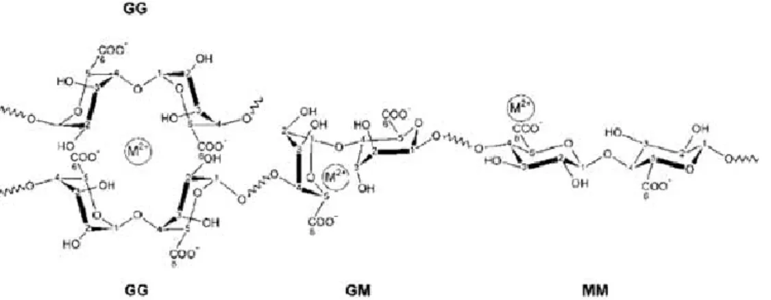
Alginates are extracted from three species of brown Algae such as Laminaria hyperborean, Ascophyllum nodosum, Macrocystis pyrifera and generally sold as sodium salt. Alginate is a water-soluble linear polysaccharide and consists of a mixture of -D-mannuronic acid (M) and -L-guluronic acid (G) residues. The ratio of M to G blocks can vary significantly depending upon the source of the raw materials used in alginate manufacture. Due to the shape of the monomers and their various ways to link, the geometries of the G-block and M-block regions, are substantially different. In detail, G-blocks have a bucked shape while the M- block tends to be as an extended ribbon (see Fig. 2)
Fig. 2. Chemical structure of alginate constituted of 2 guluronic acid (G) monomers and 2 mannuronic acid (M) monomers, with (1–4) linkages.
Two G-blocks aligned side by side, result in the formation of a hole with specific dimension able to bind selectively divalent ions in a cooperative fashion. On dissolution in an aqueous medium, alginate forms a hydrocolloid, which gels ionotropically following the addition of divalent cations, including
indeed this ability to form gels under extremely mild conditions without the use of chemicals or a particular pH.
.
Fig. 3. Mechanism of ionotropic gelation of alginate based polymer in the presence of divalent cations (M2+).
The polymer cross-linking occurs following the exchange of sodium ions from the guluronic acids with the divalent cations resulting in a chain-chain association that constitutes the junction zones of the so-called “egg box model” (Fig.4).
Fig. 4. The egg-box model after binding of divalent cations to homopolymeric G-blocks.
The buckled chain of guluronic acid units is shown as a two-dimensional analogue of a corrugated egg-box with interstices in which the divalent ions may pack and be coordinated. Since hydrogel formation occurs following selectively linkage between the carboxylic moieties on the G blocks of alginate and cations, high ratio of G:M results in stiff gels.
molecular weight and, for this reason, the molecular weight is not one unique value.
Polymer molecular weight is important because it determines many physical properties. If molecular weight is too low, the mechanical properties will generally be too low for the polymer material to have any useful practical and commercial applications.
If the Ni is the number and Wi is the weight of polymer molecules having a specific molecular weight Mi, the total number and weight of the polymer is
The fraction is called the polydispersity index and it has been reported to be between 1.4 and 6.0 for alginates, relating to the various purification processes.
The molecular weight distribution has a significant impact on some of the alginate gel properties including biocompatibility, stability, mechanical resistance, permeability, biodegradability and most important the gel formation [28].
Alginate is used extensively in food industry as a thickener, emulsifier and as a stabilizer. Furthermore, being an anionic polymer with carboxyl end groups, it has found application as pharmaceutical excipient to promote the mucoadhesive properties towards mucosal tissues. After the early study of Lim in 1980s, describing the use of alginate for cell microencapsulation, this polymer became the most widely used biomaterial for cell entrapment.
Although the suitability of other natural and synthetic polymers is currently under investigation, none has reached the same level of performance as alginates. For their ability to form gels in very mild conditions, alginate cross-linked with Ca2+ or Ba2+ ions has been used successfully to encapsulate cells maintaining a good viability and functions also during long-term culture [29]. Among biomaterials, alginate hydrogels are thought to be inert because they
i i i n i i N M M N 2 i i i i i i w i i i i i w M Nw M M w N M
/
n wM M
lack native ligands that could allow interaction with mammalian cells, providing numerous advantages for tissue engineering including the possibility of minimally invasive injection of hydrogel/cell microcapsules. Alginate gels have a defined pore size and a narrow pore-size distribution; in addition, they are mechanical and chemical stabile and contain low amount of toxic, pyrogenic and immunogenic substances.
Unfortunately, all the above features do not pertain at all the commercially available alginates; for biomedical procedures (i.e. cell encapsulation) only ultrapure alginates should be considered.
Alginate has been extensively characterized in terms of purity, biocompatibility and in vivo performances. In fact, as a natural derived material, alginate may contain impurities such as are polyphenols, proteins and lipopolysaccharide (LPS) (endotoxin) that cause a reduction in its biocompatibility. Even commercially available ultrapure alginates have been found to contain residual contaminants that can limit their use for the production of microcapsule. Inflammatory responses due to impurities can influence the performance and function of the devices. For instance, NO, produced form macrophages stimulated by alginate contaminants, thanks to small molecular dimensions can diffuse rapidly through the gel matrix causing severe cytotoxic effects on the encapsulated cells.
In this respect, an efficient purification process, designed to remove contaminants, including potentially immunogenic compounds, represents a mandatory step for tissue engineering application. Nevertheless, it is important that the purification process does not alter the polymer features (especially number and weight average molecular weights) that are critical for the gelation process and therefore the microcapsule formation.
Alginate beads are generally prepared by dripping a sodium alginate solution containing the desired cells into a divalent crosslinking solution such as Ca2+, Sr2+, or Ba2+ (gelling bath). Ca2+ ions are preferred for gelation of alginate; especially, for the encapsulation procedures, because of the high biocompatibility. After preparation, calcium gelified beads are usually coated by a cationic polyelectrolyte. The coating is performed to slow down the
swelling and in vivo degradation of the microcapsules, however it may cause immunological reactions and fibrotic growth; this latter can subsequently decrease the therapeutic efficacy of the entrapped cells sharply reducing the diffusive properties of the alginate capsule [30].
In this respect, the use of the positively charged polyelectrolytes for the capsular coating (i.e. poly-L-lysine) can induce a more intense overgrowth compared to negative ones. This funding was attributed to activated macrophages that preferentially adhere to positive charged surfaces.
Other cross-linking ions as an alternative for calcium have been used as well as Ba2+. It has been proved that Ba2+ ions provide stronger gels [30] allowing the transplantation of microcapsules without the need of coating procedure. As drawback, barium is known to be toxic and concerns have been raised about its use as crosslinking agent. Recent studies have shown that when using low concentrations of barium, short time of gelling incubation and intensive rinsing of the obtained barium beads, no barium leakage from the microcapsules was observed [31]. For this reason, the alginate gelation with barium is preferred since it forms stronger crosslinks with alginate, which results in stronger gels than with calcium.
1.3. Scaffold design and their characteristics
The essential function of a hydrogel based scaffold for cell therapy is to provide a temporary 3D structure for cells. The main criteria for scaffold design include controlled biodegradability, suitable mechanical strength and appropriate surface chemistry. Another important role of the scaffold is its porosity; this property greatly influences indeed the diffusion of nutrients, therapeutic proteins and catabolites.
A large variety of immobilization devices of different geometries have been proposed for cell transplantation purposes and they are generally classified in macrodevices and microdevices (Fig 5) [32].
Macrodevices, usually characterized by dimensions comprised between 0.5-1.5 mm in diameter and 1-10 mm in length are able to entrap thousands to
millions of cells within their wall. The most common geometric configurations for macrodevices are vascular chambers, hollow fibres and flat membranes (sheets). An example of the use of macrodevices for cell transplantation is represented by cells seeded in a special compartment chamber, directly anastomosed to blood vessels, usually as arterio-vein shunts. In this way, the cells are continuously perfused by blood ultrafiltrate, which seemingly facilitated biochemical exchange. The membrane, in contact with the blood stream, is associated with an appropriate molecular weight cut off (commonly below 100 kD) to avoid that immune cells or antibodies would cross the cell containing chamber. However, the requirement of a surgery for implantation and/or retrieval as well as the risk of blood clotting have resulted in the lack of interest for these devices over the last years.
Fig. 5 General scheme of hydrogel based scaffolds suitable for in vivo cells transplantation protocols: vascular chamber, hollow fibre (cylinder) and microcapsule containing embedded cells.
Moreover, the bigger the grafted encapsulated tissue volume, the more frequent is the incidence of adverse side effects in the recipients.
Among microdevices, microcapsules represent the most widely studied immunoisolating systems [33]. In this respect, in the current PhD thesis, a number of new or revisited technologies for the production of
immunoisolating microdevices are investigated and optimized.
The spherical shape of microcapsules is considered advantageous since provide an optimal surface-to-volume ratio for protein and nutrient diffusion, improving the cell viability and functions compared to other scaffold geometries. Furthermore the small microcapsule dimensions, usually with a diameter comprised between 250 to 750 μm, facilitate the implantation in different body sites through small diameter catheters without the need for invasive surgery.
Fig.6. Schematic representation of a microcapsular device for cell encapsulation protocol.
The cell encapsulation in immmunoisolatig microcapsule, schematized in Fig. 6, can result in a number of potential advantages: (a) the physically isolation of cells from the external environment within a 3-D semipermeable membrane barrier, (b) the transplantation of cells without needing of the lifetime systemic administration of immunosuppressive drugs required to prevent the host rejection process, (c) the possibility to co-encapsulate (within the same device) cells from different sources and with distinct characteristics, including genetically engineered cells, (d) compared with encapsulation of proteins, cell immobilization allows a controlled release of produced therapeutic products giving rise to more physiological blood concentrations and (e) in case of mechanical failure of the encapsulation device, the possible toxic effects of a rapid and massive release of the therapeutic products, is avoided compared to macrodevices [34].
Microcapsule surrounding membranes are expected to permit the diffusion of nutrients and molecules such as oxygen and growth factors essential for
cellular metabolism, proliferation, differentiation and morphogenesis, while excluding the entry of all high molecular weight molecules such as immunoglobulins and immune system cells [33].
Microcapsules are generally constituted of hydrogels that offer highly hydrated microenvironment with a high degree of diffusion for low-molecular mass (Mr) biochemical and physical stimuli for cellular processes. Additionally, gels can be prepared by mild and rapid procedures, leading to a uniform distribution of cells into the gel matrix and high biocompatibility associate to minimal host-cell adhesion and protein adsorption phenomena. Nevertheless, the success of therapeutic approaches based on cell encapsulation require a detailed characterization of the biomaterials employed and of the cell–material and material-host tissue interactions, with special regard to the biocompatibility and immunogenicity of the cell-microcapsule assembly.
In conclusion, many requisites have to be considered for the clinical success of microcapsule, including dimensional, mechanical and biocompatibility properties; many of them are below discussed.
1.3.1. Dimensional, permeability and morphological properties
Dimension and morphology represent a critical characteristic that deeply influence many practical aspects related to the microcapsule use, including: cell loading capacity, diffusive properties (later discussed), method of implantation (gauge of the catheter) and finally biocompatibility.
It has been demonstrated that the microcapsule diameter can affect the host immune response, revealing that small dimensions resulted in a cellular reaction.
The success of cell-encapsulating microcapsules is also determined by their abilities to facilitate the transport of biological molecules into and from the encapsulated cells by the microcapsule pores. Extensive efforts are being made to control the pore size in order to prevent the entry of immunogenic molecules and cells. Specifically, the alginate microcapsules calcium cross-linked alginate hydrogel have generally pores ranging from 5 to 20 nm, able
to prevent the diffusion of large molecules, but allowing the diffusion of small ones. As already discussed, the pore size is further controlled by the deposition of polyelectrolyte complex layers on the surface by the exposure of alginate hydrogels to polycations such as poly-L-lysine.
Microcapsular shape also influences the in vivo performances, irregular geometries, such as the presence of fused or partially fused microparticles (a phenomenon usually named as “coalescence”) or tear shaped microparticles (phenomenon usually named as “tail”) result in the formation of capsular fibrotic overgrowth [35]. Moreover, microcapsular defects, in term of cracks or surface fissures, together with an irregular (rough or waved) surface, often cause a marked immunological in vivo response. Immediately following implantation, the surface of the material is coated with plasma proteins that further direct cellular adhesion and activation. The growth of host cells on the biomaterial device surface is considered to have negative effects because of reduced diffusion of oxygen and nutrients to the encapsulated graft resulting in necrosis of the enveloped cells.
Noteworthy, the surface roughness of the device has recently been described to provoke a strong tissue response in rats when alginate microcapsules were implanted with a strong surface roughness as visualized by atomic force microscopy [30].
1.3.2. Mechanical properties
The mechanical properties of cell-encapsulating microcapsules are highly important to ensure the therapeutic efficacy of transplanted cells. Some hydrogels do not provide an optimal mechanical stiffness (resistance to deformation) and toughness (resistance to fracture) able to structurally protect the embedded cells usually exposed to many different mechanical stresses during the processing, post processing and in vivo implantation procedures.
Cell encapsulation devices may also lose their mechanical and structural integrity, sometimes due to the action of the host immune system. The structural failure is indeed critical when the scaffold is in vivo implanted, in
which high mechanical stress is exerted. In this respect, the mechanical properties are generally studied and determined by specific test such as compression analysis between two flat surfaces resulting in force– displacement and force–time plots by which it is possible to obtain mechanical property parameters. The determination of the mechanical and elastic properties can be used to tune the microcapsule by adjusting the polymer source and concentration, processing methods, formulation and gelling conditions (i.e. the ionic strength of the gelation medium and the type of divalent cations). For example, increasing the polymer and the cross-linking agent concentrations led to an increase of the mechanical stiffness and toughness [30].
1.3.3. Biocompatibility properties
Biocompatibility of the microcapsules and the biomaterial constituents is the most critical issue for their end applications. Microcapsule biocompatibility is usually referred to different specific properties related to the host reaction to the encapsulation device; for instance the potentiality to, in vivo, elicits immune responses [36]. Following the implantation of the cell-encapsulating device, the host immune response is activated by the adsorption of proteins on the surface, which will stimulate the recruitment of immune cells. The immune cells directly destroy the transplanted cells or causes fibrotic overgrowth around the capsule limiting the efflux of nutrients or the bioactive molecules secreted by the entrapped cells. These cascade events are reduced by with the use of biocompatible materials, which present minimal amounts of toxins and prevent protein adsorption.
Other critical issues related to microcapsule performances are the body site and the implantation methods used. Both factors can indeed affect the type and the intensity of host response towards the implanted microcapsules.
1.4. References
[1] A. Atala. Regenerative medicine strategies. J Pediatr Surg, 47(1), 17, 2012.
[2] W.W. Minuth, L. Denk, A. Glashauser. Cell and drug delivery therapeutics for controlled renal parenchyma re generation. Adv Drug Del Rev, 62, 841, 2010.
[3] D. Sheyn, O. Mizrahi, S. Benjamin et al. Genetically modified cells in regenerative medicine and tissue engineering. Adv Drug Del Rev, 62, 683, 2010.
[4] P.Woźniak, M. Bil, J. Ryszkowska et al. Candidate bone-tissue-engineered product based on human-bone-derived cells and polyurethane scaffold. Acta Biomat, 6, 2484, 2010.
[5] Y. Huang, J. Yang, C. Wang et al. Dental Stem Cells and Tooth Banking for Regenerative Medicine. J Experim Clinic Med, 2, 111, 2010.
[6] D. Sheyn, O. Mizrahi, S. Benjamin et al. Genetically modified cells in regenerative medicine and tissue engineering. Adv Drug Del Rev, 62, 683, 2010.
[7] A. M. Davidoff, C. C. Ng, P. Brown et al. Bone marrow-derived cells contribute to tumor neovasculature and when modified to express an angiogenesis inhibitor can restrict tumor growth in mice. Clin Cancer Res, 7, 2870, 2001.
[8] G. Basta, P. Montanucci, G. Luca et al. Long-term metabolic and immunological follow-up of nonimmunosuppressed patients with type 1 diabetes treated with microencapsulated islet allografts: four cases. Diabetes
Care, 34(11), 2406, 2011.
[9] J. Koo, T.M.S. Chang. Secrecation of erythropoietin from microencapsulated rat-kidney cell- Preliminary results. Int Art Org, 16, 557, 1993.
[10] F. Bautz, S. Rafii, L. Kanz, R. Mohle. Expression and secretion of vascular endothelial growth factor-A by cytokine-stimu- lated hematopoietic progenitor cells: Possible role in the hematopoietic microenvironment. Exp Hematol, 28, 700, 2000.
[11] P. Lu, L.L. Jones, E.Y. Snyder et al. Neural stem cells constitutively secrete neurotrophic factors and promote extensive host axonal growth after spinal cord injury. Exp Neurol, 181, 115, 2003.
[12] J. Yang, M. Yamato, K. Nishida et al. Cell delivery in regenerative medicine: the cell sheet engineering approach. J Control Rel, 116(2), 193, 2006.
[13] J. Hubbel. Biomaterials in tissue engineering. Nature Biotech 13, 565, 1995.
[14] J.T. Wilson, E. L. Chaikof. Challenges and emerging technologies in the immunoisolation of cells and tissues. Adv Drug Del Rev, 60, 124, 2008.
[15] B.V. Slaughter, S.S. Khurshid, O.Z. Fisher et al. Hydrogels in Regenerative Medicine. Adv. Mater, 21, 3307, 2009.
[16] A.S. Hoffman. Hydrogels for biomedical applications. Adv Drug Del Rev 54, 3, 2002.
design variables and applications. Biomat, 24, 4337, 2003.
[18] N.A. Peppas, P. Bures, W. Leobandung, H. Ichikawa. Hydrogels in pharmaceutical formulations. Eur J Pharm and Biopharm, 50, 27, 2000.
[19] W. Friess. Collagen–biomaterial for drug delivery. Eur J Pharm Biopharm, 45, 113, 1998.
[20] R.J. Mullins, C. Richards, T. Walker. Allergic reactions to oral, surgical and topical bovine collagen. Anaphylactic risk for surgeons. Austr New Zealand J Ophthalmol, 24(3), 257, 1996.
[21] S. Young, M. Wong, Y. Tabata et al. Gelatin as a delivery vehicle for the controlled release of bioactive molecules. J Control Rel, 109, 256, 2005.
[22] H. Shinmoto, K. Yamagishi, T. Kimura et al. Antigenicity estimation of collagenase-treated gelatins by a novel assay system using human antibodies secreted by B-lymphoblastoid cells. Food Sci Technol Res, 7, 331, 2001.
[23] S.E. Noorjahan, T.P. Sastry. Hydrogels based on physiologically clotted fibrin–gelatin
Composites. J Polymer Science A, 42, 2241, 2004.
[24] J. Necas, L. Bartosikova, P. Brauner et al. Hyaluronic acid (hyaluronan): a review. Veterinarni Medicina, 53, 397, 2008.
[25] S. Stokols, M. H. Tuszynski. Freeze-dried agarose scaffolds with uniaxial channels stimulate and guide linear axonal growth following spinal cord injury. Biomat, 27, 443, 2006.
differentiation of adipose-derived adult stem cells in agarose, alginate, and gelatin scaffolds. Bioamt, 25, 3211, 2004.
[27] I. Ghidoni, T. Chlapanidas, M. Bucco et al. Alginate cell encapsulation: new advances in reproduction and cartilage regenerative medicine. Cytotech, 59, 49, 2008.
[28] P. De Vos, B.J. De Haan, R. Van Schilfgaarde. Effect of the alginate composition on the biocompatibility of alginate-polylysine microcapsules. Biomat, 18, 273, 1997.
[29] G. Luca, M. Calvitti, C. Nastruzzi et al. Encapsulation, in vitro characterization, and in vivo biocompatibility of Sertoli cells in alginate-based microcapsules. Tissue Eng, 13(3), 641, 2007.
[30] P. de Vos, M.M. Faas, B. Strand et al. Alginate-based microcapsules for immunoisolation of pancreatic islets. Biomaterials, 27, 5603, 2006.
[31] B. Thu, P. Bruheim, T. Espevik et al. Alginate polycation microcapsules. I. Interaction between alginate and polycation. Biomat, 17, 1031, 1996.
[32] E.F. Nafea, A. Marson, L.A. Poole-Warren et al. Immunoisolating semi-permeable membranes for cell encapsulation: focus on hydrogels. J Control Rel 154, 110, 2011.
[33] G.J Lim, S. Zare, M. Van Dyke et al. Cell microencapsulation. Adv Exp Med Biol, 670, 126, 2010.
[34] J.M. Rabanel, X. Banquy, H. Zouaoui et al. Progress technology in microencapsulation methods for cell therapy. Biotechnol Prog, 25, 946, 2009.
[35] S. Mazzitelli, A. Tosi, C. Balestra. Production and characterization of alginate microcapsules produced by a vibrational encapsulation device. J Biomat Appl, 23, 123, 2008.
[36] H. Auchincloss, D.H. Sachs. Xenogeneic transplantation. Annu Rev Immunol, 16, 433, 1998.
CHAPTER 2
Bioencapsulation for cell therapy:
experimental procedures for the production and
characterization of microcapsules
This chapter is extracted from the articles:
2. “Induction by TNF-α of IL-6 and IL-8 in cystic fibrosis bronchial IB3-1 epithelial cells encapsulated in alginate microbeads”
M. Borgatti, S. Mazzitelli, G. Breveglieri, R. Gambari and C. Nastruzzi, Journal of Biomedicine and Biotechnology, 2010, Article ID 907964 (Borgatti and Mazzitelli equal contribution).
2. “Encapsulation of mesenchymal stem cells from Wharton's jelly in alginate microbeads”
L. Penolazzi, E. Tavanti, R. Vecchiatini, E. Lambertini, F. Vesce, R. Gambari, S. Mazzitelli, F. Mancuso, G. Luca, C. Nastruzzi, R. Piva, Tissue Eng Part C Methods, 2010, 16(1), 141-55.
3. “Preparation and characterization of polysaccharidic microbeads by a microfluidic technique: application to the encapsulation of Sertoli cells”
L. Capretto, S. Mazzitelli, G. Luca and C. Nastruzzi, Acta Biomaterialia, 2010, 6(2), 429-35 (Capretto and Mazzitelli equal contribution).
2.1. Cell encapsulation in microparticles: methods of preparation
Although the concept of cell therapy is very appealing, in practice a great deal of technology and know-how is needed for the production of long-term functional cell based devices.
Many types of natural and synthetic polymers have been explored for microencapsulation protocols. As reported in Chapter 1, the capsular material must be biocompatible, mechanically stable and provide an environment assuring the cell survival and the secretion of therapeutic products [1].
As already discussed, alginate is the most used encapsulation polymer for the immunoisolation of allogeneic and xenogeneic cells. Alginate based microcapsules are usually produced by the extrusion of cell containing alginate droplet into a gelling bath, followed by a polycation coating, that assures the control of molecular weight cut-off of the microcapsular structure [2].
The production of alginate microcapsules can be performed by a few alternatives including coaxial bead generator, jetcutter and electrostatic droplet generation technologies (Fig. 1) [3].
Fig. 1. Microcapsule production by coaxial bead generator (left), electrostatic droplet generation (center) and jetcutter technologies (right).
Irrespectively of the preparation strategy, the microcapsule formation consists of three main steps, as below described.
1. The suspension of viable cells in the aqueous colloidal alginate dispersion (the polymer is usually employed at a concentration ranging from 1 to 3 %, w/v).
2. The generation of alginate droplets, representing the precursors of the microcapsules; this step is preferably achieved by a controlled size procedure.
3. The consolidation of the droplets by a gelation process, resulting in the final formation of soft hydrogel based microparticles.
Specifically, the formation of uniform alginate microdroplets (step 2) represents the most critical step of many preparation processes described in the literature.
The underlying principle of the droplet generation is that a liquid, when forced through a nozzle, is extruded initially as individual droplets.
However, with an increasing flow rate, the liquid forms a laminar jet that breaks into small droplets either naturally (by gravity) or with the aid of various means. Generally, the alginate droplet break-off, at the nozzle tip, is achieved by an air-jet directed to the forming droplet, electrostatic forces, a mechanical cutting of the alginate flow or the vibration of the micronozzle. Varying different experimental settings such as the nozzle diameter, the alginate pumping rate and the applied air-flow or electric field, the droplet diameter can be adjusted.
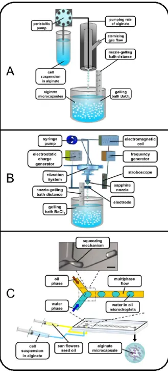
In Fig. 2 are reported the general scheme of the currently employed procedures for the production of alginate microcapsules.
One of the main aim of the PhD thesis was the development of new or more performing encapsulation procedures based on a variety of physical principles and instrumentations, including coaxial bead generator (Fig. 2 A), vibrating-nozzle procedure (Fig. 2 B) and microfluidics. (Fig. 2 C). The studied and well-validated encapsulation procedures were thereafter applied to the entrapment of different cell systems including bronchial epithelial cell line, mesenchymal stem cells and Sertoli cells.
Fig. 2. Schematic representations of different encapsulation protocols: coaxial bead generator (A), vibrating-nozzle procedure (B) and microfluidic based approach (C).
3.2. Cell encapsulation by coaxial bead generator
The encapsulation procedure based on the use of a coaxial bead generator (Fig. 2 A), represents one of the most used instrumentation for the production of polysaccharidic microcapsules, intended for cell encapsulation. Various encapsulation systems are commercially available for the production of
alginate microbeads in a controllable manner (i.e. Coaxial Airflow Induced Dripping VAR J1 from Nisco Engineering Inc, Switzerland, see Fig. 3).
The general principle of the instruments is based on a coaxial air stream that blows polymer droplets from a needle tip into a gelling bath.
Fig. 3. Pictures of the Coaxial Coaxial Airflow Induced Dripping VAR J1 from Nisco Engineering Inc. The instrument is connected with hoses providing alginate solution and air, respectively.
In our laboratory, we have recently designed and produced a new model of coaxial bead generator, named “gas driven mono-jet device”. The entire project was developed with the aim to improve some instrumental characteristics and performances of coaxial bead generators on the market available including the connectivity (to alginate-cell suspension and gas/air generator) and the possibility to change the internal diameter of the nozzle, in order to obtain microcapsules with different diameters.
Fig. 4. Schematic and 3-D representation of the gas driven mono-jet device showing the two lateral and top inlets for the alginate feeding (1) and the atomizing gas (2).
In this respect, our device is equipped with two lateral and one top standard rapid connectors based on female luer lock (as air and alginate inlets) and an internal nozzle (commercially available blunt end needles) that is
easily interchangeable, depending on the dimensions of the final droplets required. The complete encapsulation system is composed of a gas driven mono-jet device connected to a precision peristaltic pump (for the alginate feeding) and to a gas flask (usually nitrogen) equipped with a flow meter (providing the gas for the atomization of the alginate). The generated microdroplets are then consolidated to give microparticles by a gelation procedure generally based on calcium or barium ion solutions. Typically, the cell suspension is continuously mixed by a magnetic stirrer to prevent cell clumping, which could lead to inhomogeneous cell distribution within the microparticles.
2.2.1. Application of a coaxial bead generator to IB3-1 cells
Cystic fibrosis (CF) is an autosomal recessive disorder caused by mutations of the CF transmembrane conductance regulator (CFTR) gene, which encodes a transmembrane protein present on a variety of cell types and organelles [4]. The CF lung is characterized by chronic bacterial infection of the airways, thickened airway mucous, and bronchiectasis [5]. The excess of mucus is largely caused by the influx of neutrophils, attracted to the site by the increased expression of chemokines such as interleukin-6 (IL-6) and interleukin-8 (IL-8), by bacterial products and inflammatory cytokines [6]. In particular, IL-8, that is potent chemokine, is induced transcriptionally by a wide variety of stimuli including tumor necrosis factor-alpha (TNF-a), hyperosmotic shock and bacterial [7]. In order to possibly study in detail the mechanism(s) of activation of IL-8 in CF, the IB3-1 cell system has been recently proposed [8]. IB3-1 is a bronchial epithelial cell line, derived from a CF patient with a CFTR genotype of F508del/W1282X, therefore carrying the associated cystic fibrosis mutation. This cell line can be induced to high expression of proinflammatory proteins, following infection with Pseudomonas aeruginosa or by treatment with TNF-a, for at least 24 h. In order to develop a specific system to possibly study the mechanism of bacterial activation of IB3-1 cells as well as the eff ect of the secreted
chemokines on target cell populations, in co-culture experiments (eg. Pseudomonas aeruginosa or polymorphonuclear cells) alginate microcapsules containing IB3-1 cells were produced. The co-culture experiments could be performed in the presence/absence of a semipermeable membrane embedding the IB3-1 cells, representing a physical barrier to cell/cell interactions but allowing the cross-talking among the diff erent cells mediated by soluble factors.
2.2.1.1. Materials and methods
Cell cultures
IB3-1 cells were obtained from LGC Promochem (Teddington, Middlesex, UK) and were grown in LHC-8 basal medium (Biofluids, Rockville, MD, USA), supplemented with 5% FBS in the absence of gentamycin. All culture flasks and plates were coated with a solution containing 35 mg/mL bovine collagen (Becton-Dickinson Italia, Milan, Italy), 1 mg/mL bovine serum albumin (Sigma, St. Louis, MO, USA), and 1mg/mL human fibronectin (Becton-Dickinson).
Encapsulation of IB3-1 cells
Before encapsulation, confluent monolayers of IB3-1 were scraped off by 0.05% trypsin/EDTA (Gibco, Grandisland, NY, USA) (2min), washed with PBS, counted by hemocytometric analysis, and assayed for viability by double staining with propidium iodide (PI) and Calcein-AM (Sigma), following the manufacturer‟s indications. Briefly, IB3-1 cells were suspended in a 1.5% (w/v) aqueous solution of highly purified sodium alginate (Inotech, Dottikon, Switzerland), further purified by a multistep filtration process, at a concentration of 8–12 × 106 cells/ml. The resulting cell suspension was continuously aspirated by a syringe pump and extruded through the gas driven mono-jet, developed in our laboratory, under sterile conditions. The generated microdroplets were hardened by an ionotropic gelling process into a 1.2% (w/v) barium chloride solution that resulted in the production of barium alginate microbeads. After 3 min of incubation into the gelling bath,
the microbeads were washed twice with saline and placed in LHC-8 basal medium (Biofluids), supplemented with 5% FBS at 37◦C in an humidified atmosphere of 5% CO2.
Dimensional and morphological characterization of alginate microbeads The morphology of barium alginate microbeads was evaluated by optical microscopy and stereomicroscopy (Nikon microscopes, Tokyo, Japan). Microbead size was determined by photomicrograph analyses (Eclipsenet version 1.16.5; Laboratory AU5c Imaging s.r.o. for Nikon B.V.). Samples of 200–400 beads were considered.
Viability determination of encapsulated IB3-1 cells
After encapsulation and after different lengths of time, the viability of IB3-1 cells was analysed by double staining with propidium iodide (PI) and Calcein-AM, following manufacturer‟s instructions. For the propidium iodide (PI) and Calcein-AM analysis, cells were visualized under a fluorescence microscope (Nikon, Optiphot-2, Nikon Corporation, Japan). Viable cells were stained in green while the dead ones were stained in red.
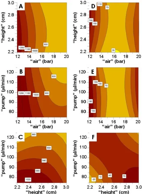
Experimental Design and Statistical Analysis
To study the effect and the influence of different experimental parameters on the size and size distribution of alginate microbeads, a randomized central compositive face centered design (CCF) consisting of 17 runs was used. The experimental design and the evaluation of the experiments were performed by the PC software MODDE 8.0 (Umetrics AB, Sweden), followed by multiple linear regression (MLR) algorithms. The following experimental parameters (“factors”) were considered: the atomizing air flow (“air”), the alginate pumping rate (“pump”), and the distance between the nozzle of gas driven mono-jet device and the surface of the gelling bath (“height”).
Treatment of monolayers
IB3-1 cells were seeded at the initial concentration of 30,000 cells/cm2 and the cell number/ml determined after 3 days of culture. Cell number/ml was determined after trypsin treatment by using a model ZBI Coulter Counter (Coulter Electronics, Hialeah, FL, USA). Treatment of monolayers with 80 ng/ml TNF- (PeProTech EC, London, UK) was performed on 70% confluent
cells for 24 hours. Treatment of encapsulated cells: equal quantity (20x106 cells) of free and encapsulated cells (derived from the same flask, previously cultured for 3 days and successively detached by trypsin) were treated with 80 ng/ml TNF- for 24 hours.
Cytokine profiles
Cytokines in tissue culture supernatants released from the cells under analysis, were measured by Bio-Plex cytokine assay (Bio-Rad Laboratories, Hercules, CA) [9] as described by the manufacturer. The Bio-Plex cytokine assay is designed for the multiplexed quantitative measurement of multiple cytokines in a single well using as little as 50 µl of sample. In our experiments, the premixed multiplex beads of the Bio-Plex human cytokine 7-plex which included seven cytokines (IL-1r , IL-6, IL-8, G-CSF, MCP-1 (MCAF), RANTES, VEGF) were used. 50 μl of cytokine standards or samples (supernatants recovered from treated cells) were incubated with 50 μl of anti-cytokine conjugated beads in 96-well filter plates for 30 min at room temperature with shaking. Plates were then washed by vacuum filtration three times with 100 μl of Bio-Plex wash buffer, 25 μl of diluted detection antibody were added, and plates were incubated for 30 min at room temperature with shaking. After three filter washes, 50 μl of streptavidin-phycoerythrin was added, and the plates were incubated for 10 min at room temperature with shaking. Finally, plates were washed by vacuum filtration three times, beads were suspended in Bio-Plex assay buffer, and samples were analysed on a Bio-Rad 96-well plate reader using the Bio-Plex Suspension Array System and Bio-Plex Manager software (Bio-Rad Laboratories, Hercules, CA).
Quantification of IL-8 and IL-6 transcripts
Total RNA was isolated (High Pure RNA isolation kit, Roche), retro transcribed (Promega Corporation, Madison, USA) and the resulting cDNA was quantified by relative quantitative real-time PCR. The sequences of the oligonucleotides used for amplification of IL-8 mRNA were: 5‟-GTG CAG TTT TGC CAA GGA GT-3‟ (forward) and 5‟-TTA TGA ATT CTC AGC CCT CTT CAA AAA CTT CTC-3‟ (reverse); for IL-6 mRNA: 5‟-AGG AGA CTT GCC
TGG TGA AA-3‟ (forward) and 5‟-CAG GGG TGG TTA TTG CAT CT-3‟ (reverse); for GAPDH mRNA: 5‟-AAG GTC GGA GTC AAC GGA TTT-3‟ (forward); 5‟-ACT GTG GTC ATG AGT CCT TCC A-3‟ (reverse). For PCR, 0,5/20 μl aliquots of cDNA were used for each Sybr Green real-time PCR reaction to quantify the relative tissue expression of IL-8 and IL-6 transcripts. Each 25 μl of total reaction volume contained 0.5 μl of cDNA, 10 pmol of primers, 1 x iQ™ SYBR® Green Supermix (Bio-Rad Laboratories, Hercules, CA). Real-time PCR reactions were performed for a total of 40 cycles (denaturation, 95°C for 10 s; annealing, 68°C for 30 s for IL-8, 65°C for 30 s for IL-6; elongation, 72°C for 60 s) using an iCycler IQ® (Bio-Rad Laboratories, Hercules, CA). The relative proportions of each template amplified were determined based on the threshold cycle (Tc) value for each PCR reaction. The ΔΔCt method was used to compare gene expression data. Each sample was quantified in duplicate from at least two independent experiments. Mean ± S.D. values were determined for each fold difference. Amplification of human GAPDH cDNA served as internal standards (housekeeping gene). Duplicate negative controls (no template cDNA) were also run with every experimental plate to assess specificity and indicate potential contamination.
Data Analysis and statistics
Statistical analysis was performed by one-way analysis of variance followed by the Student‟s t test. A P value <0.05 was considered statistically significant.
2.2.1.2. Results
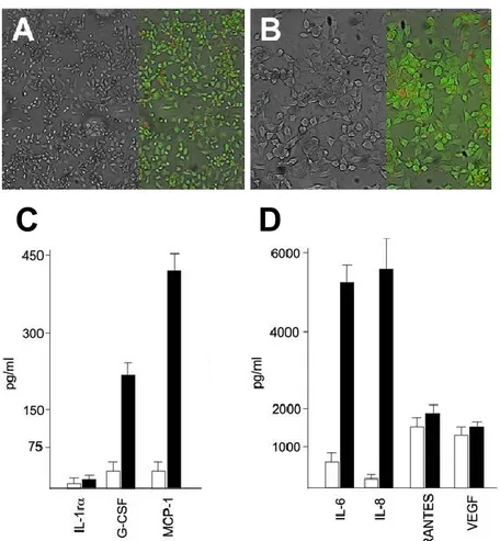
Release of pro-inflammatory proteins by IB3-1 cells exposed to TNF- : a Bio-Plex analysis
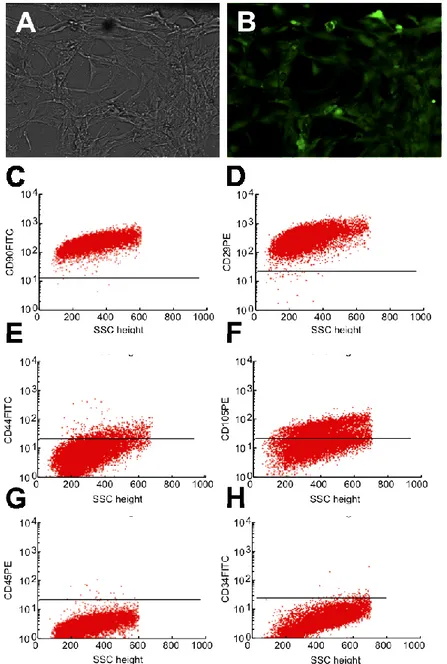
IB3-1 cystic fibrosis cell monolayers (see Figure 5 A-B) were treated, after 3 days cell culture, for 24 hours in the presence of 80 ng/ml of
TNF-thereafter, the conditioned media, from treated and control cell populations, were analysed for presence of pro-inflammatory cytokines by