RESEARCH
Electroencephalographic features in patients
undergoing extracorporeal membrane
oxygenation
Lorenzo Peluso
1*, Serena Rechichi
1,2, Federico Franchi
1,2, Selene Pozzebon
1,2, Sabino Scolletta
2,
Alexandre Brasseur
1, Benjamin Legros
3, Jean‑Louis Vincent
1, Jacques Creteur
1, Nicolas Gaspard
3,4†and Fabio Silvio Taccone
1†Abstract
Background: Neurologic injury is one of the most frequent causes of death in patients undergoing extracorporeal membrane oxygenation (ECMO). As neurological examination is often unreliable in sedated patients, additional neu‑ romonitoring is needed. However, the value of electroencephalogram (EEG) in adult ECMO patients has not been well assessed. Therefore, the aim of this study was to assess the occurrence of electroencephalographic abnormalities in patients treated with extracorporeal membrane oxygenation (ECMO) and their association with 3‑month neurologic outcome.
Methods: Retrospective analysis of all patients undergoing venous–venous (V–V) or venous–arterial (V–A) ECMO with a concomitant EEG recording (April 2009–December 2018), either recorded intermittently or continuously. EEG background was classified into four categories: mild/moderate encephalopathy (i.e., mostly defined by the pres‑ ence of reactivity), severe encephalopathy (mostly defined by the absence of reactivity), burst‑suppression (BS) and suppressed background. Epileptiform activity (i.e., ictal EEG pattern, sporadic epileptiform discharges or periodic discharges) and asymmetry were also reported. EEG findings were analyzed according to unfavorable neurological outcome (UO, defined as Glasgow Outcome Scale < 4) at 3 months after discharge.
Results: A total of 139 patients (54 [41–62] years; 60 (43%) male gender) out of 596 met the inclusion criteria and were analyzed. Veno–arterial (V–A) ECMO was used in 98 (71%); UO occurred in 99 (71%) patients. Continuous EEG was performed in 113 (81%) patients. The analysis of EEG background showed that 29 (21%) patients had severe encephalopathy, 4 (3%) had BS and 19 (14%) a suppressed background. In addition, 11 (8%) of patients had seizures or status epilepticus, 10 (7%) had generalized periodic discharges or lateralized periodic discharges, and 27 (19%) had asymmetry on EEG. In the multivariate analysis, the occurrence of ischemic stroke or intracranial hemorrhage (OR 4.57 [1.25–16.74]; p = 0.02) and a suppressed background (OR 10.08 [1.24–82.20]; p = 0.03) were independently associated with UO. After an adjustment for covariates, an increasing probability for UO was observed with more severe EEG background categories.
© The Author(s) 2020. Open Access This article is licensed under a Creative Commons Attribution 4.0 International License, which permits use, sharing, adaptation, distribution and reproduction in any medium or format, as long as you give appropriate credit to the original author(s) and the source, provide a link to the Creative Commons licence, and indicate if changes were made. The images or other third party material in this article are included in the article’s Creative Commons licence, unless indicated otherwise in a credit line to the material. If material is not included in the article’s Creative Commons licence and your intended use is not permitted by statutory regulation or exceeds the permitted use, you will need to obtain permission directly from the copyright holder. To view a copy of this licence, visit http://creat iveco mmons .org/licen ses/by/4.0/. The Creative Commons Public Domain Dedication waiver (http://creat iveco mmons .org/publi cdoma in/zero/1.0/) applies to the data made available in this article, unless otherwise stated in a credit line to the data.
Open Access
*Correspondence: [email protected]
†Nicolas Gaspard and Fabio Silvio Taccone have equally contributed as senior author
1 Department of Intensive Care, Erasme Hospital, Université Libre de Bruxelles (ULB), Route de Lennik, 808, 1070 Brussels, Belgium Full list of author information is available at the end of the article
Background
In the last decade, the number of patients treated with extracorporeal membrane oxygenation (ECMO) for car-diopulmonary and respiratory failure has significantly increased [1, 2]. The use of ECMO can lead to a number of major complications [3]. Among them, neurological complications, such as ischemic and hemorrhagic stroke and seizures, are associated with longer hospital stay and increased risk of poor outcome [4]. Several studies docu-mented a high occurrence of neurological complications during ECMO treatment among infants and children, while more recent studies described such complica-tions also in adult patients [4–6]. As patients undergoing ECMO support usually requires sedation, neurological examination is not always reliable and neuromonitoring becomes important to detect rapidly such neurological complications. There has been increasing interest in the application of different neuromonitoring tools in ECMO patients, including electroencephalography (EEG), neu-roimaging, near-infrared spectroscopy (NIRS) and chem-ical biomarkers [7]; the available data on their usefulness in adult ECMO patients remain scarce [8].
EEG is a noninvasive tool that measures cortical elec-trical activity, with a good spatial and temporal resolu-tion and sensitivity to changes in both brain structure and function [9]. In critically ill patients, EEG is widely used to recognize or assess different neurological com-plications, such as seizure and encephalopathy [10, 11], or to prognosticate neurological outcome after an acute brain injury [12, 13]. Nevertheless, the use of EEG as neuromonitoring tool in adult ECMO patients has also been reported only in small case-series [8, 14].
The aim of this study was therefore to evaluate the occurrence of EEG abnormalities and their prognostic value in adult ECMO patients.
Methods
Study design and patient selection
This retrospective study was performed in the Depart-ment of Intensive Care at Erasme Hospital, Brussels (Bel-gium). The local Ethical Committee (Comité d’Ethique Hospitalo-Facultaire Erasme-ULB) approved the study but waived the need for informed consent because of its retrospective nature (Protocol 2018/263). From our institutional database (April 2009–December 2018), all patients undergoing veno–arterial (V–A) and veno– venous (V–V) ECMO and who had a concomitant EEG
monitoring were considered eligible for the study. EEG was recorded either as continuous or discontinuous. Exclusion criteria were: ECMO missing data; EEG arti-facts that made EEG records unreadable; death before 24 h from ICU admission, as the occurrence of neurolog-ical dysfunction could not be evaluated and EEG record-ing was quite limited.
Patients’ care and monitoring
All patients were monitored using an arterial catheter and a central venous catheter. A deep sedation status during mechanical ventilation was initially obtained using a combination of sedatives (i.e., midazolam or propofol) and analgesics (i.e., morphine or sufentanil) and then adjusted according to clinical needs. Advanced hemodynamic monitoring was used (PiCCO, Pulsion, Munich, Germany) and the assessment of cardiac func-tion by repeated trans-esophageal and/or trans-thoracic echocardiography. Mean arterial pressure was main-tained at > 65–70 mmHg using volume resuscitation, noradrenaline and/or dobutamine, whenever needed, or by adjusting the ECMO blood flow in veno-arterial (V-A) configuration. Blood flow, FiO2 and gas flow of
the ECMO were adapted to maintain PaO2 between 60
and 150 mmHg and PaCO2 between 35 and 45 mmHg,
with a prior adaptation of the ventilator for a protective ventilation associated to the lowest FiO2. Blood glucose
was kept between 110 and 150 mg/dl using continuous insulin infusion. Enteral nutrition was initiated as soon as possible and continued thereafter according to gastric tolerance. Management of ECMO is reported in Addi-tional file 1.
Data collection and definition
We collected data on demographics, comorbidities, indi-cations and duration of ECMO support, as well as ICU length of stay and hospital mortality. We also collected Glasgow Coma Score during ECMO and major compli-cations during ECMO therapy (i.e., cerebral ischemic stroke; intracranial hemorrhage—ICH), brain death, major bleeding, i.e., reduction of at least 2 g/dL in hemo-globin levels requiring ≥ 4 RBC units to be transfused over 24 h), ECMO configuration, the use of red blood cells transfusions (RBCT), sedative, analgesic and/or antiepileptic drugs. Also, the results from brain imaging were collected, whenever available during the entire ICU stay. We also recorded mechanical ventilation settings
Conclusions: In adult patients treated with ECMO, EEG can identify patients with a high likelihood of poor outcome. In particular, suppressed background was independently associated with unfavorable neurological outcome.
and ECMO parameters for the first 2 days of EEG moni-toring. For the final analysis, only the worst of these val-ues were considered.
Neurological evaluation at 3 months after ECMO insertion was assessed using the Glasgow Outcome Scale (GOS; 1 = Death or severe injury without recovery of consciousness; 2 = Persistent vegetative state; 3 = Severe injury with permanent need for help with daily living; 4 = Moderate disability with no need for assistance in everyday life; 5 = Low disability with minor neurological and psychological deficits) [15]. The GOS evaluation was assessed during follow-up visits or by telephone inter-view with the general practitioner [16]. Favorable neu-rological outcome (FO) was considered as a GOS 4–5, unfavorable outcome (UO) as GOS 1–3.
EEG recording and definition
The EEG recording was started by medical indication and was either discontinuous (i.e., 30 min) or continuous (i.e., more than 12 h), depending on clinical patient’s status and medical decisions. Twenty-one EEG electrodes were placed on the scalp, according to the International 10–20 system (Software: BrainRT, OSG Inc., Rumst, Belgium). A clinical neurophysiologist (NG), blinded to patients’ clinical features, reviewed all EEG recordings according to the definitions of the American Clinical Neurophysiol-ogy Society’s Standardized Critical Care EEG Terminol-ogy [17]. These EEG were subsequently classified into four categories based on background: mild/moderate encephalopathy (i.e., defined by the presence of reactiv-ity), severe encephalopathy (i.e., defined by the absence of reactivity), burst-suppression (BS) and suppressed back-ground (Additional file 1 for detail).
Reactivity was tested at least once daily by trained EEG technicians using a stimulation protocol comprising auditory, tactile and nociceptive stimulus. The presence of reactivity was assessed by the clinical neurophysiolo-gists and defined as a clear and reproducible change in background amplitude or frequency, including attenu-ation of activity. Stimulus-induced period, rhythmic or ictal discharges (SIRPIDs) were not considered as the presence of reactivity if they were the only form of reac-tivity observed in the recording. No effort was made to decrease sedation prior to reactivity testing. Asymmetry was defined as the presence of a consistent asymmetry in amplitude between hemispheres or of a consistent asym-metry in frequency of ≥ 0.5 Hz, present for the majority (≥ 50%) of the recording. In addition, the occurrence of seizures and periodic discharges was also recorded. Outcome of the study
The primary outcome of this study was to report the occurrence of EEG abnormalities and assess their
association with UO. Secondary outcome included the occurrence of EEG abnormalities in survivors versus non-survivors, in V–A versus V–V ECMO patients and in patients with cardiac arrest (CA) versus those without CA.
Statistical analysis
Discrete variables were expressed as count (percentage) and continuous variables as mean ± standard deviation (SD) or median [25th–75th percentiles]. The Kolmogo-rov–Smirnov test was used, and histograms and normal-quantile plots were examined to verify the normality of distribution of continuous variables. Demographics, clin-ical and EEG patterns differences between groups (FO vs UO; survivors vs. non-survivors; V–V vs. V–A ECMO; CA vs. non-CA patients) were assessed using the chi-square test, Fisher’s exact test, Student’s t-test or Mann– Whitney U-test, as appropriate.
Multivariable logistic regression analysis with UO as the dependent variable was performed; collinearity between variables (i.e., a linear correlation coefficient higher than 0.3) was excluded prior to modeling; only variables associated with UO in the univariate analysis (p < 0.1) and the presence of cardiac arrest for clinical rel-evance were included in the multivariate model. We did not add in the model the variable “brain death” as it is part of the definition of death itself and was considered redundant with regard to study outcomes. Odds ratios (OR) with 95% confidence intervals (CI) were computed using an enter model. A similar approach was used to perform the multivariate analysis with hospital mortal-ity as the dependent variable. We tested the fitness of the model using Hosmer and Lemeshow goodness-of-fit test. In case of no patients in the control group, we added in such group one patient to calculate OR, as differently it was not possible to obtain the value. The ORs of EEG background category were calculated using mild/mod-erate encephalopathy as reference. All statistical tests were two-tailed, and a p value < 0.05 was considered as statistically significant. Data were analyzed using IBM SPSS Statistics for Macintosh 25 (Armonk, NY, USA) and GraphPad PRISM version 8.0 (San Diego, CA, USA).
Results
Study population
Of 596 ECMO run in 458 patients over the study period (main characteristics in Additional file 2), 141 had con-comitant EEG monitoring; 2 were excluded because EEG data were not interpretable, and 139 (median age 54 [41– 62] years, 60 [43%] male gender) met the inclusion crite-ria and were included in the final analysis (Fig. 1). V–A ECMO was used in 98 (71%) of patients to treat either CA (n = 74) or cardiogenic shock (n = 24); V–V ECMO
was used in 41 (29%) patients to treat acute respiratory distress syndrome (n = 33), as a bridge to transplant in decompensated end-stage lung diseases (n = 6) or for severe pneumonia (n = 2). A total of 73 cerebral CT-scan and 4 cerebral magnetic resonance imaging (MRI) were performed during the ICU stay; ischemic stroke or ICH were observed in 26 patients. The overall ICU length of stay was 9 [3–23] days; hospital mortality occurred in 91 (65%) patients and UO at 3 months was observed in 99 (71%) patients. Patients’ characteristics are summarized in Table 1 and Additional file 3.
EEG monitoring
Continuous EEG was performed in 113 (81%) patients. The analysis of EEG background showed that 29 (21%) patients had severe encephalopathy, 4 (3%) had BS, and 19 (14%) a suppressed background. In addition, 11 (8%) of patients had seizures or status epilepticus, 10 (7%) had generalized periodic discharges (GPDs) or lateralized periodic discharges (LPDs), and 27 (19%) had asymmetry on EEG (Table 1). The median time from ECMO place-ment to EEG recording was 1 [0–2] days.
EEG findings, neurological outcome and mortality
Patients with UO presented more frequently neurological complications (ischemic stroke, ICH or brain death) and had a higher ECMO blood flow, lactate levels as well as a lowest worst GCS during ECMO than those with FO (Table 1 and Additional file 3). On EEG analysis, patients with UO had more frequently a suppressed background than patients with FO (Table 1 and Additional file 3). In the multivariate analysis, after an adjustment for covariates (age, presence of cardiac arrest and high lac-tate levels) and considering all the EEG backgrounds, we observed that the occurrence of ischemic stroke or ICH (OR 4.57 [1.25–16.74]; p = 0.02) and suppressed
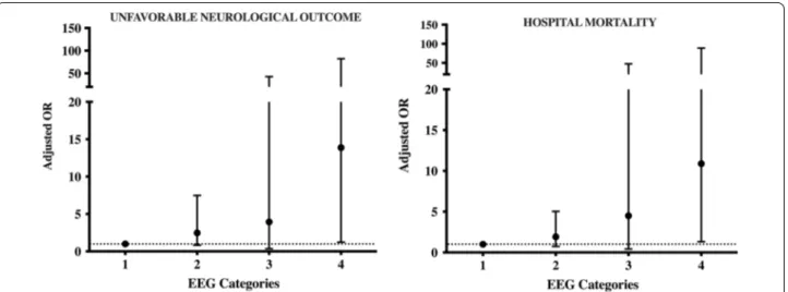
background (OR 10.08 [1.24–82.20]; p = 0.03) were inde-pendently associated with UO (Fig. 2 and Table 2).
Performing a secondary multivariate analysis and dichotomizing EEG according to reactivity, we observed that stroke or ICH (OR 3.85 [1.05–14.15]; p = 0.04) and presence of unreactive EEG (OR 5.39 [1.86–15.62];
p < 0.01) were independently associated with UO
(Addi-tional file 4). Equally, reactive EEG (OR 5.39 [1.86–15.62];
p < 0.01) and the absence of ischemic stroke or ICH (OR
3.85 [1.05–14.15]; p = 0.04) were independent predictors of favorable outcome (Additional file 5).
Non-survivors (15/91 had a diagnosis of brain death) had a higher ECMO blood flow and lactate levels than survivors. On EEG analysis, non-survivors had also more frequently a suppressed EEG background than survi-vors (Additional file 6). In the multivariate analysis, high lactate (OR 1.13 [1.02–1.24]; p = 0.02) and suppressed background (OR 10.88 [1.33–88.67]; p = 0.03) were inde-pendently associated with hospital mortality (Additional file 7).
EEG findings and subgroup analyses
Patients on V–A ECMO had more frequently chronic heart failure and CA than those on V–V ECMO; also, lower gas flow, PaCO2, venous oxygen saturation and
body temperature as well as higher lactate and glucose levels were observed in V–A than in V–V ECMO patients (Additional file 8). Despite V–A ECMO patients experi-enced more frequently brain death and major bleeding, ICU and hospital mortality was not significantly different than V–V ECMO patients. On EEG analysis, no differ-ences were observed between the two groups.
Patients on ECMO after CA were more frequently on V–A configuration and on continuous EEG monitor-ing than others (Additional file 9); also, lower pH and body temperature as well as higher lactate and glucose levels were observed in CA patients when compared to the others. Despite the higher rate of brain death in CA patients, ICU and hospital mortality was not significantly different from non-CA patients. On EEG analysis, sup-pressed background was more frequently observed in CA patients compared to others (16 [19%] vs. 3 [6%]). Exam-ples of one ECMO patient with EEG asymmetry associ-ated with an ischemic stroke and another patient with seizures (before and after treatment) are shown in Addi-tional files 10 and 11.
Discussion
In this study evaluating EEG findings in a large cohort of adult ECMO patients, we observed that 38% of patients presented a severe EEG background abnormality (i.e., severe encephalopathy, BS or suppressed background) and 15% seizures or periodic discharges. The presence Fig. 1 Flowchart of the study
of suppressed EEG background was significantly associ-ated with both UO and hospital mortality. The use of an EEG classification based on background analysis could be helpful to assess prognosis of adult ECMO patients, in particular for suppressed background. To the best of our
knowledge, this is the first study reporting the independ-ent prognostic role of EEG in a large ECMO population.
The literature is sparse on the use of EEG monitor-ing in adult patients on ECMO. In one study, 2 of the 13 adult patients had seizures and required specific Table 1 Characteristics of the study population, according to neurological outcome at 3 months (complete data are presented in Additional file 2: Additional Table 1)
UO unfavorable outcome, FO favorable outcome, EEG electroencephalography, V-A ECMO veno-arterial extracorporeal membrane oxygenation, COPD chronic obstructive pulmonary disease, NYHA New York Heart Association, ICH intracranial hemorrhage, ICU Intensive Care Unit, GOS Glasgow Outcome Scale, SE status epilepticus, GPDs generalized periodic discharges, LPDs lateralized periodic discharges
ALL (n = 139) UO(n = 99) FO(n = 40) p value Age (years) 54 [41–62] 56 [44–65] 48 [37–60] 0.06 Male gender, n (%) 60 (43) 45 (45) 15 (37) 0.45 Continuous EEG, n (%) 113 (81) 84 (85) 29 (72) 0.10 Cardiac arrest, n (%) 86 (62) 63 (64) 23 (57) 0.56 V–A ECMO, n (%) 98 (71) 74 (75) 24 (60) 0.10 Year of ECMO, n (%) 0.16 2009–2012 34 (25) 20 (20) 14 (35) 2013–2015 50 (36) 39 (40) 11 (28) 2016–2018 55 (39) 40 (40) 15 (37) Comorbidities COPD/Asthma, n (%) 18 (13) 13 (13) 5 (12) 1.00 Chronic hemodialysis, n (%) 18 (13) 14 (14) 4 (10) 0.59 Cirrhosis, n (%) 6 (4) 5 (5) 1 (2) 0.67
Heart failure (NYHA III‑IV), n (%) 33 (24) 26 (26) 6 (15) 0.19
Immunosuppression, n (%) 22 (16) 15 (15) 7 (17) 0.80 Cancer, n (%) 7 (5) 9 (7) – 0.19 Therapies Sedative drugs, n (%) 132 (95) 94 (95) 38 (95) 1.00 Analgesic drugs, n (%) 137 (99) 97 (98) 40 (100) 1.00 Antiepileptic drugs, n (%) 21 (15) 12 (12) 9 (22) 0.19 Complications Stroke/ICH, n (%) 26 (19) 23 (23) 3 (7) 0.03 Brain death, n (%) 15 (11) 15 (15) – 0.01 Systemic bleeding, n (%) 33 (24) 26 (26) 7 (17) 0.38 Outcome variables
ICU stay, days 9 [3–23] 7 [2–12] 26 [11–31] < 0.01
Hospital stay, days 12 [3–51] 7 [2–15] 59 [40–86] < 0.01
ICU death, n (%) 90 (65) 90 (91) – < 0.01 Hospital death, n (%) 91 (65) 91 (92) – < 0.01 GOS at 3 months 1 [1–4] 1 [1–1] 5 [5–5] < 0.01 EEG findings Seizures /SE 11 (8) 9 (9) 2 (5) 0.51 GPDs/LPDs 10 (7) 8 (8) 2 (5) 0.72 Asymmetry 27 (19) 19 (19) 8 (20) 1.00 Background categories < 0.001 Mild/moderate encephalopathy 87 (62) 52 (53) 35 (87) Severe encephalopathy 29 (21) 24 (24) 5 (13) Burst‑suppression 4 (3) 4 (4) 0 (0) Suppressed background 19 (14) 19 (19) 0 (0)
antiepileptic medications; all EEGs had a background of predominant theta and delta frequencies and severe encephalopathy (i.e., poor variability and absence of reactivity) was associated with unfavorable neuro-logic outcome [8]. In another study including 25 adult patients on V-A ECMO, 95% of patients had a diffuse EEG slowing and 59% had discontinuous or an unreac-tive background; more severe findings were more fre-quently observed in patients with poor neurological outcome [14]. In a larger prospective cohort of V–A ECMO patients, low background frequency and an unreactive EEG were independently associated with poor outcome at 28 days [18]. Very low Bispectral Index (BIS) values, which is derived by a two-channel frontal EEG and suggest suppressed background or BS, were observed in adult patients undergoing urgent V–A ECMO cannulation for cardiac arrest and subsequently developing brain death [19]. In patients undergoing
V–A ECMO for refractory cardiac arrest, malignant EEG patterns occurring within 96 h from admission were independently associated with poor neurological outcomes [20]. Our findings add important information on the role of EEG monitoring in this setting; although suppressed background was mainly observed in CA patients, this pattern may appear also in other ECMO patients and might be useful to perform further investi-gation to explain its etiology (i.e., diffuse injury, intrac-ranial lesions and sedation). A further interesting and clinically relevant finding was related to the presence of epilepsy or status epilepticus (8% in our cohort) that in absence of EEG could not be detected and eventu-ally treated. Moreover, severe EEG abnormalities were independently associated with UO and this was not related only to cerebrovascular complications, which are usually reported in ECMO patients [21]. Finally, EEG abnormalities were observed in both V–A and Fig. 2 Adjusted odds ratio for unfavorable neurological outcome at 3 months and hospital mortality for the different categories of EEG background,
using mild/moderate encephalopathy as reference (= 1); 2 = severe encephalopathy; 3 = burst‑suppression; 4 = suppressed background
Table 2 Univariate and multivariate analysis to unfavorable neurological outcome at 3 months
Hosmer and Lemeshow goodness-of-fit test: p = 0.04
Univariate Multivariate
Unadjusted OR [CI 95%] p value Adjusted OR [CI 95%] p value
Age 1.02 [0.99–1.04] 0.10 1.02 [1.00–1.05] 0.10 Cardiac arrest 1.44 [0.70–3.00] 0.32 1.20 [0.52–2.73] 0.67 Lactate 1.11 [1.02–1.21] 0.02 1.08 [0.98–1.19] 0.14 Stroke/ICH 3.93 [1.11–13.91] 0.03 4.57 [1.25–16.74] 0.02 Background categories Mild/moderate encephalopathy 1 1 Severe encephalopathy 3.23 [1.13–9.27] 0.03 2.48 [0.82–7.48] 0.11 Burst‑suppression 2.69 [0.29–25.10] 0.39 3.94 [0.36–42.75] 0.26 Suppressed background 12.79 [1.64–99.94] 0.02 10.08 [1.24–82.20] 0.03
V–V configurations, suggesting a wide applicability of such monitoring in all ECMO patients.
Severe encephalopathy, BS and suppressed background have already been reported as reliable predictors of UO in different subgroups of critically ill patients. Azabou et al. showed that the absence of EEG reactivity (i.e., severe encephalopathy) was independently associated with the occurrence of delirium and ICU mortality in septic patients [11]. In comatose patients admitted for a hypoxic ischemic brain injury following CA, the pres-ence of highly malignant EEG patterns (i.e., persistent suppressed background, BS or severe encephalopathy) was associated with a poor neurological outcome, with a high positive predictive value (94–97%) [13]. In mechani-cally ventilated patients, even short time in BS was inde-pendently associated with the occurrence of post-coma delirium and a delayed resolution of delirium, even after adjustment for covariates [22]. In both patients with trau-matic brain injury or subarachnoid hemorrhage, neuro-logical outcome was poor in most of the patients with absent EEG reactivity [23, 24]. Importantly, considering the requirement for a dedicated neurophysiologist and available devices, as well as the complexity of EEG inter-pretation, we advise caution on the wide use of EEG to prognosticate adult ECMO patient outcome until larger studies would validate this association with UO.
Our population showed a high rate of hospital mortality and poor neurological outcome; hence, comparison with previous studies remains difficult. Indeed, we enrolled many patients treated either with V–V or V–A ECMO that presented different diagnosis on ICU admission, as EEG monitoring has been implemented as neuromoni-toring tool of critically ill patients in clinical practice. In previous studies, the rate of poor outcome ranged from 10 to 60%, as EEG was used in much selected cases, when a neurological complication was suspected. As neuro-logical examination in ECMO patients is still limited by the concomitant use of sedatives, as these patients usu-ally require sedation, EEG could be considered as a valu-able tool to obtain relevant information about residual brain function, the occurrence of EEG abnormalities and changes in EEG background over time. Interestingly, the incidence of epileptiform EEG patterns in adult ECMO patients was quite limited and not associated with poor outcome, as antiepileptic drugs were rapidly initiated to limit these events.
This study presents some limitations. First, its retro-spective design and subgroups analysis could provide important biases to the data interpretation. Second, we did not standardize the timing to initiated EEG monitor-ing in all our patients (except for patients suffermonitor-ing from CA who were monitored since day 1); as a consequence, we could have not missed early EEG abnormalities in
some patients, which were potentially associated with poor outcome. Third, we used continuous EEG moni-toring only in 81% of the patients; as a consequence, the incidence of electrographic seizures in patients with non-continuous EEG monitoring could have been under-estimated. Fourth, we did not specifically analyze the morphology of BS, which may have influence the prog-nostic value of EEG, at least in CA patients [25]. Fifth, the role of sedation on EEG abnormalities still remains an open question; we did not specifically report cumulative sedative drugs, although the effects of sedation on EEG background are also dependent on other factors [26]. Moreover, patients on V–A ECMO patients were more frequently hypothermic during the first 24 h (i.e., tem-perature management after CA) and most of V–V ECMO patients deeply sedated in the early phase of therapy. Prospective studies evaluating the changes in EEG back-ground due to sedation withdrawal in ECMO patients would be useful to quantify the impact of the sedative drugs on EEG abnormalities. Sixth, we did not specifi-cally report reasons for death (e.g., withdrawal of life sus-taining therapies, WLST), as these may impact on the prognostic role of monitoring tools. Similarly, it remains difficult to evaluate whether the EEG had contributed to decisions of WLST decisions, modifications of specific therapies or simply reflected underlying severe brain injury. Seventh, we did not compare EEG patterns with brain imaging or biomarkers of brain injury and this anal-ysis might have been interesting to assess the correlation between anatomical and functional brain dysfunction in such patients. Finally, we did not observe a prognos-tic role for BS; however, only few patients presented this EEG abnormality.
Conclusions
The use of EEG monitoring can be a useful tool to assess brain dysfunction and prognosis of patients undergoing ECMO support. Future studies will help to validate such findings and better describe EEG abnormalities of inter-est in this setting.
Supplementary information
Supplementary information accompanies this paper at https ://doi. org/10.1186/s1305 4‑020‑03353 ‑z.
Additional file 1 ECMO management and EEG definitions.
Additional file 2 Main characteristics of ECMO patients, according to EEG monitoring.
Additional file 3 Characteristics of the study population, according to neurological outcome at 3 months.
Additional file 4 Univariate and multivariate analyses to predict unfavora‑ ble neurological outcome at 3 months, according to EEG reactivity.
Additional file 5 Univariate and multivariate analyses to predict favorable neurological outcome at 3 months, according to EEG reactivity.
Additional file 6 Characteristics of the study population, according to hospital mortality.
Additional file 7 Univariate and Multivariate analysis to hospital mortality. Additional file 8 Characteristics of the study population, according to the ECMO configuration.
Additional file 9 Characteristics of the study population, according to the occurrence of cardiac arrest.
Additional file 10 Description of a case of early diagnose of stroke. Additional file 11 Description of a case of Nonconvulsive Status Epilepti‑ cus treated with success.
Abbreviations
ECMO: Extracorporeal membrane oxygenation; EEG: Electroencephalography; NIRS: Near‑infrared spectroscopy; V–A: Veno–arterial; V–V: Veno–venous; ICU: Intensive Care Unit; FiO2: Fraction of inspired oxygen; PaO2: Partial pres‑ sure of oxygen in arterial blood; PaCO2: Partial pressure of oxygen in arterial blood; ICH: Intracranial hemorrhage; RBC: Red blood cells; RBCT: Red blood cells transfusion; GOS: Glasgow outcome scale; FO: Favorable outcome; UO: Unfavorable outcome; BS: Burst‑suppression; CA: Cardiac arrest; SD: Standard deviation; OR: Odds ratio; CI: Confidence interval.
Acknowledgements Not applicable. Authors’ contributions
LP, NG and FST conceived the study; LP, AB and FST selected the population; NG reviewed all EEG recordings; SR, FF, BL collected the data; LP and FST con‑ ducted the statistical analysis and wrote the first draft of the paper; LP, BL, SS, JLV, JC, NG, FST revised the text for intellectual content. All authors read and approved the final manuscript.
Funding
No funding was obtained to this study. Availability of data and materials
The datasets used and/or analyzed during the current study are available from the corresponding author on reasonable request.
Ethics approval and consent to participate
The study protocol was approved by the local ethics committee (P2018/263) and the informed written consent was waived for the retrospective design of the study.
Consent for publication Not applicable. Competing interests
The authors declare that they have no competing interests. Author details
1 Department of Intensive Care, Erasme Hospital, Université Libre de Bruxelles (ULB), Route de Lennik, 808, 1070 Brussels, Belgium. 2 Department of Medi‑ cal Biotechnologies, Anesthesia and Intensive Care Unit, University of Siena, Via Bracci 1, 53100 Siena, Italy. 3 Department of Neurology Erasme Hospital, Université Libre de Bruxelles, Route de Lennik, 808, 1070 Brussels, Belgium. 4 Department of Neurology, Yale University Medical School, 15, York Street, New Haven, CT 06510, USA.
Received: 7 July 2020 Accepted: 21 October 2020
References
1. Schmidt M, Tachon G, Devilliers C, et al. Blood oxygenation and decar‑ boxylation determinants during venovenous ECMO for respiratory failure in adults. Intensive Care Med. 2013;39:838–46.
2. Nasr DM, Rabinstein A. Neurologic complications of extracorporeal mem‑ brane oxygenation. J Clin Neurol. 2015;11(4):383–9.
3. Zangrillo A, Landoni G, Biondi‑Zoccai G, et al. A meta‑analysis of compli‑ cations and mortality of extracorporeal membrane oxygenation. Crit Care Resusc. 2013;15(3):172–8.
4. Mehta A, Ibsen LM. Neurologic complications and neurodevelopmen‑ tal outcome with extracorporeal life support. World J Crit Care Med. 2013;2:40–7.
5. Graziani LJ, Gringlas M, Baumgart S. Cerebrovascular complications and neurodevelopmental sequelae of neonatal ECMO. Clin Perinatol. 1997;24:655–75.
6. Hervey‑Jumper SL, Annich GM, Yancon AR, et al. Neurological complica‑ tions of extracorporeal membrane oxygenation in children. J Neurosurg Pediatr. 2011;7:338–44.
7. Bembea MM, Felling R, Anton B, Salorio CF, Johnston MV. Neuromonitor‑ ing during extracorporeal membrane oxygenation: a systematic review of the literature. Pediatr Crit Care Med. 2015;16(6):558–64.
8. Cho SM, Ziai W, Mayasi Y, Gusdon AM, Creed J, Sharrock M, Stephens RS, Choi CW, Ritzl EK, Suarez J, Whitman G, Geocadin RG. Noninvasive neu‑ rological monitoring in extracorporeal membrane oxygenation. ASAIO J. 2020;66(4):388–93.
9. Claassen J, Taccone FS, Horn P, et al. Recommendations on the use of EEG monitoring in critically ill patients: consensus statement from the neurointensive care section of the ESICM. Intensive Care Med. 2013;39(8):1337–51.
10. Rittenberger JC, Popescu A, Brenner RP, et al. Frequency and timing of nonconvulsive status epilepticus in comatose post‑cardiac arrest subjects treated with hypothermia. Neurocrit Care. 2012;16:114–22.
11. Azabou E, Magalhaes E, Braconnier A, Yahiaoui L, Moneger G, Heming N, Annane D, Mantz J, Chrétien F, Durand MC, Lofaso F, Porcher R, Sharshar T, Groupe d’Explorations Neurologiques en Réanimation (GENER). Early standard electroencephalogram abnormalities predict mortality in septic intensive care unit patients. PLoS One 2015; 10(10):e0139969.
12. Rosenthal ES, Biswal S, Zafar SF, O’Connor KL, Bechek S, Shenoy AV, Boyle EJ, Shafi MM, Gilmore EJ, Foreman BP, Gaspard N, Leslie‑Mazwi TM, Rosand J, Hoch DB, Ayata C, Cash SS, Cole AJ, Patel AB, Westover MB. Con‑ tinuous electroencephalography predicts delayed cerebral ischemia after subarachnoid hemorrhage: a prospective study of diagnostic accuracy. Ann Neurol. 2018;83(5):958–69.
13. Lamartine Monteiro M, Taccone FS, Depondt C, Lamanna I, Gaspard N, Ligot N, Mavroudakis N, Naeije G, Vincent JL, Legros B. The prognostic value of 48‑h continuous EEG during therapeutic hypothermia after cardiac arrest. Neurocrit Care. 2016;24(2):153–62.
14. Sinnah F, Dalloz MA, Magalhaes E, Wanono R, Neuville M, Smonig R, Radjou A, Mourvillier B, Bouadma L, Timsit JF, d’Ortho MP, Rouvel‑Tallec A, Sonneville R. Early electroencephalography findings in cardiogenic shock patients treated by venoarterial extracorporeal membrane oxygenation. Crit Care Med. 2018;46(5):e389–94.
15. Jennett B, Bond M. Assessment of outcome after severe brain damage. Lancet. 1975;1(7905):480–4.
16. Wilson JT, Pettigrew LE, Teasdale GM. Structured interviews for the Glasgow Outcome Scale and the extended Glasgow Outcome Scale: guidelines for their use. J Neurotrauma. 1998;15(8):573–85. https ://doi. org/10.1089/neu.1998.15.573.
17. Hirsch LJ, LaRoche SM, Gaspard N, et al. American Clinical Neurophysiol‑ ogy Society’s standardized critical care EEG terminology: 2012 version. J Clin Neurophysiol. 2013;30:1–27.
18. Magalhaes E, Reuter J, Wanono R, et al. Early EEG for prognostication under venoarterial extracorporeal membrane oxygenation. Neurocrit Care. 2020. https ://doi.org/10.1007/s1202 8‑020‑01066 ‑3.
19. Jouffroy R, Lamhaut L, Guyard A, Philippe P, An K, Spaulding C, Baud F, Carli P, Vivien B. Early detection of brain death using the Bispectral Index (BIS) in patients treated by extracorporeal cardiopulmonary resuscitation (E‑CPR) for refractory cardiac arrest. Resuscitation. 2017;120:8–13. 20. Kim YO, Ko RE, Chung CR, et al. (2020) Prognostic value of early intermit‑
tent electroencephalography in patients after extracorporeal cardiopul‑ monary resuscitation. J Clin Med. 2020;9(6):E1745.
•fast, convenient online submission
•
thorough peer review by experienced researchers in your field
• rapid publication on acceptance
• support for research data, including large and complex data types
•
gold Open Access which fosters wider collaboration and increased citations maximum visibility for your research: over 100M website views per year
•
At BMC, research is always in progress. Learn more biomedcentral.com/submissions Ready to submit your research
Ready to submit your research ? Choose BMC and benefit from: ? Choose BMC and benefit from:
21. Lorusso R, Gelsomino S, Parise O, Di Mauro M, Barili F, Geskes G, Vizzardi E, Rycus PT, Muellenbach R, Mueller T, Pesenti A, Combes A, Peek G, Frenckner B, Di Nardo M, Swol J, Maessen J, Thiagarajan RR. Neurologic injury in adults supported with veno‑venous extracorporeal membrane oxygenation for respiratory failure: findings from the extracorporeal life support organization database. Crit Care Med. 2017;45(8):1389–97. 22. Andresen JM, Girard TD, Pandharipande PP, Davidson MA, Ely EW, Watson
PL. Burst suppression on processed electroencephalography as a predic‑ tor of postcoma delirium in mechanically ventilated ICU patients. Crit Care Med. 2014;42(10):2244–51.
23. Claassen J, Hirsch LJ, Frontera JA, Fernandez A, Schmidt M, Kapinos G, Wittman J, Connolly ES, Emerson RG, Mayer SA. Prognostic significance of continuous EEG monitoring in patients with poor‑grade subarachnoid hemorrhage. Neurocrit Care. 2006;4(2):103–12.
24. Hebb MO, McArthur DL, Alger J, Etchepare M, Glenn TC, Bergsneider M, Martin N, Vespa PM. Impaired percent alpha variability on continuous
electroencephalography is associated with thalamic injury and predicts poor long‑term outcome after human traumatic brain injury. J Neuro‑ trauma. 2007;24(4):579–90.
25. Hofmeijer J, Tjepkema‑Cloostermans MC, van Putten MJ. Burst‑suppres‑ sion with identical bursts: a distinct EEG pattern with poor outcome in postanoxic coma. Clin Neurophysiol. 2014;125(5):947–54.
26. Purdon PL, Pavone KJ, Akeju O, et al. The ageing brain: age‑dependent changes in the electroencephalogram during propofol and sevoflurane general anaesthesia. Br J Anesthesia. 2015;115(Suppl 1):i46–57.
Publisher’s Note
Springer Nature remains neutral with regard to jurisdictional claims in pub‑ lished maps and institutional affiliations.