Endocrine Side Effects Induced by Immune
Checkpoint Inhibitors
Salvatore Maria Corsello, Agnese Barnabei, Paolo Marchetti, Liana De Vecchis, Roberto Salvatori, and Francesco Torino
Endocrinology Unit (S.M.C.), Università Cattolica, 00168 Rome, Italy; Endocrinology Unit (A.B.), Regina Elena National Cancer Institute, 00144 Rome, Italy; Department of Clinical and Molecular Medicine (P.M.), Medical Oncology Division, Sant’Andrea Hospital, Sapienza University of Rome, 00189 Rome, Italy; Department of Systems Medicine (L.D.V., F.T.), Chair of Medical Oncology, Tor Vergata University of Rome, 00133 Rome, Italy; and Pituitary Center (R.S.), Division of Endocrinology, Johns Hopkins University School of Medicine, Baltimore, Maryland 21287
Context: In recent years, progress has been made in cancer immunotherapy by the development
of drugs acting as modulators of immune checkpoint proteins, such as the cytotoxic T-lymphocyte antigen-4 (CTLA4) and programmed death-1 (PD-1), two co-inhibitory receptors that are expressed on T cells upon activation. These molecules play crucial roles in maintaining immune homeostasis by down-regulating T-cell signaling, thereby preventing unbridled T-cell proliferation while main-taining tolerance to self-antigens, such as tumor-associated antigens. CTLA4 blockade through systemic administration of the CTLA4-blocking antibody ipilimumab was shown to confer signif-icant survival benefit and prolonged stable disease in patients affected by advanced cutaneous melanoma. Other immune checkpoint inhibitors are under clinical evaluation. However, immune checkpoint blockade can lead to the breaking of immune self-tolerance, thereby inducing a novel syndrome of autoimmune/autoinflammatory side effects, designated as “immune-related adverse events,” mainly including rash, colitis, hepatitis, and endocrinopathies.
Data Acquisition: We searched the medical literature using the words “hypophysitis,”
“hypopi-tuitarism,” “thyroid,” “adrenal insufficiency,” and “endocrine adverse events” in association with “immune checkpoint inhibitors,” “ipilimumab,” “tremelimumab,” “PD-1,” and “PD-1-L.”
Evidence Synthesis: The spectrum of endocrine disease experienced by patients treated with
ipilimumab includes most commonly hypophysitis, more rarely thyroid disease or abnormalities in thyroid function tests, and occasionally primary adrenal insufficiency. Hypophysitis has emerged as a distinctive side effect of CTLA4-blocking antibodies, establishing a new form of autoimmune pituitary disease. This condition, if not promptly recognized, may be life-threatening (due to secondary hypoadrenalism). Hypopituitarism caused by these agents is rarely reversible, and pro-longed or lifelong substitutive hormonal treatment is often required. The precise mechanism of injury to the endocrine system triggered by these drugs is yet to be fully elucidated.
Conclusions: Although reports of endocrine side effects caused by cancer immune therapy are
abundant, their exact prevalence and mechanism are unclear. Well-designed correlative studies oriented to finding and validating predictive factors of autoimmune toxicity are urgently needed.
(J Clin Endocrinol Metab 98: 0000 – 0000, 2013)
ISSN Print 0021-972X ISSN Online 1945-7197 Printed in U.S.A.
Copyright © 2013 by The Endocrine Society
Received December 3, 2012. Accepted February 6, 2013.
Abbreviations: aCM, advanced cutaneous melanoma; anti-CTLA4-mAbs, anti-CTLA4 mAbs; anti-CTLA4-H, hypophysitis induced by anti-CTLA4-mAbs; anti-CTLA4-T, thyre-opaty induced by anti-CTLA4-mAbs; AE, adverse event; CTLA4, cytotoxic T-lymphocyte antigen-4; E-IRAE, endocrine IRAE; G, grade; IRAE, immune-related AE; mAb, monoclonal antibody; mCRPC, metastatic castration-resistant prostate cancer; MHC, major histocom-patibility complex; MRI, magnetic resonance imaging; PAI, primary adrenal insufficiency; PD-1, programmed death-1; PD-1-L, PD-1 ligand; PSA, prostate-specific antigen; RCC, renal cell cancer; T-regs, T-regulatory cells.
S P E C I A L F E A T U R E
R e v i e w
doi: 10.1210/jc.2012-4075 J Clin Endocrinol Metab jcem.endojournals.org 1
T
he appearance of cancer reveals that the host immunity failed to control tumor progression due to ineffec-tiveness or to acquired tolerance (1). Several mechanisms are proposed to explain cancer immune escape, including the presence of immunoregulatory cell types and the pro-duction of immunosuppressive factors by leukocytes or by cancer cells themselves (2). Anticancer immunotherapy aims to improve the ability to immunologically reject the tumor by generating an adequate immune response, breaking tumor-induced immune tolerance. To attain these goals, several approaches have been developed, in-cluding immunization with cancer cells or molecules and adoptive T-cell transfer (3). The administration of immu-nostimulatory agents such as interferons and IL-2, which induce a less specific activation of the immune system, can be effective in few hematological malignancies, with less marked efficacy in solid tumors (ie, melanoma and renal cell cancer [RCC]) (3). Current research has led to the development of immune regulatory antibodies that inhibit immunological checkpoints, such as the cytotoxic T-lym-phocyte antigen-4 receptor (CTLA4), the programmed death-1 (PD-1) receptor pathway, and others (4). These agents enhance immunological antitumor activity by breaking down tumor immune tolerance (5, 6). Particu-larly, a new class of immunomodulating antibodies di-rected against CTLA4 (anti-CTLA4-mAbs) has been ex-tensively tested in clinical trials (7). One of these agents, ipilimumab, demonstrated for the first time improvement of overall survival in advanced cutaneous melanoma (aCM) and was found to be active against other tumor types (7, 8). However, inhibition of CTLA4 induces side effects defined as “immune-related adverse events” (IRAEs) (9). Autoimmunity is the suggested mechanism sustaining these toxicities (10, 11). IRAEs mainly include colitis/diarrhea, dermatitis, hepatitis, and endocrinopa-thies (12). Among endocrine toxicities, hypophysitis has emerged as a distinctive side effect (9, 13). This condition, because of secondary adrenal insufficiency, may be life-threatening if not promptly recognized (14). Monoclonal antibodies (mAbs) blocking PD-1 or one of its ligands (PD-1-L) are at an earlier phase of clinical development (6, 15). Endocrine toxicity has been reported with these agents as well.Herein, we analyze in parallel the available findings that characterize “classic” and anti-CTLA4-induced hy-pophysitis, highlighting common features and some dif-ferences. In addition, clinical and pathogenic aspects of the other endocrine IRAEs (E-IRAEs) are scrutinized.
Endocrine Toxicities Induced by Anti-CTLA4 mAbs
Ipilimumab and tremelimumab are mAbs directed against CTLA4, a receptor expressed on T cells that exerts a
sup-pressive effect on the immune response after T-cell/anti-gen-presenting cell interaction (5, 16). Blocking the recep-tor, an increased T-cell activation and antitumor effects are obtained (17). In 2 large phase III trials, ipilimumab was shown to confer significant survival benefit and pro-longed stable disease in patients affected by aCM (8, 18). Similar results were not obtained by tremelimumab (19). In 2011, the US Food and Drug Administration and the European Medicines Agency licensed ipilimumab for ad-vanced melanoma (20, 21). The approved dose is 3 mg/kg administered as an iv infusion every 3 weeks for a total of 4 doses. In some patients, maintenance therapy may con-tinue with additional infusions at longer intervals. In clin-ical trials, the dose ranged from 0.3 to 10 mg/kg. Objective clinical responses and an overall survival benefit were demonstrated with 3 mg/kg (8, 22), but not with the lowest dose. The toxicity profile worsens in a dose-dependent manner. In a pooled analysis of 325 patients treated with 10 mg/kg ipilimumab every 3 weeks for 4 doses, IRAEs of any grade (G) were observed in 72.3%. G3– 4 IRAEs were observed in 25.2%, mainly in the gastrointestinal tract (12%), liver (7%), skin (3%), and endocrine system (3%) (23). Anti-CTLA4-IRAEs are managed through adher-ence to specific guidelines (24 –26), including the admin-istration of systemic glucocorticoids or other immunosup-pressants. Retrospective analysis suggests that patients who experience G3– 4 IRAEs may be more likely to benefit from anti-CTLA4 therapy (9, 10, 27).
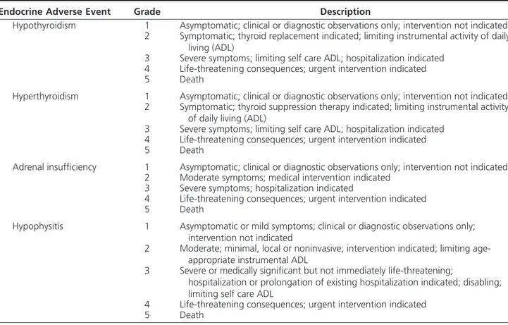
The spectrum of E-IRAEs includes hypopituitarism caused by hypophysitis and, more rarely, thyroid disease or abnormalities in thyroid function tests. Primary adrenal insufficiency (PAI) has been reported occasionally as well (for toxicity grading, see Table 1).
Hypophysitis induced by anti-CTLA4-mAbs
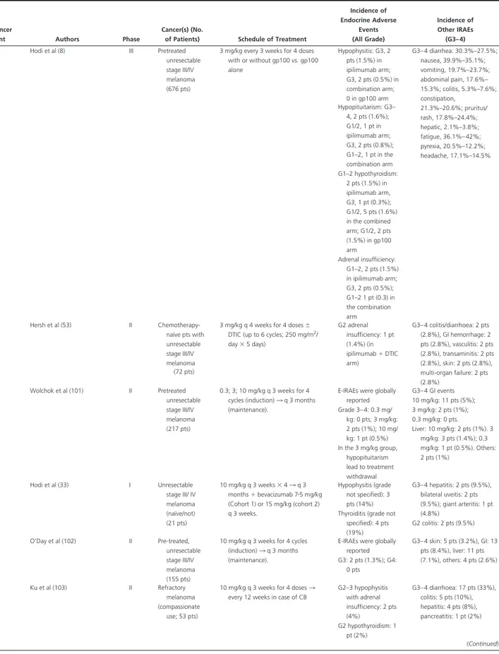
In initial trials, the incidence of hypophysitis induced by anti-CTLA4-mAbs (anti-CTLA4-H) varied considerably (0 –17%) (13). In more recent larger trials, the incidence seemed not to exceed 5% (Table 2). Although most data are from patients affected by aCM, anti-CTLA4-H has been reported in patients with different tumor types.
The incidence of anti-CTLA4-H seems to be dose-de-pendent (Table 2). At the lower ipilimumab dose (1–3 mg/kg), it occurred in 1.8 –3.3% of cases (10, 11). Only in 1 study (28) of 139 melanoma patients receiving 1–3 mg/ kg⫾ a peptide vaccine, G3–4 hypophysitis was diagnosed in 9% of patients. When the dose exceeds 3 mg/kg, hy-pophysitis varies from 4.9 to 17% (29, 30). In a recent phase I dose-escalation trial, 30 patients with metastatic castration-resistant prostate cancer (mCRPC) received ip-ilimumab (1–10 mg/kg) in combination with an anti-pros-tate-specific antigen (PSA) vaccine. Hypophysitis
oc-curred in 4 (13.3%) patients with higher doses (5 mg/kg, 2 patients G2; 10 mg/kg, 1 patient G2 and 1 patient G3) (31). In another study of mCRPC evaluating the combi-nation of ipilimumab (0.3 to 5 mg/kg) with prostate cancer cell vaccine, G2–3 hypophysitis was diagnosed in all 3 patients at the third dose level (3 mg/kg), in 2 of 16 (13%) patients of the 3 mg/kg expansion cohort, and in 2 of 3 patients at the higher dose (5 mg/kg) (32). Higher rates of hypophysitis (14%) are associated with the administra-tion of ipilimumab in combinaadministra-tion with bevacizumab, an agent with antiangiogenic and putative immunogenic ac-tivities (33).
The 10 mg/kg ipilimumab dose was not associated with hypophysitis when combined with chemotherapy in all studies (18, 34, 35) but one (36). Similarly, hy-pophysitis was not reported in 72 patients affected by aCM with brain radiotherapy-pretreated metastases (37). These data suggest that cytotoxic chemotherapy and radiotherapy may prevent anti-CTLA4-H,
presum-ably through immune cell depletion. Tremelimumab has been reported to induce hypophysitis in 0 –2.5% of patients (Table 2).
Patients with anti-CTLA4-H were almost all male. They present with nonspecific symptoms such as fatigue, weakness, headache, nausea, vertigo, behavior change, vi-sual impairments such as diplopia (less frequent than in classic lymphocytic autoimmune hypophysitis [LAH]), confusion, memory loss, loss of libido (29, 38), anorexia, insomnia, hallucinations, temperature intolerance, and subjective sensation of fever and chills (39).
The onset of anti-CTLA4-H symptoms occurs at an average of 6 –12 weeks after initiation of therapy. Patients who received 3 mg/kg ipilimumab developed symptoms at a median time of 11 weeks (ie, before the fourth dose), suggesting a possible cumulative effect (40). In a trial of mCRPC (10 mg/kg), hypophysitis symptoms occurred af-ter the first infusion (4 wk) in 1 patient and afaf-ter the fourth (16 wk) in another (39).
Table 1. Toxicity Grading of Endocrine Adverse Events Associated to Administration of Immune Checkpoint
Inhibitors, Such as Hypothyroidism, Hyperthyroidism, Adrenal Insufficiency, Hypophysitis, According to Common Terminology Criteria for Adverse Events (CTCAE) of National Institutes of Health – National Cancer
Institute (95)
Endocrine Adverse Event Grade Description
Hypothyroidism 1 Asymptomatic; clinical or diagnostic observations only; intervention not indicated
2 Symptomatic; thyroid replacement indicated; limiting instrumental activity of daily living (ADL)
3 Severe symptoms; limiting self care ADL; hospitalization indicated
4 Life-threatening consequences; urgent intervention indicated
5 Death
Hyperthyroidism 1 Asymptomatic; clinical or diagnostic observations only; intervention not indicated
2 Symptomatic; thyroid suppression therapy indicated; limiting instrumental activity of daily living (ADL)
3 Severe symptoms; limiting self care ADL; hospitalization indicated
4 Life-threatening consequences; urgent intervention indicated
5 Death
Adrenal insufficiency 1 Asymptomatic; clinical or diagnostic observations only; intervention not indicated
2 Moderate symptoms; medical intervention indicated
3 Severe symptoms; hospitalization indicated
4 Life-threatening consequences; urgent intervention indicated
5 Death
Hypophysitis 1 Asymptomatic or mild symptoms; clinical or diagnostic observations only;
intervention not indicated
2 Moderate; minimal, local or noninvasive; intervention indicated; limiting age-appropriate instrumental ADL
3 Severe or medically significant but not immediately life-threatening;
hospitalization or prolongation of existing hospitalization indicated; disabling; limiting self care ADL
4 Life-threatening consequences; urgent intervention indicated
5 Death
Hypothyroidism is defined as a disorder characterized by a decrease in production of thyroid hormone by the thyroid gland. Hyperthyroidism is defined as a disorder characterized by an excessive levels of thyroid hormone in the body. Adrenal insufficiency is defined as a disorder that occurs when the adrenal cortex does not produce enough of the hormone cortisol and in some cases, the hormone aldosterone. It may be due to a disorder of the adrenal cortex as in Addison’s disease or primary adrenal insufficiency. General toxicity grading for endocrine adverse event is applicable to hypophysitis. No specific definition of hypophysitis is available yet.
In most cases, magnetic resonance imaging (MRI) re-veals enlargement of the pituitary gland (up to 60 –100% of baseline size), with thickening of the stalk (29). The height of the gland in the sagittal view increases from 3.4 – 6 to 7.7–11.8 mm (29, 41). In some cases, the MRI is normal. The pituitary gland may enhance homogeneously or appear heterogeneous. In general, MRI changes appear to be of lesser magnitude than sporadic LAH (40). At fol-low-up, pituitary decreases gradually on glucocorticoid treatment in a variable period of time (4 –12 wk), despite
a more rapid reduction of symptoms (40, 42, 43). Levels of ACTH, cortisol, TSH and free T4, GH, prolactin, IGF-I, FSH, LH, and testosterone (in males) were variably al-tered, indicating different degrees of hypopituitarism (44). In anti-CTLA4-H, ACTH and TSH seem to be invariably lost (29, 39, 40). Most male patients (83– 87%) had hypogonadotropic hypogonadism. Some of these findings may be confounded by sickness-induced hypogonadism and sick euthyroid syndrome. In a case series (42), 3 of 5 patients had low serum IGF-I.
Pro-Table 2. The Incidence of Endocrine Adverse Events in Studies on New Immunoregulatory Anticancer Agents
Anticancer
Agent Authors Phase
Cancer(s) (No.
of Patients) Schedule of Treatment
Incidence of Endocrine Adverse Events (All Grade) Incidence of Other IRAEs (G3– 4) Ipilimumab Attia et al (10) I Pretreated stage
IV melanoma
3 mg/kg every 3 weeks, or 3 mg/kg 3 doses reduced to 1 mg/kg every 3 weeks⫹ vaccination with modified HLA-A*0201-restricted peptides
from gp100 MAA G3– 4 hypophysitis: 1 pt (1.8%) G3– 4 colitis: 7 pts (13%), dermatitis: 4 pts (7%), uveitis: 1 pt (1.8%), enterocolitis: 1 pt (1.8%), hepatitis: 1 pt (1.8%) (56 pts) Maker et al (44) I Naïve-metastatic melanoma
1–3 mg/kg⫹ interleukin-2 No endocrine toxicity was reported
G3– 4 colitis: 4 pts (11%), uveitis: 1 pt (2.8%), arthritis: 1 pt (2.8%)
(36 pts) Maker et al (30) I-II Pretreated stage
IV melanoma (46 pts) 3 3 9 mg/kg (intra-patient dose escalation) G3– 4 hypophysitis: 8 pts (17%); (5 mg/ kg: 1 pt), (9 mg/ kg: 7 pts) G3– 4 colitis/diarrhoea: 6 pts (13%), uveitis: 1 pt (2.8%), arthritis: 1 pt (2.8%), dermatitis: 1 pt (2.8%), hepatitis: 1 pt (2.8%), nephritis: 1 pt (2.8%) G3– 4 hypothyroidism: 1 pt (2.2%) Downey et al (28) I-II Pretreated stage
IV melanoma
3 mg/kg⫹ peptide vaccinations or intra-patient dose escalation 3 peptides (HLA-A*0201status)
G3– 4 hypophysitis: 13 pts (9%) G3– 4 enterocolitis: 24 pts (17%), dermatitis: 8 pts (6%), hepatitis: 2 pts (3%), uveitis: 3 pts (2%) (139 pts) G1–2 hypothyroidism: 3 pts (2%) Phan et al (43) II Pretreated stage
IV melanoma
3 mg/kg⫹ peptide vaccinations (two modified HLA-A*0201-restricted
peptides from gp100) G3/4 hypophysitis: 1 pt (7.1%) G3– 4 dermatitis: 3 pts (21.4%), enterocolitis/colitis: 2 pts (14䡠3), hepatitis: 1 pt (7.1%) (14 pts) Blansfield et al (29) R.S. Pretreated stage
IV melanoma
3 mg/kg every 3 weeks G3– 4 hypophysitis: 8/163 pts (4.9%), 6/113 melanoma pts (5%), 2/50 RCC pts (4%)
NR. The report focused on clinical aspects of patients who developed anti-CTLA4-IH (113 pts) and RCC (50 pts) Royal et al (96) II Metastatic pancreas ADC (27 pts)
3 mg/kg every 3 weeks⫻ 4 for a maximum 2 courses.
G2–3 hypopituitarism: 1 pt (3.7%)
G3– 4 colitis: 1 pt (3.7%); encefalitis: 1 pt (3.7%) Weber et al (97) I Stages IIIC/IV
melanoma
3 mg/kg every 8 weeks for 12 months ⫹ MART-1/gp100/tyrosinase peptides
G2–3 hypopituitarism (DLT): 1 pt (4%)
No G4 toxicity; G2–3 DLT were GI toxicity: 2 (8%) and skin toxicity: 2 (8%). (25 pts)
Fong et al (98) I mHRPC Escalating doses (0.5, 1.5, 3 mg/kg) every 3 weeks for 4 cycles⫹ GM-CSF
No endocrine toxicity was reported
No G4 toxicity; G3 skin (DLT) (18 pts)
Small et al (99) I mPC 3 mg/kg single dose No endocrine toxicity was reported G3– 4 asthenia: 1 pt (7.1%), pain: 1 pt (7.1%), rash: 1 pt (7.1%) (14 pts) Yang et al (11) I mRCC (61 pts) 3 mg/kg 3 1 mg/kg or all doses at 3 mg/kg q 3 weeks
G3– 4 primary adrenal insufficiency: 1 pt (1.6%) G3– 4 hypopituitarism: 2 pts (3.3%) (All doses) G3– 4 enteritis/colitis: 17 pts (28%), skin: 1 pt (1.6%), arthralgia: 1 pt (1.6%), aseptic meningitis: 1 pt (1.6%)
Weber et al (52) I-II Unresectable. stage III or IV melanoma (88 pts)
Ipilimumab transfectoma -derived 2.8 mg/kg or 5 mg/kg for 3 doses; or ipilimumab hybridoma -derived 3 mg/kg for 3 doses G3– 4 adrenal insufficiency: 1 pt (1.2%) (All doses) G3– 4 colitis: 3 pts (3.4%), diarrhoea: 4 pts (45%), GI perforation: 1 pt (1.1%) Ansell et al (100) I Relapsed/ Refractory B-Cell NHL 3 mg/kg 3 monthly 1 mg/kg⫻ 3 months (dose level 1), 3 escalation to 3 mg/kg monthly⫻ 4 months (dose level 2) G1–2 hypophysitis: 1 pt (6%) No G4 toxicity; G3 diarrhoea: 5 pts (28%), fatigue: 1 pt (6%), neutropenia: 1 pt (6%) (18 pts) (Continued)
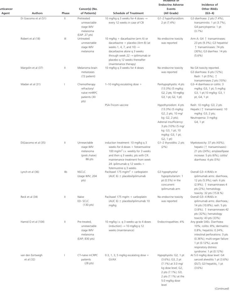
Table 2. Continued
Anticancer
Agent Authors Phase
Cancer(s) (No.
of Patients) Schedule of Treatment
Incidence of Endocrine Adverse Events (All Grade) Incidence of Other IRAEs (G3– 4) Hodi et al (8) III Pretreated
unresectable stage III/IV melanoma (676 pts)
3 mg/kg every 3 weeks for 4 doses with or without gp100 vs. gp100 alone Hypophysitis: G3, 2 pts (1.5%) in ipilimumab arm; G3, 2 pts (0.5%) in combination arm; 0 in gp100 arm G3– 4 diarrhea: 30.3%–27.5%; nausea, 39.9%–35.1%; vomiting, 19.7%–23.7%; abdominal pain, 17.6%– 15.3%; colitis, 5.3%–7.6%; constipation, 21.3%–20.6%; pruritus/ rash, 17.8%–24.4%; hepatic, 2.1%–3.8%; fatigue, 36.1%– 42%; pyrexia, 20.5%–12.2%; headache, 17.1%–14.5% Hypopituitarism: G3– 4, 2 pts (1.6%); G1/2, 1 pt in ipilimumab arm; G3, 2 pts (0.8%); G1–2, 1 pt in the combination arm G1–2 hypothyroidism: 2 pts (1.5%) in ipilimumab arm, G3, 1 pt (0.3%); G1/2, 5 pts (1.6%) in the combined arm; G1/2, 2 pts (1.5%) in gp100 arm Adrenal insufficiency: G1–2, 2 pts (1.5%) in ipilimumab arm; G3, 2 pts (0.5%); G1–2 1 pt (0.3) in the combination arm Hersh et al (53) II Chemotherapy-naïve pts with unresectable stage III/IV melanoma
3 mg/kg q 4 weeks for 4 doses⫾ DTIC (up to 6 cycles; 250 mg/m2/
day⫻ 5 days) G2 adrenal insufficiency: 1 pt (1.4%) (in ipilimumab⫹ DTIC arm) G3– 4 colitis/diarrhoea: 2 pts (2.8%), GI hemorrhage: 2 pts (2.8%), vasculitis: 2 pts (2.8%), transaminitis: 2 pts (2.8%), skin: 2 pts (2.8%), multi-organ failure: 2 pts (2.8%) (72 pts) Wolchok et al (101) II Pretreated unresectable stage III/IV melanoma (217 pts) 0.3; 3; 10 mg/kg q 3 weeks for 4 cycles (induction) 3 q 3 months (maintenance).
E-IRAEs were globally reported Grade 3– 4: 0.3 mg/ kg: 0 pts; 3 mg/kg: 2 pts (1%); 10 mg/ kg: 1 pt (0.5%) In the 3 mg/kg group, hypopituitarism lead to treatment withdrawal G3– 4 GI events 10 mg/kg: 11 pts (5%); 3 mg/kg: 2 pts (1%); 0.3 mg/kg: 0 pts. Liver: 10 mg/kg: 2 pts (1%). 3 mg/kg: 3 pts (1.4%); 0.3 mg/kg: 1 pt (0.5%). Others: 2 pts (1%) Hodi et al (33) I Unresectable stage III/ IV melanoma (naïve/not) (21 pts) 10 mg/kg q 3 weeks⫻ 4 3 q 3 months⫹ bevacizumab 7䡠5 mg/kg (Cohort 1) or 15 mg/kg (cohort 2) q 3 weeks. Hypophysitis (grade not specified): 3 pts (14%) Thyroiditis (grade not
specified): 4 pts (19%) G3– 4 hepatitis: 2 pts (9.5%), bilateral uveitis: 2 pts (9.5%); giant arteritis: 1 pt (4.8%) G2 colitis: 2 pts (9.5%) O’Day et al (102) II Pre-treated, unresectable stage III/IV melanoma (155 pts)
10 mg/kg q 3 weeks for 4 cycles (induction) 3 q 3 months (maintenance).
E-IRAEs were globally reported G3: 2 pts (1.3%); G4: 0 pts G3– 4 skin: 5 pts (3.2%), GI: 13 pts (8.4%), liver: 11 pts (7.1%), others: 4 pts (2.6%) Ku et al (103) II Refractory melanoma (compassionate use; 53 pts)
10 mg/kg q 3 weeks for 4 doses 3 every 12 weeks in case of CB
G2–3 hypophysitis with adrenal insufficiency: 2 pts (4%) G2 hypothyroidism: 1 pt (2%) G3– 4 diarrhoea: 17 pts (33%), colitis: 5 pts (10%), hepatitis: 4 pts (8%), pancreatitis: 1 pt (2%) (Continued)
Table 2. Continued
Anticancer
Agent Authors Phase
Cancer(s) (No.
of Patients) Schedule of Treatment
Incidence of Endocrine Adverse Events (All Grade) Incidence of Other IRAEs (G3– 4) Di Giacomo et al (51) II Pretreated unresectable stage III/IV melanoma
10 mg/kg q 3 weeks for 4 doses 3 every 12 weeks in case of CB
G1–2 hypothyroidism: 2 pt (7.4%) G3 diarrhoea: 2 pts (7.4%), transaminitis: 1 pt (3.7%), G4 pancytopenia: 1 pt (3.7%) (EAP: 27 pts) Robert et al (18) III Untreated
unresectable stage III/IV melanoma
10 mg/kg⫹ dacarbazine (arm A) or dacarbazine⫹ placebo (Arm B) (at weeks 1, 4, 7, and 10) 3 dacarbazine alone q 3 weeks through week 22 3 ipilimumab or placebo q 12 weeks thereafter (maintenance therapy) No endocrine toxicity was reported Arm A: G4 1 transaminases: 23 pts (9.3%); G3 hepatitis/ 1 transaminases: 74 pts (30%); G3 diarrhea: 14 pts (5.6%)
Margolin et al (37) II Melanoma brain metastases (72 patient)
10 mg/kg q 3 weeks for 4 doses No endocrine toxicity was reported No G4 toxicity reported. G3 diarrhoea: 6 pts (12%); Rash: 1 pt (5%); 1 transaminases 2 pts (10%) Madan et al (31) I Chemotherapy refractory/ naïve mHRPC patients (30 pts)
1–10 mg/kg escalating dose⫹ Panhypophysitis: 4 pts (13.3%) (5 mg/kg: G2, 2 pts; 10 mg/kg: G3, 1 pt, G2, 1 pt) G3– 4 diarrhoea or colitis: 3 mg/kg: G3, 1 pt; 5 mg/kg: G3, 1 pt;10 mg/kg: G3, 1 pt, G4, 1 pt
PSA-Tricom vaccine Hypothyroidism: 4 pts (13.3%) (5 mg/kg: G2, 2 pts; 10 mg/ kg: G2, 2 pts); Adrenal insufficiency: 3 pts (10%) (5 mg/ kg: G3, 1 pt; 10 mg/kg: G3, 1 pt; G2, 1 pt) Rash: 10 mg/kg: G3, 2 pts Hepatic (1 transaminases): 10 mg/kg: G3, 2 pts; Neutropenia: 3 mg/kg: G4, 1 pt DiGiacomo et al (35) II Unresectable stage III/IV melanoma (pretr./naïve) induction treatment: 10 mg/kg q 3 weeks for 4 doses⫹ fotoemustine 100 mg/m2i.v. weekly for 3 weeks
and then q 3 weeks; pts with CR: maintenance treatment from week 24: ipilimumab q 12 weeks⫹ fotemustine q 3 weeks G1–2 thyroiditis: 2 pts (2%) Myelotoxicity: 37 pts (43%); hepatic (1 transaminases): 21 pts (24%); amylase/lipase increase: 5 pts (6%); colitis/ diarrhoea: 4 pts (5%) 86 pts Lynch et al (36) IIb NSCLC (stage III/IV; 204 pts) Paclitaxel 175 mg/m2⫹ carboplatin (AUC 6)⫾ placebo/ipilimumab G3 hypophysitis/ hypopituitarism: 1 pt (0.5%) in the concurrent ipilimumab arm Overall G3– 4 IRAEs in ipilimumab arms: diarrhoea, 12 pts (5.9%); rash: 6 pts (2.9%); 1 transaminases 4 pts (2%); hematology toxicity: 32 pts (15.8.%) Reck et al (34) II Naïve ED- SCLC Paclitaxel 175 mg/m⫹ carboplatin (AUC 6)⫾ placebo/ipilimumab 10 mg/kg No endocrine toxicity was reported Overall G3– 4 IRAEs in ipilimumab arms: diarrhoea,: 14 pts (10.8%); rash: 5 pts (3.8%); 1 transaminases 42 pts (32%); hematology toxicity: 43 pts (33%) (130 pts) Hamid O et al (104) II Pre-treated, unresectable stage III/IV melanoma (EAP; 830 pts)
10 mg/kg i.v. q 3 weeks up to 4 doses (induction) 3 10 mg/kg q 12 weeks (maintenance)
Endocrinopathies: 4% Any grade SAEs. Diarrhoea: 10%; colitis: 8%; dermatitis: 0.8%; hepatitis: 0.24%; intestinal perforations: 3 pts (0.36%); multi-organ failure: 1 pt (0.12%); acute respiratory distress syndrome: 1 pt (0.12%) van den Eertwegh
et al (32) I CT-naïve mCRPC patients 0.3, 1, 3, 5 mg/kg escalating dose⫹ GVAX Hypophysitis: G2, 1 pt (3.6%); G3, 2 pt (7.1%) at 3.0 mg/ kg dose level; G2, 2 pts (7.1%); G3, 2 pts (7.1%) at the 5.0 mg/kg dose level At 5.0 mg/kg dose level: G4 sarcoid alveolitis 1 pt (3.6%) (DLT); G3 hepatitis, 1 pt (3.6%) (28 pts) (Continued)
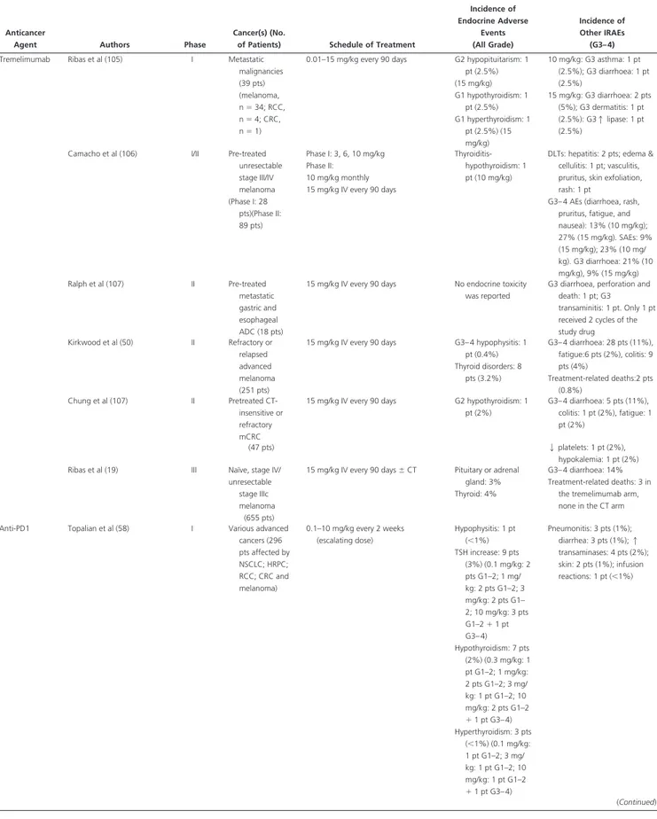
Table 2. Continued
Anticancer
Agent Authors Phase
Cancer(s) (No.
of Patients) Schedule of Treatment
Incidence of Endocrine Adverse Events (All Grade) Incidence of Other IRAEs (G3– 4) Tremelimumab Ribas et al (105) I Metastatic
malignancies (39 pts) (melanoma, n⫽ 34; RCC, n⫽ 4; CRC, n⫽ 1)
0.01–15 mg/kg every 90 days G2 hypopituitarism: 1 pt (2.5%) (15 mg/kg) G1 hypothyroidism: 1 pt (2.5%) G1 hyperthyroidism: 1 pt (2.5%) (15 mg/kg) 10 mg/kg: G3 asthma: 1 pt (2.5%); G3 diarrhoea: 1 pt (2.5%) 15 mg/kg: G3 diarrhoea: 2 pts (5%); G3 dermatitis: 1 pt (2.5%): G31 lipase: 1 pt (2.5%)
Camacho et al (106) I/II Pre-treated unresectable stage III/IV melanoma (Phase I: 28 pts)(Phase II: 89 pts) Phase I: 3, 6, 10 mg/kg Phase II: 10 mg/kg monthly 15 mg/kg IV every 90 days Thyroiditis-hypothyroidism: 1 pt (10 mg/kg)
DLTs: hepatitis: 2 pts; edema & cellulitis: 1 pt; vasculitis, pruritus, skin exfoliation, rash: 1 pt
G3– 4 AEs (diarrhoea, rash, pruritus, fatigue, and nausea): 13% (10 mg/kg); 27% (15 mg/kg). SAEs: 9% (15 mg/kg); 23% (10 mg/ kg). G3 diarrhoea: 21% (10 mg/kg), 9% (15 mg/kg) Ralph et al (107) II Pre-treated metastatic gastric and esophageal ADC (18 pts)
15 mg/kg IV every 90 days No endocrine toxicity was reported
G3 diarrhoea, perforation and death: 1 pt; G3
transaminitis: 1 pt. Only 1 pt received 2 cycles of the study drug Kirkwood et al (50) II Refractory or relapsed advanced melanoma (251 pts)
15 mg/kg IV every 90 days G3– 4 hypophysitis: 1 pt (0.4%) Thyroid disorders: 8 pts (3.2%) G3– 4 diarrhoea: 28 pts (11%), fatigue:6 pts (2%), colitis: 9 pts (4%) Treatment-related deaths:2 pts (0.8%) Chung et al (107) II Pretreated CT-insensitive or refractory mCRC
15 mg/kg IV every 90 days G2 hypothyroidism: 1 pt (2%) G3– 4 diarrhoea: 5 pts (11%), colitis: 1 pt (2%), fatigue: 1 pt (2%) (47 pts) 2 platelets: 1 pt (2%), hypokalemia: 1 pt (2%) Ribas et al (19) III Naïve, stage IV/
unresectable stage IIIc melanoma
(655 pts)
15 mg/kg IV every 90 days⫾ CT Pituitary or adrenal gland: 3% Thyroid: 4%
G3– 4 diarrhoea: 14% Treatment-related deaths: 3 in
the tremelimumab arm, none in the CT arm Anti-PD1 Topalian et al (58) I Various advanced
cancers (296 pts affected by NSCLC; HRPC; RCC; CRC and melanoma) 0.1–10 mg/kg every 2 weeks (escalating dose) Hypophysitis: 1 pt (⬍1%) TSH increase: 9 pts (3%) (0.1 mg/kg: 2 pts G1–2; 1 mg/ kg: 2 pts G1–2; 3 mg/kg: 2 pts G1– 2; 10 mg/kg: 3 pts G1–2⫹ 1 pt G3– 4) Hypothyroidism: 7 pts (2%) (0.3 mg/kg: 1 pt G1–2; 1 mg/kg: 2 pts G1–2; 3 mg/ kg: 1 pt G1–2; 10 mg/kg: 2 pts G1–2 ⫹ 1 pt G3–4) Hyperthyroidism: 3 pts (⬍1%) (0.1 mg/kg: 1 pt G1–2; 3 mg/ kg: 1 pt G1–2; 10 mg/kg: 1 pt G1–2 ⫹ 1 pt G3–4) Pneumonitis: 3 pts (1%); diarrhea: 3 pts (1%); 1 transaminases: 4 pts (2%); skin: 2 pts (1%); infusion reactions: 1 pt (⬍1%) (Continued)
lactin levels may be elevated or low in approximately 25% of patients (29, 30). Only 1 case has been associ-ated with diabetes insipidus (39).
Similar to classic LAH, the treatment has been gluco-corticoids. Almost all patients experienced resolution of acute symptoms a few days after withdrawal of the drug and the start of high-dose glucocorticoids, levothyroxine, and sex hormone replacement (29). However, pituitary function may be impaired for a longer period of time de-spite glucocorticoid therapy. The time needed for resolu-tion of symptoms and duraresolu-tion of replacement therapy with physiological hydrocortisone dosages (mean, 20 wk) may be considerably longer or even lifelong, considering the limited survival of these patients (23, 38). In fact, anti-CTLA4-H is the only possibly irreversible IRAE (45). Recovery of pituitary-thyroid function has been re-ported in 37–50% of patients (39, 40, 42), whereas gonadal axis recovered in 57% of men (29). Conversely, very few patients were able to discontinue glucocorti-coid replacement, due to persistent secondary adrenal insufficiency (9, 39).
The protective role of glucocorticoids in reducing the incidence/severity of anti-CTLA4-H remains to be ex-plored. Re-treatment with ipilimumab after interruption due to G1–2 hypophysitis seems to be safely possible (46). Importantly, high-dose glucocorticoid treatment (and re-placement therapy) do not appear to decrease the antitu-mor effects of CTLA4 blockade (46, 47).
Thyroid side effects by anti-CTLA4-mAbs
Thyroid is the second most frequent endocrine organ involved in anti-CTLA4-mAb toxicity. However, the available clinical details are very limited. Damage to the thyroid induced by these agents presents as thyroiditis as-sociated with antithyroglobulin and anti-thyro
peroxi-dase antibody positivity and hypothyroidism, or transient hyperthyroidism. Rare cases of Graves’ ophthalmopathy have also been reported, with elevation of TSH-receptor antibodies but normal thyroid function (48, 49).
The incidence of thyreopathy induced by anti-CTLA4-mAbs (anti-CTLA4-T) varies between 0 and 4% (Table 2). In 2 studies, tremelimumab was associated with 4% thy-reopathy (19, 50). With ipilimumab, the incidence seems lower (0 –2%) (Table 2), with subclinical or mild (G1–2) hypothyroidism being the most frequent event. In the large phase III trial (8) leading to approval of ipilimumab, the drug was associated with thyroid disorders or abnormal thyroid function tests in approximately 2% of patients. Similar incidence of G1–2 thyroiditis is reported in trials evaluating ipilimumab as a single agent or in combina-tion with chemotherapy in aCM (35) or other malig-nancies (Table 2). In a small report on 27 patients with refractory melanoma receiving a high dose of ipili-mumab (10 mg/kg for 4 doses, followed by further doses in case of clinical benefit), G2 hypothyroidism occurred in 7% of cases (51).
Two cases of thyroiditis induced by ipilimumab in combination with bevacizumab have been reported (48). In a recent phase I study on pretreated or naive aCM patients (ipilimumab, 10 mg/kg every 3 wk for 4 doses, then every 3 mo; bevacizumab, 7.5 or 15 mg/kg every 3 wk), 19% (4 of 21) developed thyroiditis (33). Conversely, no cases of endocrine adverse events (AEs) were reported in 36 patients who received ipilimumab (0.1–3 mg/kg) combined with IL-2 (720 000 IU/kg ev-ery 8 h), a drug known to be associated with autoim-mune thyroiditis (30). In patients with mCRPC who received ipilimumab (1–10 mg/kg escalating dose) in combination with an anti-PSA vaccine, G2
hypothy-Table 2. Continued
Anticancer
Agent Authors Phase
Cancer(s) (No.
of Patients) Schedule of Treatment
Incidence of Endocrine Adverse Events (All Grade) Incidence of Other IRAEs (G3– 4) Anti-PD1-L Brahmer et al (59) I Various advanced
cancers (207 pts, not, including melanoma) 0.3–10 mg/kg every 2 weeks (escalating dose) Hypophysitis: 0 Hypothiroidism: 6 pts (3%): (3 mg/kg: 1 pt G1–2; 10 mg/ kg: 5 pts G1–2) G1–2 autoimmune thyroiditis: 2 pts (1%) Adrenal insufficiency: 3 pts (1.5%): (3 mg/kg: 1 pt G1–2 ⫹ 1 pt G3–4; 10 mg/kg: 1 pts G1–2) G3– 4 toxicity⬍5% (skin, hepatitis, infusion reactions)
Abbreviations: CRC, colorectal cancer; CT, cytotoxic chemotherapy; DLT, dose-limiting toxicity; EAP, expanded access program; ED-SCLC, extensive-disease-small-cell lung cancer; G, toxicity grade according to CTC-NCI criteria; HRPC, hormone resistant prostate cancer; NSCLC, non-small cell lung cancer; Pretr, pretreated; Pt(s), patient(s).
roidism was diagnosed in 4 (13.3%) cases at higher dose levels (5 or 10 mg/kg) (31).
The onset of anti-CTLA4-T seems to be faster com-pared with nonendocrine IRAEs, occurring after 2 to 4 infusions, but similar to the time of onset of anti-CTLA4-H. Most cases have a subclinical course or may be transient, consistent with a silent autoimmune thyroiditis. Alternatively, it may evolve into permanent hypothyroid-ism, requiring thyroid hormone supplementation (9). The administration of anti-CTLA4-mAbs did not seem to worsen previous thyroid disease.
Other endocrinopathies induced by anti-CTLA4-mAbs
PAI has been rarely reported (0.3–1.5%) with anti-CTLA4-mAbs alone or in combination with chemotherapy (8, 19, 52, 53). Yang et al (11), in a phase II study of ipili-mumab in RCC, reported a case of PAI in a patient with metastasis in his residual adrenal gland who received 12 doses at 3 mg/kg. Conversely, in a phase I trial of mCRPC with combination of ipilimumab (1–10 mg/kg escalating dose) and anti-PSA vaccine, PAI was diagnosed in 3 (10%) at higher dosage (5 mg/kg, 1 patient G2; 10 mg/kg, 1 patient G2 and 1 patient G3) (31). No clinical details regarding these patients are available.
Endocrine Toxicities Induced by Anti-PD-1 and Anti-PD-1L mAbs
PD-1 is another inhibitory receptor expressed on activated T cells. PD-1L, one of its ligands, is broadly expressed on antigen-presenting cells, nonimmune tissues, and tumor cells, and its expression correlates with an unfavorable prognosis in multiple types of cancer (54). Sustained ex-pression of PD-1 on tumor reactive T cells is associated with a functionally exhausted phenotype (55–57). Phar-macological interference with PD-1 or its ligand PD-1L increases antitumor immunity, enhances immunity in vitro, and mediates antitumor activity in preclinical mod-els. Phase I trials with anti-PD-1 and anti-PD-1L mAbs have yielded encouraging results with durable objective responses and an acceptable safety profile.
BMS-936558, an antibody that specifically blocks PD-1 (0.1 to 10.0 mg/kg every 2 wk), was evaluated in 296 patients affected by various cancers, including aCM, non-small-cell lung cancer, mCRPC, RCC, or colorectal can-cer. Cumulative response rates ranged between 18 and 28%. G3– 4 drug-related AEs occurred in 14% of patients, with 3 deaths from pulmonary toxicity. Hypophysitis was observed in 1 case (⬍1%). No patient experienced PAI. Thyroid disease or abnormalities in thyroid function tests
were rare or sporadic. TSH increased in 9 patients (3%). Clinical hypothyroidism was diagnosed in 7 patients (2%) and hyperthyroidism in 3 patients (⬍1%) (58).
In another phase I trial of 207 patients with various advanced cancers (melanoma not included), BMS-936559, an anti-PD-1L (0.3–10 mg/kg at escalating doses), induced durable tumor regression (objective re-sponse rate, 6 –17%) and prolonged stabilization of dis-ease (12– 41% at 24 wk). G3– 4 drug-related AEs occurred in 9% of patients (59). No patient developed hypophysitis. Six (3%) patients showed hypothyroidism, and 2 (1%) had G1–2 autoimmune thyroiditis. Adrenal insufficiency was diagnosed in 3 patients (1.5%). All these side effects were reported in patients who received higher doses (3–10 mg/kg) (Table 2).
Practical Clinical Approaches
Patients on anti-CTLA4-mAbs with symptoms suggesting hypophysitis should promptly undergo pituitary MRI and pituitary function assessment. If anti-CTLA4-H is con-firmed, the drug should be held, and methylprednisolone (1–2 mg/kg iv) should be given for a few days. This should be followed by oral prednisone (1–2 mg/kg), with gradual tapering over 4 weeks (25). An alternative high-dose ste-roid regimen is 4 mg dexamethasone every 6 hours for 7 days, followed by a gradual tapering to 0.5 mg/d and then a change to hydrocortisone at replacement dose (13, 60). Once hypophysitis resolves with appropriate treatment and adequate hormone replacement has been tailored, re-challenge with the anticancer treatment should be consid-ered. Clearly, this decision should be made on an individ-ual case basis. If the agent is restarted, close monitoring of pituitary function should be done (13, 60). The diagnosis and treatment of hypothyroidism is more straightforward. The ipilimumab package insert recommends testing of thyroid function and serum chemistries at baseline and before each dose. However, some experts have recom-mended a full endocrine panel (13, 25).
Discussion
In adults, with the exception of the direct toxicity of ra-diotherapy, the endocrine system is infrequently damaged by conventional anticancer treatments (61). However, cy-tokines, such as interferons and IL-2, and even targeted agents such as tyrosine kinase inhibitors, may cause en-docrine dysfunction at a variable extent (60, 62).
Endocrine side effects induced by new immunoregula-tory anticancer drugs, taken as a whole, are infrequent or rare. Particularly, the endocrine consequences of
anti-PD-1/anti-PD-1-L mAbs seem to be negligible compared with anti-CTLA4-mAbs. This might be due to the distinct roles played by immune checkpoint receptors in regulating T-cell immunity (Figure 1). CTLA4 modulates the early phases of activation of naive or memory T cells in immune response triggered by major histocompatibility complex (MHC)-peptide complexes displayed by antigen-present-ing cells (63). In contrast, the PD-1/PD-1-L pathway serves to limit the activity of T cells at the time of an immune-inflammatory response, thereby protecting normal tissues from collateral destruction (63).
Interestingly, hypophysitis, a very rare disease, has emerged as a distinctive endocrine side effect of anti-CTLA4-mAbs and, most likely, as a new form of autoim-mune pituitary disease. It has been occasionally reported in patients treated with anti-PD-1 mAbs (58). However, many aspects of anti-CTLA4-H remain to be clarified. Although the pathogenesis of this side effect is attributable to autoimmunity, the exact immunological mechanisms responsible for both anti-CTLA4-induced tumor regres-sion and IRAEs remain to be fully elucidated. It was ini-tially suggested that anti-CTLA4-mAbs may act by de-pleting T-regulatory cells (T-regs) (64). In another study,
the antitumor and autoimmune effects resulted from di-rect activation of CD4⫹CD8⫹ effector cells (30). Al-though CD8⫹cytotoxic T-lymphocytes are likely to play a major role, the specific tumor and tissue antigen(s) in-volved in the tumor response and toxicity are unknown. It is still unclear whether the effects result from T cells spe-cifically acting against antigens shared by tumor and nor-mal cells or from the concomitant activation of multiple T-cell populations with separate antihost and antitumor activities (10, 28, 64, 65). Melan-A, an antigen shared by melanoma cells and normal melanocytes, has been asso-ciated with both tumor regression and immune-related skin reactions (65). In a patient affected by aCM and treated with ipilimumab, marked melan-A-specific T-cell reactivity in tumor and skin tissue was found, with CD8⫹ T cells localized to nevi and a simultaneous increase in melan-A-specific CD8⫹T cells in peripheral blood (65). It has been hypothesized that anti-CTLA4-H may be in-duced by antibodies directed against the pituitary gland (29), but the presence of pituitary antibodies remains to be demonstrated, and the antigen(s) involved in the autoim-mune process generating anti-CTLA4-related E-IRAEs is (are) unknown.
Figure 1. Suggested mechanisms for overcoming tumor-induced immune tolerance and the onset of IRAEs triggered by immune checkpoint
inhibitors. Tumor antigens (A) are presented to T cells by antigen-presenting-cells (APCs) via the interaction of the MHC (histocompatibility leukocyte antigen) and T-cell receptors (TCRs) representing the primary signal for activating T cells. Another costimulatory signal involving interaction between B7.1 and B7.2 on APCs and CD28 on T cells is needed to complete T-cell activation and expansion. Several coreceptors act as negative modulators of immune response at different molecular checkpoints. The CTLA4 is induced in T cells at the time of their initial response to antigen. Naive and memory T cells do not express CTLA4 on their surface, being sequestered in intracellular vesicles. After the antigen-induced TCR activation, CTLA4 is transported to the cell surface proportionally to the antigen stimulation. CTLA4 binds to B7.1 and B7.2 with greater affinity than does CD28, resulting in specific T-cell inactivation. The PD-1/PD-1L pathway is not involved in initial T-cell activation. It regulates inflammatory responses in tissues and tumor microenvironment sustained by effector T cells. Activated T cells up-regulate PD-1, and inflammatory signals in the tissues/tumor microenvironment induce the expression of PD-1Ls, which down-regulate the activity of T cells, limiting tissue damage related to immune activation. mAbs that block either CTLA4 or PD-1/PD-1L, acting as immune checkpoint inhibitors, increase cytotoxic T-cell activity by expanding T-cell activation and proliferation. The IRAEs associated with these drugs are suggested to result from this
The pathogenic mechanism of classic LAH has been better studied (14, 66), with pathological findings sup-porting the autoimmune pathogenesis (67). Furthermore, LAH has been recently induced in experimental mouse models by exploiting pituitary antigens (68). To our knowledge, anti-CTLA4-H has never been histologically confirmed. This is not crucial in clinical practice (69), and the invasiveness of the procedure necessary to obtain pa-thology specimens makes it questionable in patients with poor prognosis due to metastatic cancer. Nonetheless, pa-thology would be essential to obtain information on the presence of immune cells sustaining the pathogenesis of the disease. It has been recently suggested that 2 distinct entities of classic LAH can be distinguished on the basis of the prevalence of T-regs or T17-helper lymphocytes, which are CD4⫹ T-helper effector cells involved in mul-tiple human autoimmune diseases (70). One of these en-tities, in agreement with the classical description of LAH, demonstrates an autoimmune process with T17-helper lymphocyte dominance and lack of T-regs. The other form appears as a process in which T-regs control the immune response, which may not be “self-targeted” but rather “foreign targeted” (infective agents?). Only autoimmune-sustained hypophysitis may benefit from immunosuppres-sive corticosteroid treatment (70) and presumably may be prevented by administration of corticosteroid.
Despite these uncertainties, most clinical and radiolog-ical features of anti-CTLA4-H appear consistent with LAH, including “ex juvantibus” criteria of efficacy of glu-cocorticoid. Anti-CTLA4-H seems to differ from classic LAH in a few aspects. Patients who develop this E-IRAE are almost all males, whereas classic LAH is strikingly more frequent among females, presumably due to the prevalence of postpartum LAH (14). However, the pau-city of available reports does not allow us to emphasize that anti-CTLA4-H is prevalent in males, in opposition to most autoimmune disease. Very few patients have been described with visual field defect, owing to the relatively modest enlargement of the pituitary in anti-CTLA4-H (29, 40). At onset, neither anti-CTLA4-H, nor classic LAH offers factors predicting patients who will develop tran-sient or persistent hypopituitarism. High-dose glucocor-ticoids are the standard treatment both in anti-CTLA4-H and classic LAH. However, in anti-CTLA4-H, it is not well known whether lower dosages would still be effective. Similarly, the protective role of corticosteroids in reducing the incidence/severity of anti-CTLA4-H remains to be spe-cifically tested.
The pituitary may be the site of metastasis in patients with different cancer types (71–75). Hence, in a patient receiving an immune checkpoint inhibitor who presents with hypopituitarism and MRI evidence of pituitary
en-largement, metastatic disease must be considered in the differential diagnosis. Given the rare occurrence of diabe-tes insipidus in anti-CTLA4-H and its common occurrence in pituitary metastasis, this can be a differentiating crite-rion. Nevertheless, pituitary metastasis may occur in the absence of diabetes insipidus (76). A continuing growth of the mass despite glucocorticoid therapy should alert the physician to the possibility of a pituitary metastasis, and consideration to pituitary biopsy should be given.
The endocrine system is a frequent target of autoim-mune responses and the thyroid gland is the most common organ affected by autoimmune disease (77). Consistently, older and less specific anticancer immunoregulatory agents such as interferons and IL-2 present a well-known toxicity profile, thyroid disease being the prevalent auto-immune toxicity. The incidence of thyroid abnormalities induced by these agents ranges from 5 to 50%. In addition to hypothyroidism, thyrotoxicosis and silent thyroiditis (functionally biphasic) have also been described (78 – 81). These agents may also worsen pre-existing autoimmune thyroid disorders. It has been suggested that interferons and IL-2 trigger thyroid disease by stimulating autoreac-tive lymphocytes, leading to autoimmune thyroiditis. Higher rates of thyroid autoantibody positivity (82, 83) and increased lymphocyte infiltration of the thyroid gland (84) have been found in patients treated with IL-2. Patients who underwent fine-needle aspiration had features con-sistent with autoimmune thyroiditis (81). Adrenal dys-function and pituitary disease have been occasionally re-ported with interferon-␣ for hepatitis C, but not for cancer (85– 89). Conversely, the endocrine autoimmunity in-duced by anti-CTLA4-mAbs targets mostly the pituitary rather than the thyroid. The high prevalence of pituitary autoimmunity raises some hypotheses. First, the admin-istration of anti-CTLA4-mAbs may be responsible for an autoimmune process in which a pituitary antigen (ACTH?) triggers the inflammatory damage to pituitary. This hypothesis is supported by predominant damage to ACTH-producing cells (40). This is quite different from the clinical course of hypopituitarism secondary to other causes (adenomas, craniopharyngiomas, apoplexy, etc.), where adrenal insufficiency is a late consequence of pitu-itary damage. On the other hand, anti-CTLA4-T might be purely an “off-site” side effect related to the diffuse im-mune (auto) reactivity induced by the drugs. In this case, a genetic predisposition and/or environmental factors might have a role. However, histocompatibility leukocyte antigen status does not predict activity and toxicity of ipilimumab in melanoma patients (90). Similarly, the role of CTLA4 gene polymorphisms and of other genes, which are involved in the development of autoimmunity (91),
needs to be better clarified in larger studies on patients treated with anti-CTLA4-mAbs.
Finally, it is well known that cytokines play a key role in the pathogenesis of several autoimmune endocrine dis-eases (92). In clinical studies on patients with skin and gut toxicity induced by anti-CTLA4-mAbs, the infiltration by CD4 and CD8 T cells and highly activated effector cells correlated with IRAE intensity (93). Increased serum in-flammatory cytokines, as well as rapid resolution of some IRAE symptoms with the TNF-␣ antibody infliximab, suggested that cytokine release by activated T cells may contribute to toxicities (94). However, a “cytokine pro-file” has never been correlated with activity and toxicity in patients with anti-CTLA4-induced E-IRAEs.
Anti-CTLA4-H offers the opportunity to conduct pro-spective studies in a well-defined cohort of patients. These patients can be thoroughly characterized from an immu-nological point of view because they are exposed to the known causative agent. This research model may also test the reliability and predictive value of the available (and newer) diagnostic antipituitary antibodies. These anti-bodies, if confirmed in their diagnostic potential, might be used as predictive factors of pituitary toxicity induced by anti-CTLA4-mAbs.
Conclusion
The endocrine consequences of immune checkpoint in-hibitors remain to be fully elucidated. The increasing use of ipilimumab as treatment of aCM is likely to change the epidemiology of a very rare disease such as hypophysitis. Accordingly, both oncologists and endocrinologists are obliged to be familiar with E-IRAEs induced by these new drugs, particularly with anti-CTLA4-H. This must be promptly recognized and treated. Endocrine toxicities will be more relevant if anti-CTLA4-mAbs are shown to be efficacious in the prevention of relapsing melanoma (ad-juvant treatment). The mechanisms sustaining E-IRAEs triggered by new immunoregulatory anticancer agents are still poorly elucidated and require efforts aimed at the accurate characterization of the related organ diseases. In parallel, well-designed correlative studies oriented to find and validate predictive factors of autoimmune toxicity are urgently needed.
Acknowledgments
Address all correspondence and requests for reprints to: Salvatore Maria Corsello, MD, Endocrinology Unit, Università Cattolica, Via Federico Cesi 72, I-00193 Rome, Italy. E-mail: [email protected].
This review did not receive any specific grant from any fund-ing agency in the public, commercial, or not-for-profit sector.
Disclosure Summary: The authors have nothing to disclose.
References
1. Kim R, Emi M, Tanabe K. Cancer immunoediting from immune surveillance to immune escape. Immunology. 2007;121:1–14. 2. Stewart TJ, Smyth MJ. Improving cancer immunotherapy by
tar-geting tumor-induced immune suppression. Cancer Metastasis
Rev. 2011;30:125–140.
3. Dillman RO. Cancer immunotherapy. Cancer Biother
Radiop-harm. 2011;26:1– 64.
4. Postow M, Callahan MK, Wolchok JD. Beyond cancer vaccines: a reason for future optimism with immunomodulatory therapy.
Can-cer J. 2011;17:372–378.
5. Leach DR, Krummel MF, Allison JP. Enhancement of antitumor immunity by CTLA-4 blockade. Science. 1996;271:1734 –1736. 6. Sharma P, Wagner K, Wolchok JD, Allison JP. Novel cancer
im-munotherapy agents with survival benefit: recent successes and next steps. Nat Rev Cancer. 2011;11:805– 812.
7. Tarhini A, Lo E, Minor DR. Releasing the brake on the immune system: ipilimumab in melanoma and other tumors. Cancer
Biother Radiopharm. 2010;25:601– 613.
8. Hodi FS, O’Day SJ, McDermott DF, et al. Improved survival with ipilimumab in patients with metastatic melanoma. N Engl J Med. 2010;363:711–723.
9. Di Giacomo AM, Biagioli M, Maio M. The emerging toxicity pro-files of anti-CTLA-4 antibodies across clinical indications. Semin
Oncol. 2010;37:499 –507.
10. Attia P, Phan GQ, Maker AV, et al. Autoimmunity correlates with tumor regression in patients with metastatic melanoma treated with anti-cytotoxic T-lymphocyte antigen-4. J Clin Oncol. 2005; 23:6043– 6053.
11. Yang JC, Hughes M, Kammula U, et al. Ipilimumab (anti-CTLA4 antibody) causes regression of metastatic renal cell cancer associ-ated with enteritis and hypophysitis. J Immunother. 2007;30:825– 830.
12. Fong L, Small EJ. Anti-cytotoxic T-lymphocyte antigen-4 body: the first in an emerging class of immunomodulatory anti-bodies for cancer treatment. J Clin Oncol. 2008;26:5275–5283. 13. Torino F, Barnabei A, De Vecchis L, Salvatori R, Corsello SM.
Hypophysitis induced by monoclonal antibodies to cytotoxic T lymphocyte antigen 4: challenges from a new cause of a rare dis-ease. Oncologist. 2012;17:525–535.
14. Caturegli P, Newschaffer C, Olivi A, Pomper MG, Burger PC, Rose
NR. Autoimmune hypophysitis. Endocr Rev. 2005;26:599 – 614.
15. Ascierto PA, Simeone E, Sznol M, Fu YX, Melero I. Clinical ex-periences with anti-CD137 and anti-PD1 therapeutic antibodies.
Semin Oncol. 2010;37:508 –516.
16. Thompson CB, Allison JP. The emerging role of CTLA-4 as an immune attenuator. Immunity. 1997;7:445– 450.
17. Peggs KS, Quezada SA, Allison JP. Cell intrinsic mechanisms of T-cell inhibition and application to cancer therapy. Immunol Rev. 2008;224:141–165.
18. Robert C, Thomas L, Bondarenko I, et al. Ipilimumab plus dac-arbazine for previously untreated metastatic melanoma. N Engl
J Med. 2011;364:2517–2526.
19. Ribas A, Hauschild A, Kefford R, et al. Phase III, open-label, ran-domized, comparative study of tremelimumab (CP-675,206) and chemotherapy (temozolomide [TMZ] or dacarbazine [DTIC]) in patients with advanced melanoma. J Clin Oncol. 2008;26(suppl): 9011(Abstract).
20. Ledford H. Melanoma drug wins US approval. Nature. 2011;471: 561.
21. Hanaizi Z, van Zwieten-Boot B, Calvo G, et al. The European Medicines Agency review of ipilimumab (Yervoy) for the treatment of advanced (unresectable or metastatic) melanoma in adults who have received prior therapy: summary of the scientific assessment of the Committee for Medicinal Products for Human Use. Eur J
Cancer. 2012;48:237–242.
22. O’Day SJ, Hamid O, Urba WJ. Targeting cytotoxic T-lymphocyte antigen-4 (CTLA-4): a novel strategy for the treatment of mela-noma and other malignancies. Cancer. 2007;110:2614 –2627. 23. Lebbe´ C, O’Day S, Chiarion Sileni V, et al. Analysis of the onset and
resolution of immune-related adverse events during treatment with ipilimumab in patients with metastatic melanoma. In: Proceedings from Perspectives in Melanoma XII; October 2– 4, 2008; Schev-eningen, the Hague, the Netherlands. Abstract O-015.
24. Rubin KM. Managing immune-related adverse events to ipili-mumab: a nurse’s guide. Clin J Oncol Nurs. 2012;16:E69 –E75. 25. Weber JS, Kähler KC, Hauschild A. Management of
immune-re-lated adverse events and kinetics of response with ipilimumab.
J Clin Oncol. 2012;30:2691–2697.
26. Yervoy (ipilimumab): serious and fatal immune-mediated adverse reactions. www.yervoy.com/hcp/rems.
27. Agarwala SS, Ribas A. Current experience with CTLA4-blocking monoclonal antibodies for the treatment of solid tumors. J
Immu-nother. 2010;33:557–569.
28. Downey SG, Klapper JA, Smith FO, et al. Prognostic factors related to clinical response in patients with metastatic melanoma treated by CTL-associated antigen-4 blockade. Clin Cancer Res. 2007;13: 6681– 6688.
29. Blansfield JA, Beck KE, Tran K, et al. Cytotoxic T-lymphocyte-associated antigen-4 blockage can induce autoimmune hypophy-sitis in patients with metastatic melanoma and renal cancer. J
Im-munother. 2005;28:593–598.
30. Maker AV, Yang JC, Sherry RM, et al. Intrapatient dose escalation of anti-CTLA-4 antibody in patients with metastatic melanoma.
J Immunother. 2006;29:455– 463.
31. Madan RA, Mohebtash M, Arlen PM, et al. Ipilimumab and a poxviral vaccine targeting prostate-specific antigen in metastatic castration-resistant prostate cancer: a phase 1 dose-escalation trial.
Lancet Oncol. 2012;13:501–518.
32. van den Eertwegh AJ, Versluis J, van den Berg HP, et al. Combined immunotherapy with granulocyte-macrophage colony-stimulating factor-transduced allogeneic prostate cancer cells and ipilimumab in patients with metastatic castration-resistant prostate cancer: a phase 1 dose-escalation trial. Lancet Oncol. 2012;13:509 –517. 33. Hodi FS, Friedlander PA, Atkins MB, et al. A phase I trial of
ip-ilimumab plus bevacizumab in patients with unresectable stage III or stage IV melanoma. J Clin Oncol. 2011;29:8511 (Abstract). 34. Reck M, Bondarenko I, Luft A, et al. Ipilimumab in combination
with paclitaxel and carboplatin as first-line therapy in extensive-disease-small-cell lung cancer: results from a randomized, double-blind, multicenter phase 2 trial. Ann Oncol. 2013;24:75– 83. 35. Di Giacomo AM, Ascierto PA, Pilla L, et al. Ipilimumab and
fo-temustine in patients with advanced melanoma (NIBIT-M1): an open-label, single-arm phase 2 trial. Lancet Oncol. 2012;13:879 – 886.
36. Lynch TJ, Bondarenko I, Luft A, et al. Ipilimumab in combination with paclitaxel and carboplatin as first-line treatment in stage IIIB/IV non-small-cell lung cancer: results from a randomized, dou-ble-blind, multicenter phase II study. J Clin Oncol. 2012;30:2046 – 2054.
37. Margolin K, Ernstoff MS, Hamid O, et al. Ipilimumab in patients with melanoma and brain metastases: an open-label, phase 2 trial.
Lancet Oncol. 2012;13:459 – 465.
38. Kaehler KC, Piel S, Livingstone E, Schilling B, Hauschild A,
Scha-dendorf D. Update on immunologic therapy with anti-CTLA-4
antibodies in melanoma: identification of clinical and biological response patterns, immune-related adverse events, and their man-agement. Semin Oncol. 2010;37:485– 498.
39. Dillard T, Yedinak CG, Alumkal J, Fleseriu M. Anti-CTLA-4 an-tibody therapy associated autoimmune hypophysitis: serious im-mune related adverse events across a spectrum of cancer subtypes.
Pituitary. 2010;13:29 –38.
40. Juszczak A, Gupta A, Karavitaki N, Middleton MR, Grossman AB. Ipilimumab: a novel immunomodulating therapy causing autoim-mune hypophysitis: a case report and review. Eur J Endocrinol. 2012;167:1–5.
41. Carpenter KJ, Murtagh RD, Lilienfeld H, Weber J, Murtagh FR. Ipilimumab-induced hypophysitis: MR imaging findings. AJNR
Am J Neuroradiol. 2009;30:1751–1753.
42. Min L, Vaidya A, Becker C. Association of ipilimumab therapy for advanced melanoma with secondary adrenal insufficiency: a case series. Endocr Pract. 2012;18:351–355.
43. Phan GQ, Yang JC, Sherry RM, et al. Cancer regression and au-toimmunity induced by cytotoxic T lymphocyte-associated antigen 4 blockade in patients with metastatic melanoma. Proc Natl Acad
Sci USA. 2003;100:8372– 8377.
44. Maker AV, Phan GQ, Attia P, et al. Tumor regression and auto-immunity in patients treated with cytotoxic T lymphocyte-associ-ated antigen 4 blockade and interleukin 2: a phase I/II study. Ann
Surg Oncol. 2005;12:1005–1016.
45. Weber J. Overcoming immunologic tolerance to melanoma: tar-geting CTLA-4 with ipilimumab MDX-010). Oncologist. 2008; 13(suppl 4):16 –25.
46. Boasberg P, Hamid O, O’Day S. Ipilimumab: unleashing the power of the immune system through CTLA-4 blockade. Semin Oncol. 2010;37:440 – 449.
47. Hinrichs CS, Palmer DC, Rosenberg SA, Restifo NP. Glucocorti-coids do not inhibit antitumor activity of activated CD8⫹ T cells.
J Immunother. 2005;28:517–524.
48. Min L, Vaidya A, Becker C. Thyroid autoimmunity and ophthal-mopathy related to melanoma biological therapy. Eur J
Endocri-nol. 2011;164:303–307.
49. Borodic G, Hinkle DM, Cia Y. Drug-induced Graves’ disease from CTLA-4 receptor suppression. Ophthal Plast Reconstr Surg. 2011; 27:e87– e88.
50. Kirkwood JM, Lorigan P, Hersey P, et al. Phase II trial of treme-limumab (CP-675,206) in patients with advanced refractory or relapsed melanoma. Clin Cancer Res. 2010;16:1042–1048. 51. Di Giacomo AM, Danielli R, Calabrò L, et al. Ipilimumab
expe-rience in heavily pretreated patients with melanoma in an expanded access program at the University Hospital of Siena (Italy). Cancer
Immunol Immunother. 2011;60:467– 477.
52. Weber JS, O’Day S, Urba W, et al. Phase I/II study of ipilimumab for patients with metastatic melanoma. J Clin Oncol. 2008;26: 5950 –5956.
53. Hersh EM, O’Day SJ, Powderly J, et al. A phase II multicenter study of ipilimumab with or without dacarbazine in chemotherapy-naïve patients with advanced melanoma. Invest New Drugs. 2011;29: 489 – 498.
54. Keir ME, Butte MJ, Freeman GJ, Sharpe AH. PD-1 and its ligands in tolerance and immunity. Ann Rev Immunol. 2008;26:677–704. 55. Ahmadzadeh M, Johnson LA, Heemskerk B, et al. Tumor antigen-specific CD8 T cells infiltrating the tumor express high levels of PD-1 and are functionally impaired. Blood. 2009;114:1537–1544. 56. Mumprecht S, Schurch C, Schwaller J, Solenthaler M, Ochsenbein
AF. Programmed death 1 signaling on chronic myeloid
leukemia-specific T cells results in T-cell exhaustion and disease progression.
Blood. 2009;114:1528 –1536.
57. Zhang L, Gajewski TF, Kline J. PD-1/PD-L1 interactions inhibit antitumor immune responses in a murine acute myeloid leukemia model. Blood. 2009;114:1545–1552.
58. Topalian SL, Hodi FS, Brahmer JR, et al. Safety, activity, and immune correlates of anti-PD-1 antibody in cancer. N Engl J Med. 2012;366:2443–2454.
anti-PD-L1 antibody in patients with advanced cancer. N Engl
J Med. 2012;366:2455–2465.
60. Hamnvik OP, Larsen PR, Marqusee E. Thyroid dysfunction from antineoplastic agents. J Natl Cancer Inst. 2011;103:1572–1587. 61. Yeung SC, Chiu AC, Vassilopoulou-Sellin R, Gagel RF. The
en-docrine effects of nonhormonal antineoplastic therapy. Endocr
Rev. 1998;19:144 –172.
62. Torino F, Corsello SM, Longo R, Barnabei A, Gasparini G. Hy-pothyroidism related to tyrosine kinase inhibitors: an emerging toxic effect of targeted therapy. Nat Rev Clin Oncol. 2009;6:219 – 228.
63. Topalian SL, Drake CG, Pardoll DM. Targeting the PD-1/B7– H1(PD-L1) pathway to activate anti-tumor immunity. Curr Opin
Immunol. 2012;24:207–212.
64. Beck KE, Blansfield JA, Tran KQ, et al. Enterocolitis in patients with cancer after antibody blockade of cytotoxic T-lymphocyte-associated antigen 4. J Clin Oncol. 2006;24:2283–2289. 65. Klein O, Ebert LM, Nicholaou T, et al. Melan-A-specific cytotoxic
T cells are associated with tumor regression and autoimmunity following treatment with anti-CTLA-4. Clin Cancer Res. 2009;15: 2507–2513.
66. Caturegli P, Lupi I, Landek-Salgado M, Kimura H, Rose NR. Pi-tuitary autoimmunity: 30 years later. Autoimm Rev. 2008;7:631– 637.
67. Gutenberg A, Buslei R, Fahlbusch R, Buchfelder M, Brück W. Immunopathology of primary hypophysitis: implications for pathogenesis. Am J Surg Pathol. 2005;29:329 –338.
68. Landek-Salgado MA, Tzou SC, Kimura H, Caturegli P. Induction of experimental autoimmune hypophysitis in SJL mice. J Vis Exp. 2010;17:46. doi:10.3791/2182.
69. Howlett TA, Levy MJ, Robertson IJ. How reliably can autoim-mune hypophysitis be diagnosed without pituitary biopsy. Clin
Endocrinol (Oxf). 2010;73:18 –21.
70. Mirocha S, Elagin RB, Salamat S, Jaume JC. T regulatory cells distinguish two types of primary hypophysitis. Clin Exp Immunol. 2009;155:403– 411.
71. Fasset DR, Couldwell WT. Metastases to the pituitary gland.
Neu-rosurg Focus. 2004;16:E8.
72. Moshkin O, Rotondo F, Scheithauer BW, et al. Bronchial carcinoid tumors metastatic to the sella turcica and review of the literature.
Pituitary. 2012;15:160 –165.
73. Peppa M, Papaxoinis G, Xiros N, Raptis SA, Economopoulos T,
Hadjidakis D. Panhypopituitarism due to metastases to the
hypo-thalamus and the pituitary resulting from primary breast cancer: a case report and review of the literature. Clin Breast Cancer. 2009; 9:E4 –E7.
74. Goglia U, Ferone D, Sidoti M, et al. Treatment of a pituitary me-tastasis from a neuroendocrine tumour: case report and literature review. Pituitary. 2008;11:93–102.
75. Alacaciolu A, Oztop I, Fidan F, et al. Diabetes insipidus caused by pituitary gland metastasis accompanied by iris metastasis of small cell lung cancer: case presentation and review of the literature.
Tumori. 2008;94:765–768.
76. Basaria S, Westra WH, Brem H, Salvatori R. Metastatic renal cell carcinoma to the pituitary presenting with hyperprolactinemia.
J Endocrinol Invest. 2004;27:471– 474.
77. Hasham A, Tomer Y. Genetic and epigenetic mechanisms in thy-roid autoimmunity. Immunol Res. 2012;54:204 –213.
78. Vialettes B, Guillerand MA, Viens P, et al. Incidence rate and risk factors for thyroid dysfunction during recombinant interleukin-2 therapy in advanced malignancies. Acta Endocrinol (Copenh). 1993;129:31–38.
79. Vassilopoulou-Sellin R, Sella A, Dexeus FH, Theriault RL, Pololoff
DA. Acute thyroid dysfunction (thyroiditis) after therapy with
in-terleukin-2. Horm Metab Res. 1992;24:434 – 438.
80. Sauter NP, Atkins MB, Mier JW, Lechan RM. Transient thyro-toxicosis and persistent hypothyroidism due to acute autoimmune
thyroiditis after interleukin-2 and interferon-␣ therapy for meta-static carcinoma: a case report. Am J Med. 1992;92:441– 444. 81. Pichert G, Jost LM, Zobeli L, Odermatt B, Pedia G, Stahel RA.
Thyroiditis after treatment with interleukin-2 and interferon␣-2a.
Br J Cancer. 1990;62:100 –104.
82. Weijl NI, Van der Harst D, Brand A, et al. Hypothyroidism during immunotherapy with interleukin-2 is associated with antithyroid antibodies and response to treatment. J Clin Oncol. 1993;11: 1376 –1383.
83. Schwartzentruber DJ, White DE, Zweig MH, Weintraub BD,
Rosenberg SA. Thyroid dysfunction associated with
immunother-apy for patients with cancer. Cancer. 1991;68:2384 –2390. 84. Kragel AH, Travis WD, Feinberg L, et al. Pathologic findings
as-sociated with interleukin-2-based immunotherapy for cancer: a postmortem study of 19 patients. Hum Pathol. 1990;21:493–502. 85. Sakane N, Yoshida T, Yoshioka K, Umekawa T, Kondo M,
Shi-matsu A. Reversible hypopituitarism after interferon-␣ therapy.
Lancet. 1995;345:1305.
86. Concha LB, Carlson HE, Heimann A, Lake-Bakaar GV, Paal AF. Interferon-induced hypopituitarism. Am J Med. 2003;114:161– 163.
87. Chan WB, Cockram CS. Panhypopituitarism in association with interferon-␣ treatment. Singapore Med J. 2004;45:93–94. 88. Tebben PJ, Atkinson JL, Scheithauer BW, Erickson D.
Granulo-matous adenohypophysitis after interferon and ribavirin therapy.
Endocr Pract. 2007;13:169 –175.
89. Ridruejo E, Christensen AF, Mando OG. Central hypothyroidism and hypophysitis during treatment of chronic hepatitis C with pegylated interferon␣ and ribavirin. Eur J Gastroenterol Hepatol. 2006;18:693– 694.
90. Wolchok JD, Weber JS, Hamid O, et al. Ipilimumab efficacy and safety in patients with advanced melanoma: a retrospective anal-ysis of HLA subtype from four trials. Cancer Immun. 2010;10:9. 91. Camprubí C, Monk D. Does genomic imprinting play a role in
autoimmunity? Adv Exp Med Biol. 2011;711:103–116. 92. Ganesh BB, Bhattacharya P, Gopisetty A, Prabhakar BS. Role of
cytokines in the pathogenesis and suppression of thyroid autoim-munity. J Interferon Cytokine Res. 2011;31:721–731.
93. Hodi FS, Mihm MC, Soiffer RJ, et al. Biologic activity of cytotoxic T lymphocyte-associated antigen 4 antibody blockade in previ-ously vaccinated metastatic melanoma and ovarian carcinoma pa-tients. Proc Natl Acad Sci USA. 2003;100:4712– 4717.
94. Johnston RL, Lutzky J, Chodhry A, Barkin JS. Cytotoxic T-lym-phocyte-associated antigen 4 antibody-induced colitis and its man-agement with infliximab. Dig Dis Sci. 2009;54:2538 –2540. 95. http://evs.nci.nih.gov/ftp1/ctcae. Accessed December 2012. 96. Royal RE, Levy C, Turner K, et al. Phase 2 trial of single agent
ipilimumab (anti-CTLA-4) for locally advanced or metastatic pan-creatic adenocarcinoma. J Immunother. 2010;33:828 – 833. 97. Weber JS, Targan S, Scotland R, et al. Phase II trial of extended dose
anti-CTLA-4 antibody ipilimumab (formerly MDX-010) with a multi-peptide vaccine for resected stages IIIC and IV melanoma.
J Clin Oncol. 2006;24(suppl):2510(Abstract).
98. Fong L, Kavanagh B, Rini BI, et al. Potentiating endogenous an-titumor immunity to prostate cancer through combination immu-notherapy with CTLA4 blockade and GM-CSF. Cancer Res. 2009; 69:609 – 615.
99. Small EJ, Tchekmedyian NS, Rini BI, Fong L, Lowy I, Allison JP. A pilot trial of CTLA-4 blockade with human anti-CTLA-4 in patients with hormone-refractory prostate cancer. Clin Cancer
Res. 2007;13:1810 –1815.
100. Ansell SM, Hurvitz SA, Koenig PA, et al. Phase I study of ipili-mumab, an anti-CTLA-4 monoclonal antibody, in patients with relapsed and refractory B cell non-Hodgkin lymphoma. Clin
Can-cer Res. 2009;15:6446 – 6453.
101. Wolchok JD, Neyns B, Linette G, et al. Ipilimumab monotherapy in patients with pretreated advanced melanoma: a randomised,
double-blind, multicentre, phase 2, dose-ranging study. Lancet
Oncol. 2010;11:155–164.
102. O’Day SJ, Maio M, Chiarion-Sileni V, et al. Efficacy and safety of ipilimumab monotherapy in patients with pretreated advanced melanoma: a multicenter single-arm phase II study. Ann Oncol. 2010;21:1712–1717.
103. Ku GY, Yuan J, Page DB, et al. Single-institution experience with ipilimumab in advanced melanoma patients in the compassionate use setting: lymphocyte count after 2 doses correlates with survival.
Cancer. 2010;116:1767–1775.
104. Hamid O, Hwu WJ, Richards JM, et al. Ipilimumab (Ipi) expanded access program (EAP) for patients (pts) with stage III/IV mela-noma: 10 mg/kg cohort interim results. J Clin Oncol. 2012;30: 8508(Abstract).
105. Ribas A, Camacho LH, Lopez-Berestein G, et al. Antitumor ac-tivity in melanoma and anti-self responses in a phase I trial with the anti-cytotoxic T lymphocyte-associated antigen 4 monoclonal an-tibody CP-675,206. J Clin Oncol. 2005;23:8968 – 8977. 106. Camacho LH, Antonia S, Sosman J, et al. Phase I/II trial of
treme-limumab in patients with metastatic melanoma. J Clin Oncol. 2009;27:1075–1081.
107. Ralph C, Elkord E, Burt DJ, et al. Modulation of lymphocyte reg-ulation for cancer therapy: a phase II trial of tremelimumab in advanced gastric and esophageal adenocarcinoma. Clin Cancer
Res. 2010;16:1662–1672.
108. Chung KY, Gore I, Fong L, et al. Phase II study of the anti-cytotoxic T-lymphocyte-associated antigen 4 monoclonal antibody, treme-limumab, in patients with refractory metastatic colorectal cancer.