UNIVERSITY OF CATANIA
FACULTY OF PHARMACY
DEPARTMENT OF PHARMACEUTICAL SCIENCES
INTERNATIONAL DOCTORATE IN PHARMACEUTICAL SCIENCES
XXIII Cycle
SEMMELWEIS UNIVERSITY - BUDAPEST
FACULTY OF PHARMACY
DEPARTMENT OF ORGANIC CHEMISTRY
______________________________________________________
Dr. Antonino Grillo
Design and synthesis of new vinca alkaloid derivatives as
potential sigma-2 receptor ligands
__________________
DOCTORATE THESIS
__________________
Coordinator and Supervisor:
Prof. Giuseppe Ronsisvalle
Co-supervisor:
Prof. Péter Mátyus
Table of Contents
INTRODUCTION 3
SIGMA RECEPTOR SUBCLASSES 5
ANATOMICAL DISTRIBUTION AND RELATED FUNCTIONS 8
NERVOUS SYSTEM 8
PERIPHERAL ORGANS 9
PROPOSED ENDOGENOUS LIGANDS FOR SIGMA RECEPTORS 11
SIGMA-‐1 RECEPTOR 13
CHARACTERIZATION 13
SUBCELLULAR LOCALIZATION 14
SIGNAL TRANSDUCTION MECHANISM AND MODULATORY ACTION 15
SELECTIVE SIGMA-‐1 LIGANDS AND PHARMACOPHORIC MODEL 16
SIGMA-‐2 RECEPTOR 18
CHARACTERIZATION 18
SUBCELLULAR LOCALIZATION 19
SIGMA-‐2 RECEPTORS AND THE REGULATION OF MOTOR FUNCTION 20
SIGMA-‐2 RECEPTORS AND CELL DEATH 21
SIGMA-‐2 LIGANDS AS PROBES FOR IMAGING IN VITRO AND IN VIVO 24
SIGMA-‐2 LIGANDS 27
PROPOSED PHARMACOPHORIC MODEL FOR SIGMA-‐2 RECEPTOR 33
IBOGAINE: PHARMACOLOGICAL PROFILE 35
IBOGAINE AND ITS RELATED ALKALOIDS: SAR 37
AIMS OF THE WORK AND DRUG DESIGN 39
INDIVIDUATION OF A NATURAL SCAFFOLD 39
SUPERIMPOSITION STUDY: VINCA-‐DERIVATIVES UPON IBOGAINE 41
DESIGNED LIGANDS 44
CHEMISTRY 48
RESULTS 53
FINAL REMARKS 54
EXPERIMENTAL SECTION 55
MATERIALS AND METHODS 55
MONOGRAPHIES 56
REFERENCES 79
Introduction
Sigma receptors were first proposed in the mid-1970s thanks to the studies of Martin and co-workers (1976). They demonstrated that the mania syndrome observed on animal models, after treatment with the bezomorphanic derivative (±)-N-allyl-normetazocine (code number: (±)-SKF 10,047) (Fig. 1), was due by interaction with an opioid receptor subclass until then unknown. In fact, they distinguished three opioid subclasses according to the interaction with three different agonists: morphine (µ receptor agonist), ketocyclazocine (κ receptor agonist) and (±)-SKF 10,047 (σ receptor agonist).1
Figure 1: (±)-SKF 10,047
The hypothesis of a new opioid subclass was not confirmed when enantioselectivity studies were carried out on benzomorphan derivatives. Brady et al. demonstrated that sigma receptor showed an enantiomeric selectivity for (+)-isoforms of benzomorphans2 while µ and κ opioid receptors preferred the (−)-isoforms.3 Additionally, opioid antagonists such as naloxone e naltrexone were not able, both in vivo and in vitro, to inhibit the sigma ligands effects.4 , 5 These pharmacological evidences produced the idea that the psychotomimetic effects of SKF-10047 were mediated not by an opioid-sigma receptor, as Martin proposed, but by a different receptor system.
The high affinity of (+) SKF-10,047 (Fig. 2) for the binding site of the NMDA antagonist 1-phenylcyclohexyl-piperidine (PCP) (Fig. 2), led to the co-identification of the sigma receptor with PCP binding site.6 The Sigma-PCP hypothesis fell through when the
N CH3 CH3 O H H H
antipsychotic haloperidol (Fig. 2) demonstrated the inability to displace [3H] PCP from its binding site, even though it had high affinity for [3H] (+) SKF 10,047 labelled sites (K
i = 4 nM in rat brain membranes).7 Hence it was concluded that [3H](+)-SKF10,047 bound at two different sites: the haloperidol-sensitive site and the PCP-sensitive site, respectively with high and low affinity.
N N CH3 CH3 O H N O H Cl F O PCP (+)-SKF-10,047 Haloperidol Figure 2
Even a co-relationship with dopaminergic sites was proposed because of the haloperidol and other antipsychotic drugs activity on sigma sites. 8
The confusion about the identity of sigma sites was due to the lack of selective ligands in the pharmacological studies. When new high affinity and high selectivity ligands, such as 1,3-Di(2[5-3H] tolyl) guanidine (DTG) and (+)-[3H]-3-[3-hydroxyphenyl]-N-(1-propyl)piperidine (3-PPP) (Fig.3) were discovered, many doubts about the knowledge of sigma receptors were resolved.9,10
Figure 3 N H NH NH CH3 CH3 O H N CH3 DTG 3-PPP
Sigma receptor subclasses
At the moment a number of studies confirm the existence of multiple binding sites named sigma-1 and sigma-2.11,12 The two subclasses are mainly determined by their different selectivity toward some ligands.
Although initial evidences suggested a co-identity of (+)-3-PPP and DTG binding sites, further studies indicated a diverse drug-specificity of both sites. Indeed, binding studies on C57BL/6 mouse brain membranes showed that (+)-3-PPP and haloperidol possessed an affinity 20-30 fold higher for [3H] (+)-3-PPP sites than for [3H]-DTG sites. Instead, (+)-pentazocine and (+)-SKF10047 possessed a nano-molar affinity for [3 H](+)-3-PPP sites, and respectively, micro-molar and null (with a concentration of 10 µM) for [3 H]-DTG sites (table 1).13 The same authors obtained similar results studying some of the inhibitors of mono amino oxidase (MAO). The selective MAO A inhibitor clorgiline showed an affinity for [3H](+)-3-PPP sites 159 fold higher than for [3H] DTG sites (table 2). Also Deprenyl, MAO B inhibitor, produced similar results, however its two stereoisomers differed for the affinity toward [3H](+)-3-PPP sites.14 All together these data, supported the existence of two subclasses: sigma-1 receptor corresponding to [3 H](+)-3-PPP and [3H](+)-SKF-10047 binding sites and sigma-2 receptor corresponding to [3H] DTG binding site.
Table 1. Comparison of affinity data of sigma ligands toward [3H](+)- 3-PPP e [3H]DTG sites in C57BL/6 mouse brain membranes. Ki and nH are respectively the affinity constant and Hill’s coefficient.
Table 2. Comparison of affinity constants of MAO inhibitors for [3H](+)-3-PPP, [3H](+) SKF-10047 and [3H]DTG binding sites in C57BL/6 mouse brain membranes.
The stereo-selectivity is one of the most important issues regarding the interaction ligand-receptor. As shown before, the dextrorotatory isomers of benzomorphan including (+)-pentazocine, (+)-cyclazocine, (+)-SKF-10,047 and their related, possess a higher affinity on sigma sites than levorotatory isomers, which are more selective for opioid sites. Though considering the sigma subclasses, this is valid only for sigma-1 receptor and not for sigma-2 receptor, which tends to have a slight preference for (–)-stereoisomers of benzomorphans (table 3).
Bowen and co. demonstrated, through binding assays in homogenate rat brain, that the ligand-specificity of [3H](+)-pentazocine sites, agreed with the already known sigma-1
Ligands [3H](+)-3- PPP [3H]DTG Ki (nM) nH Ki (nM) nH Ki ratio Haloperidol (+)-Pentazocine (+)-3-PPP (+)-SKF-10047 DTG (–)-Pentazocine (–)-3-PPP (–)-SKF-10047 2.2 ± 0.2 5.7 ± 0.3 9.5 ± 1.0 15 ± 2.0 53 ± 4.0 73 ± 6.0 310 ± 25 510 ± 47 1.02 0.89 0.83 0.84 0.93 0.87 0.84 0.90 73 ± 7 3689 ± 450 225 ± 20 >10 000 32 ± 3 380 ± 23 1200 ± 85 3250 ± 200 0.95 0.71 0.78 − 0.88 0.68 0.70 0.88 33 647 22 >600 0.6 5.2 4 6 Ligands [3H](+)-3-PPP [3H](+)-SKF10047 [3H]DTG Ki (nM) Ki (nM) Ki (nM) Clorgiline (+)-Deprenyl (–)-Deprenyl Harmaline Ro11-1163 Ro11-1049 Pargiline Tranilcipromine 3.2 ± 0.3 79 ± 6 287 ± 22 510 ± 45 980 ± 78 1450 ± 92 3200 ± 150 6150 ± 480 2.9 ± 0.3 82 ± 7 310 ± 30 470 ± 50 860 ± 85 1800 ± 150 3750 ± 280 6700 ± 680 505 ± 43 1880 ± 120 2100 ± 130 4100 ± 220 >50 000 >50 000 6700 ± 700 8300 ± 680
dextrallorphan, sigma-1 binding blocker, agreed with sigma-2 properties (table 3).15 Moreover the specificity profile of [3H](+)-pentazocine sites in rat brain was identical to [3H]-3-PPP sites in C57BL/6 mouse brain (table 2).
Table 3. Constants of affinity of sigma ligands for sigma-1 and sigma-2 sites in rat brain.a
Ligands [3H](+)-Pentazocine [3H]DTGb Ki (nM) Ki (nM) Ki ratio Haloperidol (+)-Pentazocine (+)-SKF-10047 (–)-Pentazocine (+)-Cyclazocine DTG (+)-3-PPP (–)-Cyclazocine (–)-SKF-10047 1.9 ± 0.3 6.7 ± 1.2 28 ± 2.8 44 ± 1.2 66 ± 10 74 ± 15 79 ± 3 472 ± 28 779 ± 128 79.8 ± 20 1361 ± 134 33654 ± 9409 108 ± 6 7718 ± 864 61 ± 13 120 ± 24 656 ± 16 2283 ± 812 42 203 1201 2.4 117 0.8 1.5 1.4 2.9
a Adapted by Bowen and co-workers (1993). b The [3H]DTG binding was carried out in presence of
dextrallorphan (1 µM).
The pharmacological classification of sigma receptor types was also determined by differences in histological and subcellular distribution.16,17
Anatomical distribution and related functions
The knowledge of sigma receptors location is very helpful in the identification and the understanding of their physiological functions. In order to individuate sigma receptors in different body areas several studies were carried out through autoradiography with labelled ligands, such as [3H]-DTG, [3H]-NE-100, [3H]-(+)-SK&F 10,047, [3H]-(+)-3-PPP, and through PET (positrons emission tomography).
Nervous System
The highest concentration of sigma receptors was found in the motor nuclei of the brain stem. High levels were also discovered in the cranial, facial, hypoglossal and trigeminal nerves. Alike the cerebellum, red nucleus and grey matter of spinal cord, were found particularly rich in sigma receptors, demonstrating their involvement in motor function.18,19,20
Confirming their involvement in the modulation of motor behaviour, moderate levels of sigma receptors were found in substantia nigra neurons through nigro-striatal dopaminergic mechanisms.21,22
The presence of sigma-1 receptors in the dentate gyrus and in the stratum pyramidale cells supports their involvement in the learning and memory processes.23
Sigma receptors are located in the olfactory bulb, in limbic and paralimbic areas, such as prefrontal cortex, hypothalamic region and amygdala. These distinct locations suggest an important role of sigma receptors in the modulation of affective states.24
Likewise the neuroendocrine areas are rich in sigma receptors, particularly the supraoptic and paraventricular nuclei, implying a role in the control of the hormonal secretion. 25
Several regions of the visual system have a significant presence of sigma receptor, such as the superior colliculus and lateral geniculate, proving an important role in visual function.24
Sigma receptors are present both in the ganglions of the dorsal radix of the spinal chord and in the central grey of the midbrain, assisting in the modulation of pain perception.20,25
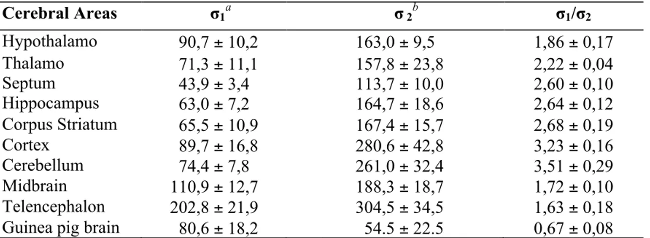
Information about the distribution in cerebral areas derived also from the binding studies illustrated in table 4.26,27
Table 4.Sigma-1 and sigma-2 binding sites in different cerebral areas (fmol/mg protein)
Cerebral Areas σ1a σ 2b σ1/σ2 Hypothalamo 90,7 ± 10,2 163,0 ± 9,5 1,86 ± 0,17 Thalamo 71,3 ± 11,1 157,8 ± 23,8 2,22 ± 0,04 Septum 43,9 ± 3,4 113,7 ± 10,0 2,60 ± 0,10 Hippocampus 63,0 ± 7,2 164,7 ± 18,6 2,64 ± 0,12 Corpus Striatum 65,5 ± 10,9 167,4 ± 15,7 2,68 ± 0,19 Cortex 89,7 ± 16,8 280,6 ± 42,8 3,23 ± 0,16 Cerebellum 74,4 ± 7,8 261,0 ± 32,4 3,51 ± 0,29 Midbrain 110,9 ± 12,7 188,3 ± 18,7 1,72 ± 0,10 Telencephalon 202,8 ± 21,9 304,5 ± 34,5 1,63 ± 0,18
Guinea pig brain 80,6 ± 18,2 54.5 ± 22.5 0,67 ± 0,08
a Sigma-1 sites labelled with [3H]-(+)-Pentazocine. b Sigma-2 sites labelled with [3H]-DTG in presence of
dextrallorphan (1µM) Peripheral organs
The heart contains high levels of sigma receptors, which 80% consist of sigma-1 type. In myocites sigma receptors influence contractility, calcium influx and cardiac rhythm. 28,29 In intracardiac neurons both subtypes regulate the excitability, modulating calcium and potassium channels.30,31
The highest concentration of sigma receptors in the body has been found in the liver, where both subtypes are present. However, the function of sigma receptors in the liver, as well as in the kidney, is yet unknown.32
Another part of the body that contains high levels of sigma-1 receptor is the spleen. Moreover the presence of sigma-1 receptor both in the spleen and immune cells would explain their role in immune system regulation.33,34
Recently, sigma-1 receptors were found in the ciliary body and retina of the eye, justifying the effects of sigma agonists in reducing ocular pressure and in retina protection against cell death. 35,36
Significant levels of sigma receptors are found in highly proliferative cells such as blood and tumour cells. Blood cells expressing sigma receptors are: granulocytes, lymphocytes, and natural killer cells.37 Tumour cells overexpress sigma receptors, especially in proliferating tumours. Indeed, sigma agonists have been shown to be useful as probes for diagnostic imaging and as anticancer agents because they are able to determine tumour cell death via apoptotic mechanism.38,39,40
Proposed endogenous ligands for sigma receptors
In spite of the significant advances achieved in the pharmacological characterization and in the knowledge of sigma receptors action mechanisms, the sigma endogenous ligands are still unknown.
Neurosteroids are the main candidates for the role of endogenous ligands of sigma receptors. Su T.P. and co. (1988) reported the nanomolar affinity of progesterone for sigma sites, because they observed that the neurosteroid inhibited the binding of both [3 H](+)-SKF10.047 (Ki = 268 nM) and [3H]-haloperidol, in cavia brain and in cavia spleen respectively.41
Subsequently, it was noted that the binding of [3H]-progesterone, used as radiolabeled ligand in different tissues, was inhibited by other sigma receptor ligands such as haloperidol, carbopentane, DTG, (+)-3-PPP and rimcazole.42,43 Different neurosteroids, possessing micromolar affinity for sigma receptors, were considered, but it is still not clarified if their physiological effects are mediated by interaction with sigma receptors.41,42 The limited functional studies only explain that some of them act as sigma-1 agonist (e. g. pregnenolone, Fig. 4) while others as sigma-1 antagonist (e. g. progesterone, Fig.4).44 Thus, the current evidences are far from identifying neurosteroids as endogenous ligands of sigma receptors. On the other hand a number of studies have identified sigma-1 receptor as a molecular target, at the structural, biochemical and physiological levels, for neuro(active)steroids actions.45
Other hypothesized endogenous ligand was the Neuropeptide Y that has been reported to have significant affinity for sigma receptors.46 Both Neuropeptide Y and peptide YY showed the ability to displace [3H](+)-SF&K10.047 from its binding sites with an IC50 around 10 nM. Unfortunately, subsequent studies did not confirm these results.47
A number of studies have shown that several divalent cations inhibit radioligands binding to sigma receptors. The cations considered include magnesium, calcium, zinc, cadmium and copper. Among these cations, some preferred the sigma-1 receptor while others the sigma-2 type.48,49 Another noteworthy finding was acquired from a binding study, performed under physiological conditions, where depolarization caused the release of zinc, from hippocampal slices, displacing [3H] DTG but not [3H](+)-pentazocine. This evidence suggested that zinc could be an endogenous ligand for sigma-2 receptor.49
Progesterone Pregnenolone
Figure 4: neurosteroids acting on sigma-1 receptor.
O H3C H3C O CH3 HO H3C H3C O CH3
Sigma-‐1 receptor
Characterization
So far, significant advances have been made in the characterization of sigma-1 receptors, not only with pharmacological methods but also in the biochemical field.
First steps were made through photoaffinity labelling with [3H] azido-DTG that showed a specific binding capacity toward a 29 KDa polypeptide found in NCB-20 cells and guinea pig cerebral membranes.50,51
McCann and Su (1991) isolated a CHAPS* solubilized sigma site complex (450 KDa), from rat liver membranes, which possessed all the pharmacological features of a neuronal sigma 1 receptor.52
The sigma-1 receptor has been recently cloned from various sources, including
guinea-pig liver,53 human brain,54 rat brain,55 mouse brain56 and human placental
choriocarcinoma cells.57The gene expressing sigma-1 receptor is 7 Kbp long and has four
exons and three introns and it is located in the human chromosome 9.54
The receptor possesses a sequence of 223 amino acidic residues and does not show homology neither with opioid receptors nor with other neurotransmitter receptors.
Sigma 1 receptor shares significant amino acid sequence similarities with the yeast sterol C8 ± C7 isomerase (ERG2 protein). Pharmacologically, but not structurally, the sigma-1 site is also related to the emopamil binding protein, the mammalian sterol C8 ± C7 isomerase.58 This was confirmed by studies on HMM-HMM® and BLAST® (Basic Local Alignment Search Tool).59,60 Recently, a significant sequence homology (35% and 45 %) was found also with another two enzymes involved in biosynthesis of steroids including
isopentenyl diphosphate isomerase and 17β-estradiol dehydrogenase.61,62 On the contrary, sigma-1 receptors do not possess sterol isomerase activity53 and the sequence homology could be explained by a conservative evolution hypothesis.
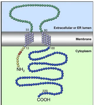
Aydar and co-workers (2004) proposed a model for sigma-1 receptor (Fig. 5) that consist of two transmembrane spanning regions, an extracellular loop with 50 amino acids, an intracellular C-terminal segment with 125 amino acids and a short intracellular N-terminal segment (10 amino acids). The C-N-terminal extremity (blue labelled in Fig. 5) has complete homology with the yeast sterol isomerase. The pair Arg-Arg (red labelled in the Fig. 5), typically found in endoplasmic reticulum (ER) receptors, is a retention signal that directs the retrieval of membrane proteins from the Golgi apparatus to the ER. 63
Figure 5: Sigma-1 receptor (Aydar and co., 2004)
Subcellular localization
are predominant in cytoplasmic areas of neuronal and retinal cell bodies, indicating an ER localization.64,65 Recently, Maurice and coworkers performed the first electron microscopic examination of sigma-1 in the adult animal, confirming that sigma-1 receptors are highly expressed in ER cistaernae. However sigma-‐1 receptors were also found in the limiting plasma membrane, in mitochondrial membrane and in postsynaptic thickening of neurons. 66
Signal transduction mechanism and modulatory action
So far, the signal transduction mechanism of sigma-1 receptors is still not well clarified, but it is clear that different cell types respond to a different action mechanism.
Recently, the most prominent and the most explored molecular action of sigma-1 receptors regards their interaction with ion channels. In fact it was discovered that the overexpression of sigma-1 ER receptors, induced by a sigma agonist administration, could cause their translocation from the ER to the sub-plasma membrane area where they could interact with ion channels.67 For instance, Hayashi and Su, proposed a model where the IP3 receptor-inhibiting protein, ankyrin, was removed from IP3 receptors when sigma-1 receptor agonists were applied to NG-108 cells, resulting in an enhancement of Ca2+ efflux from the ER into the cytosol.68 Wu and Bowen (2008) observed the same results they found that the C-terminus portion of sigma-1 receptors caused the dissociation of ankyrin from IP3 receptors in MCF-7 tumor cells.69
Aside from the already discussed functions, where sigma-1 receptor is directly involved, it is also noteworthy its regulatory activity on a number of known neurotransmitter systems including modulation of dopamine and acetylcholine synthesis and release;70 , 71 , 72 modulation of NMDA glutamate receptor electrophysiology;73 modulation of NMDA-stimulated norepinephrine release; 74 , 75 modulation of
phosphoinositol-stimulated turnover of muscarinic receptor;76 modulation of opioid analgesia;77 neuroprotective and anti-amnesiac activity;78 cocaine-induced locomotory alteration and toxicity.79,80
Selective sigma-‐1 ligands and pharmacophoric model
Recently several ligands with high sigma affinity have been synthetized. The phenylethylenediamine class is noteworthy because the ligand BD1008 and its derivatives have shown high affinity toward both sigma subtypes,81, 82 in this case the affinity was enhanced by the substitution with different lipophilic substituents. Particularly interesting, was the substitution of a phenyl group with a benzomorphanic group, which conducted to a ligand with a high sigma 1 affinity (Ki < 10 nM) and lower sigma 2 and opioid affinity.83
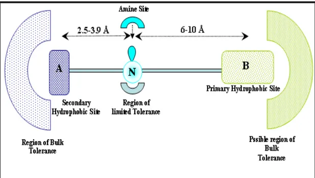
Similar to the phenylethilendiamine class are the phenylpenthylamine series, in which the scaffold contains only one nitrogen atom that is sufficient to exert a consistent sigma-1 affinity (Ki = 1 nM). Moreover this scaffold was used to hypothesize a pharmacophoric model (Fig. 6), where the two binding hydrophobic regions have to be opportunely distanced (2.5-3.9 Å and 6-10 Å) from the nitrogen atom, which could be secondary, tertiary or quaternary.84
The introduction of an ester group on the five terms chain produced the AC915 that possesses a 2000 fold greater affinity for sigma-1 versus sigma-2.85
The modifications to the haloperidol structure have led to the synthesis of E-5842, a high selectivity sigma-1 ligand.86 Regarding the selectivity significant results were obtained also with phenylacetamide class.87
Among the few synthesized antagonists, the Ne-100 possesses a high sigma-1 affinity and moderate selectivity toward sigma-2, dopaminergic D2, serotoninergic 5-HT2 and PCP receptors. It has also been proposed as an antipsychotic drug.88
Sigma-‐2 receptor
Characterization
Unlike sigma 1 subtype the sigma-2 is still not well characterized overall because it has not been cloned yet and there is no information about its sequence and conformation. Thanks to electrophoretic studies and photoaffinity labelling methods, it is known that the molecular weight of sigma-2 receptor is around 18-21.5 KDa, therefore it is smaller than sigma-1.89
Even the pharmacological characterization of sigma 2 receptors has been difficult and incomplete because of the lack of high selective sigma 2 radioligands. Usually [3 H]-DTG is used as radioligand in sigma-2 receptor binding assays, but it is also active for sigma-1 sites. As consequence, it is necessary to block sigma-1 sites with (+)-pentazocine or with dextrallorphan. On the basis of its incomplete pharmacological characterization it is possible to identify the sigma 2 site as a receptor with high affinity for haloperidol (table 3, Ki = 79.8 nm on rat brain) and DTG (table 3, Ki 61 nM on rat brain) but low affinity for (+)-benzomorphan derivatives, preferring the (−) stereoisomers.89,15 The
phenylenilmorphan CB-‐64D and CB-‐184, the alkaloid ibogaine and the ifenprodil have shown to possess a high sigma-‐2 affinity and selectivity. Unfortunately, they also have high affinity for other receptor systems: ibogaine and ifenprodil toward NMDA channel receptors and the phenylenilmorphans toward μ opioid receptors.90,91,92
The current challenge of many researchers is the development of new highly selective ligands for sigma-2 receptors. More importantly researchers have obtain tools able to properly study this subclass and to achieve a better understanding of its functions.
Subcellular localization
Early studies, regarding the subcellular localization, have shown the presence of sigma-2 and sigma-1 receptors in microsomal fractions (the richest in sigma receptors), mitochondrial and brain synaptosomal fractions of C57BL/6 mouse. In the microsomal and synaptosomal fractions the concentration ratio of the two subtypes resulted almost equal, whereas in the mitochondrial fraction the concentration of sigma-1 receptors was two times more than sigma 2 receptors.93
Unfortunately, the absence of a cloned sigma 2 gene and the lack of a purified receptor protein and related antibodies, have prevented researchers to study the sigma 2 subcellular localization through immunohistochemical techniques, as previously illustrated for sigma-1 receptor. However, recent development of highly selective sigma-2 receptor radioligands and fluorescent probes has been useful in the understanding of the subcellular distribution of sigma 2 receptor.
Bowen and co. (1996), during an attempt to purify the sigma receptor proteins, observed a low recovery of the solubilized sigma-2 receptor protein compared to sigma-1 receptor protein. They suggested a possible localization of sigma 2 receptors in detergent-resistant lipid raft domains.16 Lipid raft are microdomains of the cell membrane, enriched with cholesterol, sphingolipids, and glycosylphosphatidylinositol-linked proteins that form specialized structures termed caveolin upon the incorporation of a cholesterol binding protein. Gebreselassie and Bowen, using sucrose density centrifugation of CHAPS† extracts of rat liver P2 membranes demonstrated that [3H] DTG-binding site in protein fractions contained flotillin-2, a molecular marker of lipid rafts. 94 Subsequent in vitro binding studies revealed that [3H]-DTG binding was blocked by the sigma-2 selective
ligand CB-64D and not by the sigma-1 receptor ligand (+)-pentazocine, confirming that sigma 2 receptors are co-localized in lipid rafts. Other in vitro binding studies using [3 H]-DTG in the presence of (+)-pentazocine have shown that sigma-2 receptor binding sites are located in the mitochondria.95
Using the fluorescent probes SW107 and K05-138, Zeng and colleagues,96 lately conducted a series of confocal and two photon microscopy studies that provided a clearer picture of the localization of sigma 2 receptors in breast tumor cells. These studies were performed in EMT-6 (mouse breast tumor) or MDA-MB-435 (metastatic human tumor) using a panel of tracker dyes. Using the fluorescent probe SW107, Zeng and co. indicated that sigma 2 receptors are localized in the mitochondria, lysosomes, endoplasmic reticulum and cytoplasmic membrane. Similar results were obtained with confocal imaging studies using the fluorescent probe, K05-138, also demonstrating that ~40% of the sigma 2 receptors were internalized by receptor-mediated endocytosis, while the remaining ~60 % was internalized by other mechanisms such as passive diffusion. The rapid internalization of sigma-2 receptors via endocytosis suggests that sigma 2 selective ligands might be useful as receptor-mediated probes for delivering cytotoxic agents to solid tumors.
Sigma-‐2 receptors and the regulation of motor function
Since sigma-2 receptors are highly expressed in the brain regions regulating posture and participating in motor control, it is reasonable to think about their important contributions in the regulation of motor functions.21,22 Indeed it has been proven that microinjections of selective sigma ligands into motor regions of brain induced marked alteration in movement and posture. Microinjections of typical neuroleptics, as well as selective sigma 2 ligands, into the rat red nucleus induced acute dystonic reactions.97 While microinjections of sigma ligands into the facial nucleus or spinal trigeminal nucleus oralis, determined oral facial
dyskinesias.98 Unilateral microinjections of sigma ligands into the substanzia nigra resulted in contralateral circling.99 The effects on motor behaviour and posture were described with a pharmacological profile generally corresponding to mediations by sigma 2 receptors.99,100 Therefore, these results suggested the involvement of sigma 2 receptors in the side effects of typical antipsychotic drugs, particularly tardive dyskinesias and acute dystonias.101,102 Sigma-‐2 receptors and cell death
Some of the brain microinjection studies, described above, suggested that some sigma ligands might be neurotoxic. For example, the major haloperidol metabolite, the reduced haloperidol (potent sigma ligand) and the cyclohexane diamine, BD614, were able to cause extensive gliosis and loss of magnocellular neurons in and around the injection site.103, 104
Furthermore in vivo studies have shown that some ligands were cytotoxic to tumour cell lines of both neuronal (e.g., SK-N-SH neuroblastoma) and nonneural (e.g., C6 glioma) origin, as well as to primary cultures of rat central nervous system: cerebellar granule cells, cortical neurons, superior cervical ganglion cells. 105,106,107 Initially, sigma ligands caused damage to cell processes followed by a loss of these processes, assumption of a spherical shape (rounding) and detachment from the surface. The continue exposure to sigma ligands determined cell death, with a dose dependent effect (higher doses caused morphological changes and the subsequent cell death). Sigma 2 receptors were considered the most responsible for these effects. Indeed, sigma ligands binding both sigma-1 and sigma-2 sites, such as haloperidol, were active in evoking apoptosis, whereas sigma-1 selective ligands, such as (+)-pentazocine were inactive. Also compounds without a significant sigma affinity but having affinity for other receptors were not able to induce apoptosis.
The involvement in cell death of sigma 2 receptors was confirmed using the selective sigma-2 ligands, CB-64D and CB184 (Fig. 7), which resulted in a quite potent
cytotoxicity.90 Thus, it was clear that chronic activation of sigma 2 receptors produced morphological changes and cell death. The type of cell death induced by sigma 2 ligands, in different cell types, was widely proved to be apoptotic.108109 In fact, after treatment of SK-N-SH neuroblastoma cells or breast tumour cell lines with sigma agonists, such as CB-64D and CB184, the typical hallmarks of apoptosis were observed: inversion of phosphatidyl serine, DNA fragmentation and nuclear condensation. Similar results were observed using primary culture of rat cerebellar granular cells.108
The sigma-2 receptors determine apoptosis through a modulation of intracellular calcium, which was demonstrated in studies that used indo-1-loaded human SK-N-SH neuroblastoma cell. In these studies sigma ligands, from different structural classes, showed the ability to determine two different types of [Ca++]i (intracellular calcium concentration) enhancement. 110,111 The role of sigma 2 receptors in the mediation of the effect on [Ca++]i was clearly indicated by three considerations: the high affinity of the sigma-2 ligand CB-64D, the lower activity of CB-64L ( levo isomer), and the very low activity of selective sigma-1 ligands, such as (+)-benzomorphans, (+)-pentazocine, (±)-SKF-10047 and dextrallorphan.111 The two types of [Ca++]i rise were distinguishable temporally and by sources.111 In the first type sigma-2 ligands stimulated a rise in [Ca++]
i immediate, dose-dependent, and transient, through a release of calcium from endoplasmic reticulum. This transient rise occurred in the absence of extracellular calcium and was inhibited by the pretreatment of cells with thapsigargin, an intracellular inhibitor of ATPasi calcium pump. In the second type, the prolonged exposure of cells to sigma 2 ligands resulted in a latent and sustained rise of [Ca++]
i,, which was not affected by thapsigargin pretreatment. In this case sigma-2 ligands also induced a release of calcium from
mitochondria stores or from some other calcium store insensitive to thapsigargin (e.g. from Golgi apparatus).
Ostenfeld and co. obtained important results studying the cytotoxic effects of Lu 28-179, also known as siramesine, previously evaluated as a drug to treat anxiety and depression. Lu 28-179 has shown the capacity in killing tumor cells via a caspase-independent method, inducing cell death via a lysosomal leakage pathway and resulting in the formation of reactive oxygen species.112
Because sigma 2 receptors are overexpressed in rapid proliferation cells they could have an important role in the control of cell proliferation through the modulation of [Ca++]i.113 N CH3 O O H N CH3 Cl Cl O O H CB-64D (Ki σ2= 16,5±2,7 nM) CB-184 (Ki σ2= 13,4±2,0 nM)
Figure 7: cytotoxic sigma 2 ligands
In a recent study two structurally distinct sigma-2 receptor ligands, SV119 and WC26 (Fig. 8), were found to induce apoptosis to mice and human pancreatic cancer cells in vitro and
in vivo experiments.114 Micro-PET imaging was used to demonstrate that the sigma-2 receptor was preferentially expressed in tumor tissues as opposed to normal tissues in pancreas tumor allograft-bearing mice.Sigma-2 receptor ligands, WC26 and SV119, and the sigma-1/sigma-2 promiscuous ligand haloperidol induced apoptosis in a dose
dependent fashion in all the pancreatic cell lines tested, with a caspase-3/7 dependent mechanism.The selective sigma-1 ligand pentazocine showed minimal toxicity toward all the tested cell lines.In vivo was observed that systemic administration of WC26 did not induce apoptosis in brain, lung, kidney or spleen at any of the concentrations tested. Pancreas and liver have shown to undergo a small amount of apoptosis (<10%) while their tumors had dose-dependent increases in apoptosis (up to 50% of tumor cells were active caspase-3 positive following a single 2 mg dose of WC26, p < 0.0001). The mice appeared normal and no apparent toxicity was noted in serum biochemistry data.MoreoverWC26 significantly slowed tumor growth after a 5 day treatment compared to vehicle-injected control animals (p < 0.0001) and blood chemistry panels suggested a minimal peripheral toxicity.
Figure 8: cytotoxic ligands
Sigma-‐2 ligands as probes for imaging in vitro and in vivo
Vilner et al.,115 demonstrated that there is a higher density of sigma 2 versus sigma-1 receptors in a wide variety of human and murine tumor cells growing under cell culture conditions. An additional observation showed that MCF-7 cells, a human breast adenocarcinoma cell line, possessed a high density of sigma 2 receptors. The absence of
SV119 WC26 CH3 OCH3 N H O O N H NH2 CH3 OCH3 N H O O N N HCl
sigma-1, suggested that the sigma-2 receptor could be a potential biomarker for imaging studies of breast cancer.
Using the well-established diploid mouse mammary adenocarcinoma cell line 66, Mach and colleagues exposed that the density of sigma 2 receptors in 66P proliferative cells was about 10 times greater than the density observed in 66Q quiescent cells.116 A subsequent study demonstrated that the upregulation and the downregulation of sigma 2 receptors follow the transition of mouse mammary cells between the proliferative and quiescent states. These data suggest that this receptor is not only expressed in a single phase of the cell cycle. 117
Therefore, radiotracers having a high affinity and high selectivity for sigma-2 receptors have the potential to assess the proliferative status of human breast tumors using noninvasive imaging techniques such as Positron Emission Tomography (PET) and Single Photon Emission Computed Tomography (SPECT). This approach could be extended to assess the proliferative status of other human tumors, such as head and neck, melanoma, and lung tumors, which are known to express a high density of sigma-2 receptors.118
Although [3H]-DTG has been a very useful ligand for characterizing the sigma 2 receptor, and for identifying sigma 2 selective ligands, its rapid dissociation rate is not
ideal for in vitro binding studies. Scatchard studies in rat and human brain samples revealed that [3H]Lu 28-179 (Fig. 9) has a Kd value of 1.1 nM.119 Autoradiography studies of [3H] Lu 28-179 revealed a high density of sigma-2 receptors in the motor cortex, hippocampus, and hind brain nuclei, which was consistent with previous autoradiography studies using [3H] DTG in the presence of 1 nM dextrallorphan to mask sigma-1 binding sites. Other sigma 2 selective ligands that have been labeled with tritium include [3H]RHM-1,120 and [3H]PB28 (Fig. 9).121 [3H]-RHM-1 has the highest selectivity for
sigma-2 versus sigma-1 receptors.
A number of sigma receptor ligands have also been radiolabeled with iodine-125, a useful radionuclide in vitro binding studies. However, most 125I-labeled ligands have a high affinity for the sigma-1 and low for the sigma-2 receptor. The only sigma-2 selective ligand labeled with iodine-125 reported is the conformationally-flexible benzamide analog in figure 9. 122,123,124
In vivo, the conformationally-flexible benzamide analogs have proven to be the most useful in the development of PET radiotracers for imaging the sigma 2 receptor status of solid tumors.125 MicroPET and MicroCT imaging studies in a murine solid breast tumor EMT-6 have shown the potential of the [11C]-benzamide analogs (Fig. 9), as radiotracers for imaging the sigma 2 receptor status of breast tumors with PET.126 The short half life of carbon 11 (t1/2 = 20.4 min) is not ideal for the development of radiotracers, thus benzamide analogs have also served as lead compounds in the development of 18F-labeled sigma 2 selective radiotracers with a longer half life (t1/2 =109.8 min). The strategy chosen involved replacement of the methoxy group in the benzamide ring with a 2-fluoroethoxy group. MicroPET imaging studies indicate that [18F] benzamides analogs, are suitable probes for imaging the sigma 2 receptor status of solid tumors. 127 Clinical studies of [18F] benzamide analog in figure 9 are currently in progress in the U.S.
As discussed above, fluorescent probes SW107 and K05-138 (Fig. 9) have produced important results in the study of subcellular distribution of sigma receptors with two-photon and confocal microscopy.96
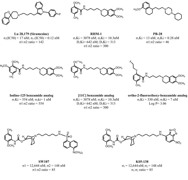
Lu 28,179 (Siramesine) σ1(IC50) = 17 nM; σ2 (IC50) = 0.12 nM σ1:σ2 ratio = 142 RHM-1 σ1Ki = 3078 nM; σ2Ki = 10.3nM D1Ki= 642 nM; D3Ki = 313 σ1:σ2 ratio = 300 PB-28 σ1Ki = 13 nM; σ2Ki = 0.28 nM σ1:σ2 ratio = 46
Iodine-125 benzamide analog σ1Ki = 554 nM; σ2ki= 1 nM σ1:σ2 ratio = 554 [11C] benzamide analog σ1Ki = 3078 nM; σ2Ki = 10.3nM D1Ki= 642 nM; D3Ki = 313 σ1:σ2 ratio = 300 ortho-2-fluoroethoxy-benzamide analog σ1Ki = 330 nM; σ2Ki = 7 nM Log P= 3.06 SW107 σ1 = 12,644 nM; σ2 = 148 nM σ1:σ2 ratio = 85 K05-138 σ1 = 12,644nM;σ2 = 148 nM σ1:σ2 ratio = 85
Figure 9: sigma 2 ligands as probes for imaging
Sigma-‐2 ligands
The development of ligands having a high affinity for the sigma 2 versus the sigma 1 receptor has not been straightforward; unfortunately at the moment only a limited number of selective sigma-2 compounds are available.
The most pharmacologically studied sigma-2 ligands are the already cited benzilidene phenilmorphan analogs, CB-64D and CB-184 (Fig. 7). The benzomorphan-7-one analog, CB-64D, was the first selective sigma 2 ligand and it was identified during a structure-activity relationship (SAR) study on ligands of µ opioid receptors.90,128 It was
N N F O OCH3 CH3 H N O N OCH3 OCH3 H3CO N N 125I OCH3 H3CO HN O N OCH3 OCH3 CH3 OC11H 3 H N O N OCH3 OCH3 CH3 H N O N OCH3 OCH3 o F CH3 OCH3 N H O O N H H N S O O N(CH3)2 CH3 OCH3 N H O O N H H N N O N NO2
observed that the adding of an (E)-benzylidene moiety into the 8-position of (−)-2-methyl-5-(3-hydroxyphenyl)morphan-7-one ring system increased affinity for sigma receptors. Specifically, the (−)-1S,5R isomer, CB-64L, showed a high affinity for sigma-1 versus sigma-2 receptors, whereas the (+)-1R,5R isomer, CB-64D, had a 185-fold higher selectivity for sigma 2 versus sigma 1 receptors. Particularly interesting was the profile of the corresponding 3,4-dichloro analog, CB-184, that was the ligand with the highest sigma-2 affinity and selectivity among those of the same series. The negative aspect of the phenylmorphan series is the interaction with the opioid receptors as shown by their high µ agonist activity in vivo, justified by the presence of the morphinanic moiety.90
Another important series of compounds having high affinity for sigma-2 receptors are the 3-(ω- aminoalkyl)-1H-indole analogs.129,130 The optimal compound of these series was the 1'-[4-[1-(4-fluorophenyl)-1H-indol-3-yl]-1-butyl]spiro[isobenzofuran-1(3H),4'-piperidine], Lu 28-179 (Fig. 9), with a 17nM (IC50) affinity on sigma 1 receptor a 0.12 nM (IC50) on sigma 2 receptor. Furthermore it exerted a very low affinity on HT1A and 5-HT2A (IC50: 21,000 nM and 2000 nM respectively) and moderate affinity on D2 and alpha-1 receptors (IC50: 800 nM and 330 nM respectively). However, the most selective sigma 2 versus sigma 1 ligand of this class was the tropane derivative 1-(4-fluorophenyl)-3-[4-[3-(4-fluorophenyl)-8-azabicyclo[3.2.1]oct-2-en-8-yl]-1-butyl]-1H-indole with a 1200 nM (IC50) affinity for sigma 1 receptor and 2.5 nM (IC50) affinity for sigma 2 receptor.
A natural compound that was found to have a high affinity for sigma 2 versus sigma 1 receptors is the hallucinogen, ibogaine, which binding studies and SAR will be discussed afterwards in the text. 131 Both CB-184 and ibogaine contain in their structure an arylpropylamine moiety that is a useful requirement for sigma-2 receptor affinity, whereas compounds with affinity for sigma-1 sites, such as Ne 100, tend to possess
phenylethylamine moiety.132 This was demonstrated by the phenylpropylpiperidine in figure 9 that has an affinity 4 fold higher for sigma-2 versus sigma-1. The modifications on the amine group led to significant increasing in sigma-2 affinity, as shown by the arylpropylpiperazine derivative in figure 10.132
Phenylpropyl-piperidine Arylpropylpiperazine derivative
(Maeda and co.)
Figure 10: sigma 2 ligands of Arylpropylamine series
Instead, Berardi and co. studied a very promising series of substituted tetralines containing a gem-dimethyl piperidine (Fig. 11).133,134 In these series they observed that when the aliphatic chain was 4 terms long the derivative showed a very high sigma 2 affinity (σ2IC50 = 0.016 nM) and a 100,000 fold higher selectivity for sigma 2 versus sigma-1 receptor. Interestingly, in the derivative with 5 terms chain (σ2IC50 = 0.016 nM) the selectivity diminished to 21 fold. The derivative without a methoxy group and with a 5 terms chain (des-methoxy, n = 5, σ2IC50= 0.008 nM) showed again a 100,000-fold higher selectivity for sigma-2 versus sigma-1.
Kawamura and co-workers, highlighted that small structural differences have a great impact on the sigma 2 affinity and selectivity.135 For example in the compound in figure 11 when R is a methyl group the derivative binds sigma-2 receptor with low affinity(Ki = 1800 nM) and possesses a 106-fold higher selectivity for sigma 2 versus sigma 1. When R
N
N N
is an ethyl group, the derivative binds sigma-2 receptor with high affinity (Ki = 13 nM) but the selectivity is lower (σ1:σ2 ratio to 3).
Substituted tetraline
(Berardi and co.)
Kawamura series
Figure 11: sigma 2 ligands
New sigma-2 ligands containig a tropane nucleus have shown to possess a sigma-2 affinity up to 5 nM and a 500-fold higher selectivity for sigma 2 versus sigma-1. The addition of an amine group in para position to the benzene ring was showed useful in increasing the sigma 2 selectivity of the un-substituted analogue (Fig. 12).136 An important tropane analogue is the (±)-SM-21 (Fig. 12) that was used as antagonist in behavioural assays where it was able to reduce the convulsive and locomotory effects of cocaine. Although early studies showed a significant sigma 2 affinity and around 14 selectivity for sigma 2 versus sigma-1 ( rat liver σ1Ki > 1000 and σ2Ki = 67,5±8) subsequent studies showed a lower sigma 2 affinity (guinea pig σ2Ki = 434 nM; σ1Ki >1000 nM), most likely because a different tissue was used in the later binding study.137,138,139
Tropane nucleus ligand
(Mach and co.)
(±)-SM-21 N (CH2)n OCH3 N N OMe O R OMe H N O O N NH2 O O O N Cl
Several SAR studies on BIMU-1 (32 nM σ2 affinity; 6,300 nM σ1 affinity) (Fig. 13) as the lead compound have conducted the identification of high affinity and high selectivity sigma 2 receptor ligands.140,141,142 Addition of a benzene ring to the N-methyl of the bridgehead nitrogen, giving the corresponding N-benzyl group, and the replacement of the urea linkage of BIMU-1 resulted in a significant increase in affinity for both sigma 2 and sigma-1 receptors, a loss of affinity for HT3 receptors and moderate affinity for 5-HT4 receptors.140 Expansion of the tropane ring to the corresponding granatane ring eliminated affinity for the 5-HT4 receptor and did not change the affinity for sigma-1 and sigma-2 receptors, relative to the tropane analogs. The most interesting analog from this initial SAR study was the granatane derivative in figure 13, which had a sigma 2 receptor affinity of ~ 3 nM and a sigma 2 versus sigma 1 selectivity of ~30. 140
Using this granatane analog as lead compound a further SAR study was started and it was observed that the substitution of benzyl group with a 2-phenethyl group resulted in a slight improvement in sigma 2 receptor affinity and increased sigma-2 selectivity (σ1:σ2 ratio to ~50).141Substitution in para position to the aromatic ring of the granatane lead compound with a dimethylamino group resulted in a further increase in the selectivity for sigma 2 receptors, because of the reduction in affinity for sigma 1 receptors.141The amino aminoalkyl group also appeared as a good substituent for assuring a high affinity for sigma-2 receptors and high sigma-1/sigma-2 selectivity ratio.142 These results were used in the design of the fluorescent probes SW107 and K05-138 (Fig. 9), useful tools in two photon and confocal microscopy studies of sigma 2 receptors (already described in the last section).
BIMU-1 Granatane analog
Figure 13: Sigma 2 ligands based on tropane and granatane analogs
Another class of compounds having a high affinity for sigma 2 receptors and excellent sigma-1/sigma-2 selectivity ratios are the conformationally-flexible benzamide analogs (Fig. 14). The analog with a 6,7-dimethoxy-1,2,3,4-tetrahydroisoqinoline ring (e.g. Fig. 14 on the left) resulted in compounds having a high affinity, excellent selectivity for sigma-2 versus sigma-1 receptors and a dramatic reduction in affinity for dopamine receptors, relative to its lead compound. 143 The removal of 6,7-dimethoxy group was critical for sigma 2 receptors affinity. Replacement of the 5-bromo group with a methyl group (Fig. 14 on the right) resulted in a further reduction in D3 receptor affinity and no change in affinity for the sigma-2 receptor. Extension of the 2 carbons spacer to the corresponding 4 carbons spacer, of RHM-1 (Fig. 9), also retained a high affinity for sigma 2 receptors. RHM-1 as discussed above was used successfully as a radio-labeled probe for sigma 2 receptors.
Analog with a 4 carbons spacer
σ1 = 3,078 nM; σ2 = 10.3 nM; σ1:σ2 ratio = 300
D3= 627 nM; D2 = 2,200 nM
Analog with a 2 carbons spacer
σ1 = 10,400 nM; σ2 = 13.3 nM; σ1:σ2 ratio = 780
D3= 3,760 nM; D2 = 2,850 nM
Figure 14: Examples of conformationally-flexible benzamide analogs with Ki values.
N HN O N CH3 N O CH3 OCH3 N H O O N H OMe Br MeO N H N OMe OMe O OMe CH3 N H N OMe OMe O
Proposed pharmacophoric model for sigma-‐2 receptor
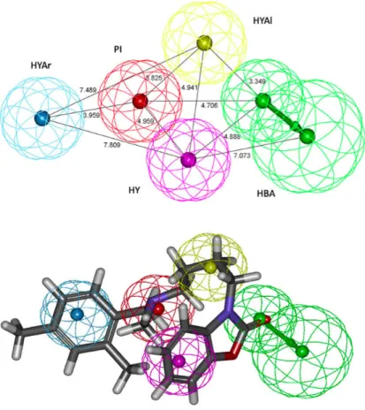
Recently, Laurini E. et al., developed a 3D-pharmacophore model for sigma 2 receptors based on a series of substituted benzo[d]oxazol-2(3H)-one derivatives.144 The best output hypothesis (Fig. 15) contained a positive ionizable atom (PI), a hydrogen bond acceptor group (HBA), a hydrophobic aromatic site (HYAr), a hydrophobic aliphatic site (HYAl), and a generic hydrophobic site (HY).
The proposed pharmacophore model for the sigma-2 receptor showed remarkable similarities but also some differences with the validated 3D-QSAR developed, by the same authors, for sigma-1 receptors. A basic amino nitrogen atom between two hydrophobic sites is pharmacophoric element required for affinity. In the sigma-2 receptors the positions and distances between these features are different with respect to the sigma 1 receptor model. The primary hydrophobic site corresponds with the hydrophobic aromatic sphere (HYAr), which is mapped by the phenyl group connected to the aliphatic spacer, at a distance of 3.96 Å from the positive ionizable feature of the amino group, in agreement with their sigma-1 receptor model (3.58 Å),145 Laggner’s model (4.1 Å),146and the optimum distance suggested by Glennon et al. (2.5–3.9 Å).147Whereas, the secondary hydrophobic site is matched by the generic hydrophobic sphere (HY) that maps to the aromatic ring of the benzooxazolone group at a shorter distance (4.96 Å) with respect to the sigma-1 receptor model (8.50 Å). The generic hydrophobic feature (HY) is mapped by the aromatic ring of the benzooxazolone in sigma-2 hypothesis. The presence of the hydrophobic aliphatic feature (HYAl) constitutes a necessary requisite for binding with high affinity to sigma-2 receptors, while it does not seem to play a major role for selectivity towards this receptor.The hydrogen bond acceptor feature (HBA) corresponds to the carbonyl oxygen of the benzooxazolone moiety.
Figure 15: Top-scoring pharmacophore model Hypo1, based on a series of substituted
benzo[d]oxazol-2(3H)-one derivatives, and mapping of a compound belonging to the same series. The hypothesis features are portrayed as mashed spheres, color-coded as follows: red, PI; light blue, HYAr; pink, HY; light green, HBA; yellow, HYAl. HBA is actually represented as a pair of spheres (the smaller sphere represents the location of the HBA atom on the ligand and the larger one the location of an HB donor on the receptor). Selected distances (Å) are labeled. Compounds are portrayed as atom-colored sticks (red, O; gray, C; blue, N; white, H).144
Ibogaine: pharmacological profile
Ibogaine (trade name: Endabuse ™), an indolic alkaloid, is contained in a number of plants but principally isolated from the shrub Tabernanthe iboga of central Africa. Ibogaine, as demonstrated by in vivo studies, is a psychoactive substance that exerts stimulatory effects on CNS (central nervous system), if administrated in low doses, whereas induces hallucinogen effects and tremors when administrated in high doses.148,149
Figure 16: ibogaine
Because of its psychotropic effects ibogaine was used in the past as an abused drug. Anecdotal reports suggested that ibogaine was able to interrupt the craving of heroine, cocaine and amphetamines in addicts.150 These observations were supported in several animal studies where ibogaine has been shown to reduce self-administration of both morphine and cocaine. 151,152,153,154 Unfortunately, the clinical use of ibogaine has found many troubles related to its neurotoxic and tremorigen effects. Specifically the treatment of rats with ibogaine 100mg/Kg determined microglia and astrocytes activation and loss of Purkinje cells in the parasagittal zones of cerebellum.155,156
The receptor sites responsible for the beneficial effects (anti-addiction) of ibogaine are not well known, but it was hypothesized that they are the result of a multiple low affinity interaction with several receptor systems. In vitro studies have showed that ibogaine possessed a micromolar affinity for σ1 receptors; µ, κ and δ opioid receptors; M1, M2 and M3, muscarinic receptors; D1 and D2 dopaminergic receptors; GABA A site and
H3CO
N H
N
benzodiazepine site in GABA receptor; 5-HT2A e 5-HT3, serotoninergic receptors; α1, α2 and β adrenergic receptors; and for PCP site in NMDA channel. In contrast ibogaine has showed a nanomolar affinity for σ2 receptor (Ki=250 nM in guinea pig brain, Ki = 90 nM in rat liver) and higher than 100-fold selectivity for sigma 2 versus sigma 1 (table 5). 157
Table 5. Affinities of ibogaine, haloperidol, haloperidol metabolite II for sigma receptors.
Ki [nM] (n = 3) a
Compound σ1b σ2(liver)c σ2(brain)d σ1 / σ2 ratio
Ibogaine 9310 ± 630 90.04 ± 10.1e 250 ± 39f 103g or 37.2h Haloperidol 1,77 ± 0.09 21.8 ± 8.5 14.2 ± 3.2 0.08g or 0.13h Haloperidol metabolite II 5.65 ± 0.26 25.4 ± 3.3 1.31 ± 0.03 0.22 g or 4.30h a Mean ± SEM. bK
i for inhibiting the binding of [3H](+)-pentazocine to guinea pig brain membranes. cKi for
inhibiting the binding of [3H]DTG to rat liver membranes. dK
i for inhibiting the binding of [3H]DTG to
guinea pig brain membranes. eN = 4. fN = 7. gK
i for σ1 / Ki for σ2 (liver). hKi for σ1 / Ki for σ2(brain).
When Bowen W. D analysed the pharmacological profile of ibogaine, he hypothesized that the anti-addiction effects of ibogaine were not related to the high sigma-2 affinity but probably due to the interactions with other receptor systems. On the contrary sigma-2 receptors could be responsible of the ibogaine-induced neurotoxicity and tremorigen effects. 158 These considerations were supported by data showing the ability of
ibogaine to induce apoptosis in nervous cells, related to the increase of [Ca2+]
i, and by the ability of sigma 2 ligands in inducing motor alteration, as already discussed (see the section: sigma 2 receptor and the regulation of motor function). Actually, the cytotoxicity in humans seems attenuated by the metabolism of ibogaine that determines the O-demethylation to noribogaine,159,160
which lacks affinity for sigma-2 receptors and produces no effects on [Ca2+
Ibogaine and its related alkaloids: SAR
Bowen and co. studied ibogaine and its related alkaloids with binding assays at sigma 1 and sigma 2 receptors and the data are summarized in table 6.161 Sigma-1 receptors, in guinea pig brain membranes were labeled with the sigma-1 selective probe, [3H](+)-pentazocine, while sigma-2 receptors in rat liver membranes were labeled with [3H]DTG in the presence of dextrallorphan to inhibit the binding to sigma-1 sites. Ibogaine exhibited moderate affinity for sigma-2 sites (Ki = 201 ± 24 nM), but had very low affinity for sigma-1 receptors (Ki = 8,554 ± 1,134 nM), resulting in 43-fold selectivity for sigma-2 sites over sigma-1. Mach and co., as previously showed, obtained similar results with the ibogaine binding at sigma receptors in guinea pig brain, whereas using rat liver the data showed a higher selectivity for sigma-2 (table 5: σ1 / σ2 ratio to 103).157
Regarding the structure-activity relationships of iboga derivatives for affinity at sigma receptors (table 6),161 (±)-Ibogamine, considered as parent compound, has an
unsubstituted indole moiety, with a sigma-2 Ki = 137 ± 13 nM and sigma 1 Ki = 1,835 ± 131 nM. Thus, the methoxy group in the 10-position (ibogaine) did not significantly change the sigma-2 affinity, but decreased the sigma-1 affinity (Ki = 8,554 ± 1,134 nM). A methoxy group in the 11-position (tabernanthine) produced no significant change in sigma-2 affinity and a small decrease in sigma-1 affinity (Ki = sigma-2,87sigma-2 ± 37 nM), resulting in 14.8-fold selectivity for sigma-2 receptors. Alike an O-t-butyl group in the 10-position did not dramatically change the sigma-2 receptor affinity or the sigma-1 affinity (Ki = 4,859 ± 682 nM), resulting in 20-fold selectivity for sigma-2 sites. A hydroxyl group in the 10-position (noribogaine) resulted in a 38-fold loss of binding affinity at sigma- 2 receptors and 8-fold loss of affinity at sigma-1 receptors (Ki = 15,006 ± 898 nM). Thus, the sigma-2 binding site did not tolerate the phenolic hydroxyl group. The presence of a carbomethoxy group in
the 16-position ((±)-coronaridine) determined complete loss of sigma-2 affinity and a 20-fold loss in sigma 1 affinity (Ki = 35,688 ± 2,858 nM) in comparison to (±)-ibogamine. Addition of a methoxy group at the 18-position of the 16-carbomethoxy analog, (±)-18- methoxycoronaridine (MC), increased the sigma-2 affinity compared to (±)-coronaridine, but still had low affinity. (±)-MC had slightly improved sigma-1 binding affinity (Ki = 28,687 ± 283 nM) compared to (±)-coronaridine.
Thus, ibogaine can be considered as a ligand with good selectivity for sigma 2 receptor over sigma-‐1 that could be used as a model in designing new sigma-‐2 agonists and sigma 2 antagonists.
Table 6. Affinities of ibogaine and related indole alkaloids at sigma 2 receptors. a
Alkaloid R1 R2 R3 R4 σ2 Ki nMb σ1 Ki nMc (±)-Ibogamine H H H H 137 ± 13 1,835 ± 131 Ibogaine OCH3 H H H 201 ± 24 8,554 ± 1,134 Tabernanthine H OCH3 H H 194 ± 10 2,872 ± 37 10-t-Butoxy-Ibogamine O-t-Bu H H H 247 ± 27 4,859 ± 682 Noribogaine OH H H H 5,266 ± 1,426 15,066 ± 898 (±)-Coronaridine H H CO2CH3 H >100,000 35,688 ± 2,858 (±)-MC H H CO2CH3 OCH3 8,472 ± 1,237 28,687 ± 283
aPortions adapted from data of Bowen and co.161 bSigma-2 receptors were labeled with [3H]DTG using rat liver membranes, in the presence of dextrallorphan to inhibit binding to sigma-1 sites. cSigma 1 receptors were labeled with the sigma-1-selective probe, [3H](+)-pentazocine, in guinea pig brain membranes. N H N R4 R3 R1 R2 10 11 16 18
Aims of the work and drug design
Focusing my research work on natural scaffolds possessing sigma 2 affinity, I have chosen the alkaloid ibogaine (Fig. 16) as reference compound of a new series of sigma-‐2 ligands. Although ibogaine has moderate affinity for sigma 2 receptors (Ki ≈ 250nM in guinea pig brain or Ki ≈ 90 in rat liver) it has a significant selectivity for sigma-‐2 versus sigma-‐1 (σ1 / σ2 ratio to 103 in rat liver).157 Therefore the main aim of the work was to design and synthesize new alkaloid-‐based ligands possessing an improved sigma-‐2 affinity and better or maintained sigma-‐2 selectivity with respect to ibogaine.
Individuation of a natural scaffold
I carried out a thorough literary investigation in order to find a natural scaffold containing the structural requirements necessary for sigma-2 receptor recognition and possessing a structure as similar to ibogaine as possible. Among the several indolic nucleus alkaloids considered, I observed that vinca-based alkaloids seem to possess the right pharmacophoric features for my purposes.
The eburnamine-vincamine alkaloids occur in the Apocynaceae plant family. The group can be divided into three major subgroups: 1. (–)-eburnamine; 2. (+)-vincamine; 3. strempeliopine. The five ring system is characteristic of these alkaloids. The “eburna” skeleton with the (20R, 21R) [(20β, 21β)] configuration belongs to the compounds termed eburnane types. Compounds with the (20S, 21S) [(20α, 21α)] configuration (“vinca” skeleton) belong to the vincane type of compunds (Fig. 17).162 Vincamine, the main alkaloid isolated from the leaves of Vinca minor,163, 164 is the mother compound of cerebrally active eburnamine derivatives. Vincamine, as well as vincanol, is a potent
blocker of the voltage-gated sodium channels.165 Vincamine reduced [3H]batrachotoxin binding (IC50 1.9 µM) in rat cortical synaptosomes, blocked the voltage-gated sodium current (IC50: 40 µM) in voltage-clamped rat cortical neurons and protected against veratridine induced cell death (IC50: 26) in cortical cultures. Vincamine and its related alkaloids (Fig. 17) including (–)-Eburnamonine (or vincamone), vindeburnol, vinpoceptine have shown modulatory effects on brain circulation and neuronal homeostasis, bearing antihypoxic and neuriprotective potencies to various degree.162,166,167 Also Vincane is pharmacologically known for its marked vasodilatatory effect (5%-10%) in cerebral blood vessels.168
Vincane: R1, R2 = H
Vincamine: R1 = COOCH3; R2 = OH
(–)-eburnamonine (or vincamone): R = O
(lactamic carbonyl at position 16)
Vinpoceptine: R1 = COOCH2CH3;R2 = H; double bond at positions 16-17
Vincanol: R1 = OH, R2 = H
Figure 17: vinca skeleton and vincane type alkaloids
N
N
H
R
1R
2 1 2 3 5 4 7 6 13 11 9 8 10 12 20 21 17 16 15 14 19 18A
B
D
E
C
N
N
H
R
R
1 Phenylpropylamine moietyN
Tertiary Nitrogen Hydrophobic aromatic moiety Hydrophobic aliphatic moietyConsidering the general structure of vincane type alkaloids (Fig. 17) it is possible to recognize four crucial requisites for the interaction with sigma-2 receptor. A positive ionisable atom is present, as described in all the sigma pharmacophoric models,144,146,147 and corresponds to the tertiary nitrogen (in blue in Fig. 18). A phenylpropylamine moiety, highlighted in red in figure 18, as in ibogaine, CB-184 and arylpropylpiperazine could be useful for sigma 2 selectivity.132 The vinca-scaffold contains a hydrophobic aromatic moiety corresponding to the indolic ring (Fig. 18), that should interact with the primary hydrophobic region of sigma-2 receptor, as well as the pharmacophoric model suggest.144 Finally, into the vinca scaffold it is possible to individuate a hydrophobic aliphatic moiety corresponding to the D ring (Fig. 17), as well as the piperidine ringin several ligands, that should be necessary for binding with high affinity to sigma 2 receptors (Fig. 18).
Superimposition study: vinca-‐derivatives upon ibogaine
The pharmacophoric considerations, discussed in the last section, were supported by a computational superimposition study where the vinca scaffold was overlaid on the ibogaine structure. The study was carried out using the software MOE (Molecular Operating Enviroment) Chemical Computing Group. Thus, I have considered the selective sigma-2 ligand ibogaine as reference compound and Vincamone and Vincane as compounds containing the scaffold. The three structures were first minimized to the lower energy of the system and subsequently overlaid, using force field MMFF94X, at standard conditions‡ and with preserved chirality (Fig. 19, 20). The superimposition resulted in an optimal structural homology of both vincamone and vincane with ibogaine (Fig. 20).
‡ Gradiente 0.00001; flexible alignment: iteration limit 500; failure limit 30; energy cutoff 15; minimization
In both superimpositions it is possible to observe complete co-planarity of the two indolic nuclei. The tertiary nitrogen (labelled in blue) occupies the same position in both iboga-structure and in vinca-structures. The ring termed D of vinca scaffold (in Fig. 17) is lifted in respect to the indole plane and seems to occupy the same volume and position of the aliphatic bicycle in ibogaine. Finally, the ethyl chain of both structures seems correctly positioned.
The main difference that occurs in vinca scaffold respect to ibogaine structure is the presence of the ring termed E (Fig. 17), in which is contained the indolic nitrogen. It would be interesting to extrapolate from binding assays information about the tolerance of sigma-2 receptor about this structural difference.
(–)-Eburnamonine (or vincamone) Vincane
Ibogaine
Figure 19: Global minimum conformers of vinca alkaloids and Ibogaine in polytube
Vicamone (yellow labelled) upon ibogaine (red labelled)
Vincane (purple labelled) upon ibogaine (orange labeled)

- 3-PPP e [ 3 H]DTG sites in C57BL/6 mouse brain membranes](https://thumb-eu.123doks.com/thumbv2/123dokorg/4480224.32302/6.892.147.810.194.412/table-comparison-affinity-sigma-ligands-sites-mouse-membranes.webp)