Neuroglobin in Breast Cancer Cells: Effect of
Hypoxia and Oxidative Stress on Protein
Level, Localization, and Anti-Apoptotic
Function
Marco Fiocchetti1, Manuela Cipolletti1, Stefano Leone1, Antonella Naldini2, Fabio Carraro2, Daniela Giordano3, Cinzia Verde1,3, Paolo Ascenzi1,3,4, Maria Marino1,3*
1 Department of Science, Roma Tre University, Viale Guglielmo Marconi 446, I-00146 Roma, Italy,
2 Department of Molecular and Developmental Medicine, University of Siena, Via Aldo Moro 2, 53100 Siena, Italy, 3 Biosciences and BioResources Institute—CNR, Via Pietro Castellino 111, 80131 Napoli, Italy, 4 Interdepartmental Laboratory of Electron Microscopy, Roma Tre University, Via della Vasca Navale 79, I-00146 Roma, Italy
Abstract
The over-expression of human neuroglobin (NGB), a heme-protein preferentially expressed in the brain, displays anti-apoptotic effects against hypoxic/ischemic and oxidative stresses enhancing neuron survival. As hypoxic and oxidative stress injury frequently occurs in fast proliferating neoplastic tissues, here, the effect of these stressors on the level, localization, and anti-apoptotic function of NGB in wild type and NGB-stable-silenced MCF-7 breast can-cer cells has been assessed. The well-known endogenous NGB inducan-cer 17β-estradiol (E2) has been used as positive control. The median pO2 present in tumor microenvironment of breast cancer patients (i.e., 2% O2) does not affect the NGB level in breast cancer cells,
whereas hydrogen peroxide and lead(IV) acetate, which increase intracellular reactive oxy-gen species (ROS) level, enhance the NGB levels outside the mitochondria and still activate apoptosis. However, E2-induced NGB up-regulation in mitochondria completely reverse lead(IV) acetate-induced PARP cleavage. These results indicate that the NGB level could represent a marker of oxidative-stress in MCF-7 breast cancer cells; however, the NGB abil-ity to respond to injuring stimuli by preventing apoptosis requires its re-allocation into the mitochondria. As a whole, present data might lead to a new direction in understanding NGB function in cancer opening new avenues for the therapeutic intervention.
Introduction
Neuroglobin (NGB) is a relatively recent discovered monomeric heme-protein so named because of its preferential expression in the nervous system [1]. NGB over-expression, driven by transiently transfected pcDNA vector, protects cultured neurons against hypoxia [2], enhances neuron survival under anoxia or oxygen/glucose deprivation [3], and displays
neuro-a11111
OPEN ACCESS
Citation: Fiocchetti M, Cipolletti M, Leone S, Naldini A, Carraro F, Giordano D, et al. (2016) Neuroglobin in Breast Cancer Cells: Effect of Hypoxia and Oxidative Stress on Protein Level, Localization, and Anti-Apoptotic Function. PLoS ONE 11(5): e0154959. doi:10.1371/journal.pone.0154959
Editor: Partha Mukhopadhyay, National Institutes of Health, UNITED STATES
Received: February 3, 2016 Accepted: April 21, 2016 Published: May 5, 2016
Copyright: © 2016 Fiocchetti et al. This is an open access article distributed under the terms of the Creative Commons Attribution License, which permits unrestricted use, distribution, and reproduction in any medium, provided the original author and source are credited.
Data Availability Statement: All relevant data are within the paper and its Supporting Information files. Funding: This work was supported by the Associazione Italiana Ricerca sul Cancro (AIRC, IG#15221) to M.M. (www.airc.it) and the Ministero dell’Istruzione, dell’Università e della Ricerca of Italy (PRIN 20109MXHMR_001) to P.A. (www.istruzione. it). The funders had no role in study design, data collection and analysis, decision to publish, or preparation of the manuscript.
protective properties against hypoxic/ischemic and oxidative stress [4–7]. In addition, in brain-derived cell lines, hypoxia [8], H2O2injury [9], and lipopolysaccharides [10] moderately induce
NGB. This suggests that NGB could behave in neurons as a sensor of injuring stimuli including oxidative stress, hypoxia, and neurotoxicity. However, as described in detail previously, no dif-ferences in NGB levels in murine models of traumatic brain injury, experimental autoimmune encephalitis, cerebral malaria, and hypoxia have been found [11–14], thus insinuating uncer-tainty on the role of NGB as a stress sensor.
Mechanism(s) by which NGB over-expression exerts its effect in neurons are uncertain. In particular, NGB has been postulated to enhance O2supply, to scavenge reactive oxygen species
(ROS) [13,15], and to modulate nitric oxide homeostasis in active neurons [8,16]. Moreover, oxidative stress (i.e., H2O2treatment) induces the specific binding of transiently over-expressed
NGB to flotillin-1, a protein associated to the plasma membrane lipid raft micro-domains, leading to cell survival [17]. Furthermore, the sex steroid hormone 17β-estradiol (E2) increases the endogenous level of NGB in several brain-derived cells and induces the heme-protein real-location into mitochondria where, upon oxidative stress injury, it interacts with mitochondrial cytochromec avoiding its release into the cytosol and the activation of the apoptotic cascade [9,10]. Whether mitochondrial and/or cytosolic NGB mechanisms act synergistically or antag-onistically in neurons to respond to oxidative stress or hypoxia is unknown.
Besides neuronal damage, oxidative stress and hypoxia are conditions frequently occurring in fast proliferating neoplastic tissues. Indeed, cancer cells adapt themselves to the stressful and dynamic microenvironment of solid tumors, where the redox status is imbalanced and oxygen/ nutrients availability is limited [18,19]. The adaptation is achieved by developing alternative compensatory metabolic reactions that render cancer cells insensitive to stress inducers such as chemotherapy and radiation [19]. Although a tumor suppressive function of transiently over-expressed NGB in hepatoma cancer cells has been described [20], other studies reported that NGB expression is differentially modulated by hypoxia and oxidative stress in cancer cell lines [21,22]. This suggests that NGB may be part of the defense mechanism established by cancer cells to counteract tumor environment stress condition by helping cells to survive [21,22]. In line with these last studies, it has been demonstrated that NGB up-regulation is one of the vital mechanisms triggered by E2 to increase the cell survival by preventing the apoptotic cascade of E2-dependent cancer cells (breast, hepatoma, and colon cancer cell lines) in the presence of oxidative stress [23,24]. As a whole, these results suggest that NGB could act in cancer cells, like in neurons, as a compensatory protective protein activated in response to injuring stimuli and able to prevent mitochondria-dependent apoptosis. To evaluate this hypothesis the effect of hypoxia, hydrogen peroxide (H2O2), and lead(IV) acetate (Pb(IV)) on the level, localization,
and function of NGB in wild-type and NGB stable silenced MCF-7 breast cancer cells has been assessed.
Materials and Methods
Reagents
E2, actinomycin D (Act), Pen-Strep solution, H2O2, RPMI-1640 media without phenol red,
Dulbecco’s modified Eagle medium (DMEM) without phenol red, charcoal-stripped fetal calf serum, protease inhibitor cocktail, bovine serum albumin fraction V (BSA), 2 ’,7’-dichlorofluor-escin diacetate (DCFH-DA), puromycin, staurosporine, and Pb(IV), were purchased from Sigma-Aldrich (St. Louis, MO, USA). The translational inhibitor, Cicloheximide (Ciclohex), was purchased by Tocris (Tocris Bioscience, Italy). Bradford protein assay was obtained from Bio-Rad Laboratories (Hercules, CA, USA). Short hairpin RNA (shRNA) of NGB Lentiviral Particles, Control shRNA Lentiviral Particles, anti-poly(ADP ribose) polymerase (PARP-1),
Competing Interests: The authors have declared that no competing interests exist.
Abbreviations: Act, actinomycin D; BSA, bovine serum albumin; COX-4, anti-mitochondrial cytochrome c oxidase-4; DCFH-DA, 2 ’,7’-dichlorofluorescin diacetate; DMEM, Dulbecco’s modified Eagle medium; E2, 17β-estradiol; IPTG, isopropyl-D thiogalactopyranoside; NGB, human neuroglobin; PARP-1, poly(ADP ribose) polymerase 1; Pb(IV), lead(IV) acetate; PI, propidium iodide; PP2A, cytosolic Protein Phosphatase 2A; control RNA, ScNGB; shRNA, short hairpin RNA; Tx, paclitaxel (taxol; ROS, reactive oxygen species; TRAP-1, TNF-receptor associated protein-1.
anti-NGB, anti-Bcl2 antibodies and Annexin V-FITC Apoptosis Detection Kit were obtained from Santa Cruz Biotechnology (Santa Cruz, CA, USA). The chemiluminescence reagent for Western blot super power ECL was obtained from Bio-Rad (Milan, Italy). All the other prod-ucts were from Sigma-Aldrich. Analytical or reagent grade prodprod-ucts were used without further purification.
Preparation and purification of human recombinant NGB
NGB cDNA was cloned into the pET3a vector (Novagen EMD Biosciences, Inc., Madison, WI, USA). The overexpression of NGB was induced in theEscherichia coli strain BL21(DE3)pLysS (Invitrogen, Carlsbad, California, USA) by treatment with 0.4 mM of isopropyl-D-thiogalacto-pyranoside (IPTG) in the presence of the heme-precursor aminolevulinic acid (1 mM). Soluble cell extract was loaded onto a DEAE-Sepharose Fast Flow (GE Healthcare Biosciences, Amer-sham Biosciences Ltd, UK) anion-exchange column equilibrated with 5 mM Tris-HCl, pH 8.5 and fractions were eluted with a NaCl gradient (from 0 to 300 mM). Eluted NGB was further purified by passage through a Sephacryl S-100 (GE Healthcare Biosciences, Amersham Biosci-ences Ltd, UK) gel filtration column. The protein obtained was> 98% pure on SDS-PAGE. The NGB concentration was determined spectrophotometrically, acquiring UV-visible spectra on a Cary 300 spectrophotometer (Varian, Palo Alto, CA). Five ng of recombinant NGB (final dilution: 1μg/1μl) were loaded in Western blot and the intensity of the bands was compared by densitometric analyses (see below). Note that, due to recombinant NGB purification, its migra-tion on SDS PAGE resulted faster than that of NGB present in whole cell lysates.
Cell culture
Human breast cancer cells MCF-7 (ATTC, LGC Standards S.r.l., Milano, Italy) were routinely grown in air containing 5% CO2in modified, phenol red-free, DMEM medium containing
10% (v/v) charcoal-stripped fetal calf serum, L-glutamine (2 mM), gentamicin (0.1 mg/ml) and penicillin (100 U/ml). Cells were passaged every 2 days and media changed every 2 days. The cell lines were grown as previously described [23] and used at passage 4–8. The cell line authen-tication was periodically performed by amplification of multiple STR loci by BMR genomics srl (Padova, Italy). NGB stably-silenced MCF-7 cells were obtained with short hairpin RNA (shRNA) lentivirus particles (Santa Cruz, CA, USA) as previously described [23]. The lentiviral infected MCF-7 cell line was routinely grown in media containing puromycin (0.5μg/ml). Cells were treated for 24 h with either vehicle (ethanol/PBS 1:10, v/v) or E2 (10 nM) or H2O2
(400μM) or Pb(IV) (200μM). Cells were harvested with trypsin and centrifuged 24 h after treatment.
Hypoxic treatment
MCF-7 cell lines were grown to 70% confluence in 6-well plates and stimulated with either vehicle or E2 (10 nM). After 2h of stimulation, cells were cultured in normoxia using an incu-bator (KW Apparecchi Scientifici, Siena, Italy) set at 5% CO2, 21% O2(atmospheric oxygen
~140 mmHg), and 37.0°C in a humidified environment. For the experiments under hypoxia, a water-jacketed incubator (Forma Scientific, Marietta, OH, USA) has been used to provide a customized and stable humidified environment through electronic control of CO2(5%), O2,
and temperature (37.0°C). The O2tension was set and maintained constantly at 2%
Western blot assay
Protein extraction and Western blot assay were performed as reported elsewhere [9]. Briefly, after treatment, cells were lysed and solubilized in the sample buffer containing 0.125 M Tris-HCl, pH 6.8, and 10% (w/v) SDS [9]. Total proteins were quantified using the Bradford Protein Assay. Solubilized proteins (20μg) were resolved by 7% or 15% SDS-PAGE at 100 V for 1 h at 24.0°C and then transferred to nitrocellulose with the Trans-Blot Turbo Transfer System (Bio-Rad, Hercules, CA) for 7 min. The nitrocellulose was treated with 3% (w/v) BSA in 138.0 mM NaCl, 25.0 mM Tris, pH 8.0, at 24.0°C for 1 h and then probed overnight at 4.0°C with either anti-NGB (final dilution 1:1000), anti-PARP-1 (final dilution 1:1000), and anti-Hypoxia-inducible factor-1α (HIF1α) (final dilution 1:1000) antibodies. The nitrocellulose was stripped by the Restore Western Blot Stripping Buffer (Pierce Chemical, Rockford, IL, USA), for 10 min at room temperature, and then probed with anti-β-tubulin antibody (final dilution 1:1000) to normalize protein loaded. The antibody reaction was visualized with the chemiluminescence Western blotting detection reagent (Amersham Biosciences, Little Chalfont, UK). The densito-metric analyses were performed by ImageJ software for Microsoft Windows (National Insti-tutes of Health, Bethesda, MD, USA).
Intracellular ROS measurement
Cells were seeded in clear bottom 96-well microplate with 2.5×10⁴cells per well. After allow-ing cells to adhere overnight, the medium was removed and cells washed once with serum-free medium. Then, cells were incubated with DCFH-DA (20μM) at 37°C, 30 min in the dark. After this time, the DCFH-DA solution was removed and cells washed once with serum-free medium and treated with selected compounds; background wells (untreated stained cells) as well as blank wells (medium only) were included. The microplates were read in the presence of compounds and media on a multi-label plate reader (VICTOR™ X3 Multi-label Plate Reader, PerkinElmer, Waltham, MA, USA) with excitation wavelength at 485 nm and emission wavelength at 535 nm to measure fluorescence intensity for each time interval (from 0 to 6 h). The fluorescence was registered as arbitrary units, the ratio between the sin-gle treatment induced fluorescence, and the vehicle fluorescence was plotted for each time considered.
Stress and Apoptosis Signaling Measurement
For the simultaneous detection of 19 signaling molecules that are involved in the regulation of the stress response and apoptosis, the PathScan1 Stress and Apoptosis Signaling Antibody Array Kit has been used according to the manufacturer’s instructions (Cell Signaling Technol-ogy, Danvers, MA, USA). Briefly, MCF-7 cells were grown until 80% confluence, treated with the selected compounds, and lysed in 1X Cell Lysis Buffer to collect cell lysates. The array-blocking buffer was added to each well for 15 min at room temperature. Then, 30μg of solubi-lized proteins were added to wells and incubated for 2 h at room temperature. Subsequently, the Detection Antibody Cocktail supplied with the kit was added and maintained for 1 h at room temperature. The slide was then incubated for 30 min with horseradish peroxidase-linked streptavidin solution at room temperature. Finally, the slide was covered with Lumi-GLO/Peroxide reagent (supplied with the kit) and exposed to chemiluminescence film (Amer-sham Biosciences, Little Chalfont, UK) for 2 to 60 sec. The images were then acquired and the signal intensity was measured using the ImageJ software for Microsoft Windows (National Institutes of Health, Bethesda, MD, USA).
Quantitative Real-Time Polymerase Chain Reaction
The sequences for gene-specific forward and reverse primers were designed using the Oligo-Perfect Designer software program (Invitrogen). The following primers were used: for human NGB 5’-GTCTCTCCTCGCCTGAGTTC-3’(forward) and 5’-GACTCACCCACTGTCGAG AA -3’ (reverse) and for human GAPDH, 5’-CGAGATCCCTCCAAAATCAA-3’ (forward) and 5’-TGTGGTCATGAGTCCTTCCA-3’ (reverse). Total RNA was extracted from cells using TRIzol Reagent (Invitrogen) according to the manufacturer’s instructions. To determine NGB gene expression levels, cDNA synthesis and qPCR were performed using the GoTaq two-step RT-qPCR system (Promega) in an ABI Prism 7900HT Sequence Detection System (Applied Biosystems, Foster City, CA) according to the manufacturer’s instructions. Each sample was tested in triplicate and the experiment repeated twice. All primers used were optimized for real-time amplification in a standard curve amplification (>98% for each pair of primers) and verifying the production of a single amplicon in a melting curve assay. Results were normalized to the expression of GAPDH mRNA. The relative level for NGB gene, reported in arbitrary units, was calculated using the 2-ΔΔCt method.
shRNA Lentiviral particles transduction
shRNA lentiviral particles transduction was performed using control shRNA Lentiviral particles (Santa Cruz sc-108080) and Neuroglobin shRNA lentiviral particles (Santa Cruz sc-42081-v) according to manufacturer’s instructions as previously described [23].
Apoptosis measurement
Phosphatidylserine externalization was quantified by flow cytometry by using the Annexin V-FITC Apoptosis Detection Kit including propidium iodide (PI) according to the manufac-turer’s guideline (Santa Cruz, CA, USA). Briefly, both attached and floating cells were collected after treatment(s), washed twice with cold PBS and re-suspended in the annexin-binding buffer at a concentration of ~1×10⁶cells/ml; 100μl of the cell suspension (~1×10⁵cells) were trans-ferred to a culture tube and 2.5μl of annexin V-FITC and 10 μl of PI were added. After incuba-tion in the dark (15 min at room temperature), 400μl of the binding buffer were added and cells were analyzed immediately by flow cytometry with the DAKO Galaxy flow-cytometer equipped with HBO mercury lamp. Analysis by flow cytometry used the FL1 (FITC) and FL3 (PI) laser lines; each sample was assessed using a collection of 10,000 events. Each experiment was carried out in triplicate and the fluorescence was calculated using a FloMax©Software.
Mitochondria isolation
Cell fractionation was performed using ApoAlert™ Cell Fractionation kit (Clontech Labora-tories Inc. Mountain View, CA, USA) according to manufacturer’s instructions. After stimu-lation, cells were harvested with trypsin (1%, v/v), suspended with complete medium, and centrifuged at 600g for 5 min. Pellet was suspended in Fractionation Buffer Mix containing DTT 1 mM and homogenized in a Dounce tissue grinder. Homogenate was centrifuged at 700g for 10 min. Pellet was suspended in Fractionation Buffer Mix to obtain mitochondrial fraction. The mitochondrial TNF-receptor associated protein 1 (TRAP-1) and cytosolic Pro-tein Phosphatase 2A (PP2A) markers were used as mitochondrial fraction purity indicators. Protein concentration of each fraction was determined using Bradford protein assay. Lysate of each fraction was then processed for Western Blot or used for immunoprecipitation assay.
Confocal microscopy analysis
MCF-7 cells were stained with anti-NGB (1:200) and anti-COX-4 (1:200) antibodies, respec-tively. Cells were processed and confocal analysis were performed as previously described [23]. The 8.2 IMARIS software was used to quantify NGB-COX-4 merged signals.
Statistical analysis
The statistical analysis was performed by Student’s t-test with the INSTAT software system for Windows. In all cases, only probability (p) values below 0.05 were considered significant.
Results
Effect of hypoxia on NGB levels
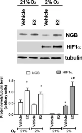
Neither 24 h (Fig 1) nor 48 h (data not shown) of physiological hypoxia (2% O2) increases
NGB protein levels in comparison to normoxia (21% O2) in breast cancer cells. However, E2
treatment (10 nM, 24 h) still induces the up-regulation of NGB and the hypoxia sensor HIF1α (Fig 1).
Fig 1. Effect of hypoxia in MCF-7 cells. NGB and HIF1α protein expression in MCF-7 cells exposed to either normoxia (21% O2;24 h) or physiological hypoxia (2% O2; 24 h), in the presence and absence of E2 (10 nM; 2h pretreatment). The amount of proteins were normalized to tubulin levels. Top panel is typical Western blot of three independent experiments. Bottom panel represents the results of the densitometric analysis. Data are means± SD of three different experiments. P<0.05 was determined with Student t-test vs. normoxia vehicle (*) and vs. hypoxia vehicle (#).
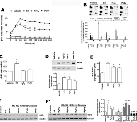
Effect of ROS-inducing compounds on NGB levels
To determine the role of NGB during oxidative stress injury, MCF-7 cells have been treated with H2O2and Pb(IV), (a pollutant which induces oxidative stress and
mitochondrial-depen-dent apoptosis) [25]. E2 treatment has been used as the positive control. Neither vehicle nor E2 (10 nM) enhance ROS level in MCF7 cells, whereas cell treatment with either H2O2(400μM)
or Pb(IV) (200μM) increased the ROS production reaching maximum levels after 30 min of treatment (14.68 ± 0.04 and 11.00 ± 1.49 fold over the control, respectively) (Fig 2A). More-over, the E2-, H2O2-, and Pb(IV)-activated signaling pathways involved in cell response to
stress and apoptosis has been evaluated by the PathScan array kit (S1 Fig). As shown inFig 2B, E2 activates the phosphorylation of AKT, SMAD2, and SAPK/JNK in line with its well-known function as activator of MCF-7 survival, proliferation, and migration [26]. On the other hand, H2O2enhances PARP-1 cleavage and AKT phosphorylation, impairing SAPK/JNK
phosphor-ylation and decreasing survivin levels; while, Pb(IV) increases the phosphorphosphor-ylation of SMAD2 and SAPK/JNK as well as the PARP-1 cleavage and survivin level (Fig 2B). Finally, the capabil-ity of E2, H2O2, and Pb(IV) to modify the level of NGB mRNA (Fig 2C) and protein (Fig 2D,
2F and 2F’) has been evaluated. E2 induces the increase of NGB mRNA 4 h after treatment (Fig 2C), whereas neither H2O2nor Pb(IV) modulate NGB mRNA levels (Fig 2C). Conversely, like
E2, both H2O2and Pb(IV) increase NGB protein levels (Fig 2D). To quantify the results
obtained by Western blot, the intensity of the NGB bands was compared with that obtained loading 5 ng of recombinant NGB. MCF-7 cells contain a very low basal level of NGB (30 ± 3.3 ng/mg protein lysate) which significantly doubles 24 h after E2 (60 ± 3.2 ng/mg protein lysate), H2O2(48 ± 2.2 ng/mg protein lysate), and Pb(IV) (46 ± 2.1 ng/mg protein lysate) treatment
(Fig 2E). In order to obtain clear evidence how NGB level could be regulated by H2O2and Pb
(IV), MCF-7 cells were treated with either the proteasomal inhibitor, MG-132 (1μM for 30 min), the lysosomal inhibitor, Chloroquine (Chloroq, 10μM for 30 min), and the translational inhibitor Cicloheximide (Ciclohex, 10μM for 30 min) before the treatment with the ROS-inducers.Fig 2F and 2F’show that NGB level is reduced by ciclohex and increased by lyso-somal degradation. H2O2and Pb(IV) treatments do not modify this trend suggesting that
ROS-inducing compounds could increase NGB levels by inhibiting lysosomal degradation and increasing NGB translation. Intriguingly, MG-132 does not modulate NGB level, but
completely impairs H2O2and Pb(IV) effect in enhancing NGB level (Fig 2F and 2F’). As a
whole, these data indicate that ROS-inducing compounds increase NGB protein levels in MCF-7 cancer cells activating specific and diverse pathways.
Effect of H
2O
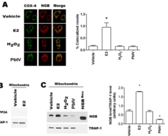
2and Pb(IV) on mitochondrial NGB localization
The NGB localization into the mitochondrial compartment is necessary to act as an anti-apo-ptotic protein in several cell lines [9,23,24]. This prompted us to verify if the selected com-pounds could modify NGB mitochondrial localization. Confocal microscopy analyses show the co-localization of NGB with the mitochondrial marker COX-4 (cytochromec oxidase-4) (Fig 3A). Although the confocal microscopy allows a purely qualitative analysis, the analysis with the 8.2 IMARIS software demonstrated that only E2 treatment (24h) significantly raises the NGB-COX-4 merged signals (Fig 3A). The increase of the NGB localization at mitochondrial level has been confirmed in isolated mitochondria by using cell fractionation kit.Fig 3B con-firms the purity of mitochondrial fraction, in fact PP2A, cytosolic marker is absent; while TRAP-1, mitochondrial marker, is evident. As expected [23], E2 (10 nM; 24 h) increases the mitochondrial NGB content; conversely, H2O2treatment (400μM; 24h) does not modify the
demonstrates that Pb(IV) (200μM, 24 h) treatment determines a significant decrease of the NGB amount in the mitochondrial fraction (Fig 3C).
Effect of H
2O
2and Pb(IV) on the NGB anti-apoptotic function
Recently, it has been demonstrated that NGB is an E2 compensatory protein, which up-regula-tion counteracts apoptosis induced by oxidative stress in several cancer cell lines [23,24]. This
Fig 2. Characterization of H2O2and Pb(IV) as MCF-7 cell stressors. (A) Cells were exposed to E2 (10 nM), H2O2(400μM), and Pb(IV) (200 μM). ROS measurement was obtained by DCFH-DA fluorescence analysis. (B) PathScan analysis of 19 target signaling proteins involved in the regulation of stress response and apoptosis in MCF-7 cells treated for 24 h with E2 (10 nM), H2O2(400μM), Pb(IV) (200 μM). Top panels are typical chemioluminescent signal of array modules of three independent experiments. Bottom panel represents the result of densitometric analysis of p-AKT, p-SMAD2, p-SAPK/JNK, cleaved PARP-1 and total Survivin proteins. Data are means± SD of three different experiments. P< 0.05 was calculated with Student’s t test vs vehicle (*). (C) NGB mRNA levels in MCF-7 cells. The NGB expression is reported as fold of induction over the vehicle (set to 100). Data represent the mean±SD of five different experiments. Significant differences (p<0.001) were determined by ANOVA followed by the Turkey-Kramer post-test with respect to unstimulated samples (*). (D) Analysis of NGB protein levels in cells treated with the above reported compounds for 24 h. The amount of protein was normalized to tubulin levels. Top panel is typical Western blot of three independent experiments. Bottom panel represents the result of densitometric analyses. Data are means± SD of three different experiments. P<0.05 was determined with Student t-test vs. vehicle (*). (E) NGB protein amount in treated cells. NGB protein cell content was quantified by comparing the Western blot band intensity of treated sample NGB with the band intensity of 5 ng NGB recombinant protein used as protein standard. Data are means± SD of three independent experiments. P<0.05 was determined with Student t-test vs. vehicle (*). (F and F’) MCF-7 cells were pre-treated for 30 min with the proteasomal inhibitor, MG-132 (1 μM), the lysosomal inhibitor, Chloroquine (Chloroq, 10μM), and the translational inhibitor Cicloheximide (Ciclohex, 10 μM) before H2O2(400μM) and Pb(IV) (200 μM) treatment for 24 h. The amount of NGB was normalized to tubulin levels. Left panels are typical Western blots of three independent experiments. Right panel represents the result of densitometric analyses. Data are means± SD of five different experiments. P<0.05 was determined with Student t-test vs. vehicle (*) or vs. non-treated samples (°).
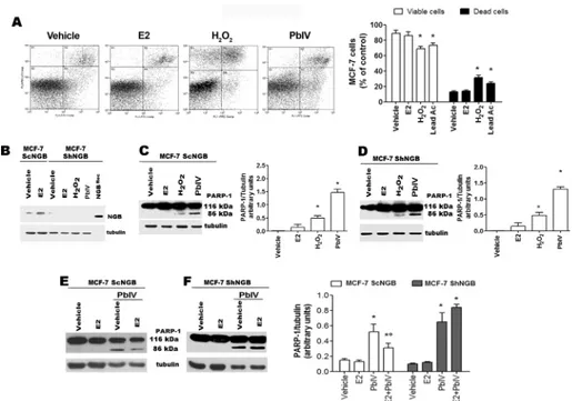
prompted us to evaluate the anti-apoptotic role of NGB in the presence of the selected ROS-inducing compounds. As reported inFig 4A, H2O2and Pb(IV), significantly reduce the
per-centage of annexin V-FITC/PI double negative MCF-7 cells (viable cells, bottom left panel), and increase the percentage of early apoptotic (annexin V-FITC positive, bottom right panel) and mid-late apoptotic (annexin V-FITC/PI double positive, top right panel) cells. This result has been further confirmed by Western blot analysis in wild type (ScNGB MCF-7) and stable NGB silenced (ShNGB MCF-7) cells (Fig 4B). Surprisingly, although both H2O2and Pb(IV)
increase NGB level, 24 h after treatment they activate the PARP-1 cleavage, another pro-apo-ptotic marker, both in the presence and in the absence of NGB (Fig 4C and 4D). This unex-pected result lead us to evaluate if E2-induced NGB over-expression in mitochondria could protect MCF-7 cells from Pb(IV)-induced apoptosis. To reach this aim, MCF-7 cells were stim-ulated with E2 for 24 h before the Pb(IV) treatment.Fig 4Eclearly indicate that E2 pre-treat-ment reduces Pb(IV)-induced PARP cleavage in ScNGB MCF-7, while this effect is completely impaired in ShNGB MCF-7 (Fig 4F).
Discussion
Here, we investigated the putative role of endogenous level of NGB in breast cancer cells as a stress sensor and as a compensatory protein, which responds to the injuring stimuli inhibiting the trigger of mitochondria-dependent apoptosis. For the first time, we showed that endoge-nous NGB is a ROS-inducible protein in MCF-7 cells.
Hypoxia is a common feature of solid tumors and the involvement of NGB in the short-term adaptation of cancer cells has been hypothesized [21]. In fact, NGB co-localizes with the hypoxia-inducible metallo-enzyme carbonic anhydrase IX in different human primary tumor specimens [21]. On the other hand, the hypoxia-dependent up-regulation of NGB mRNA has been assessed in lung cancer cells even if no information is available on the protein level [22].
Fig 3. Mitochondrial NGB localization. (A) Left, Confocal microscopy analysis of NGB and cytochromec oxidase-4 (COX-4) co-immuno-localization in MCF-7 cells treated for 24h with vehicle and/or E2 (10 nM), H2O2(400μM) and Pb(IV) (200 μM); Right, quantitative analysis of co-localization. Cells were fixed, permeabilized and stained with anti-NGB antibody (red) and co-stained with anti-mitochondrial COX-4 antibody (green) (original magnification x 63). All images are single Z-stack planes and are representative of three independent experiments. (B) Western blot analysis of PP2A (cytosolic marker) and TRAP-1 (mitochondrial marker) in mitochondrial fraction of MCF-7 cells treated with either vehicle and/or E2 (10 nM) for 24h. (C) Typical Western blot of three independent experiments of NGB expression in mitochondrial fraction of MCF7 cells treated for 24 h with the above reported compounds. Left panel is typical Western blots of three independent experiments. Right panel represents the result of densitometric analyses. The amount of proteins was normalized to the fraction marker protein TRAP-1. Data are means± SD of three different experiments. P<0.05 was determined with Student t-test vs. vehicle (*).
Although NGB is not transcriptionally regulated by HIF1α the major intracellular oxygen sen-sor [27], this globin has been proposed to be a member of the hypoxia-inducible protein family [4]. Despite this evidence, our results indicate that 2% O2, which resembles the median pO2
present in breast cancer microenvironment [28], does not up-regulate NGB levels in MCF-7 cells suggesting that NGB is not required for MCF-7 cell adaptation to hypoxic conditions. Of note, myoglobin, which is strongly up-regulated by hypoxia in MCF-7 cells [29], may attend to this function thus suggesting a cell context dependent modulation of NGB from hypoxia
As well hypoxia, oxidative stress is characteristic of both tumor development and cancer cells resistance to antitumor drugs. High ROS levels, as occur in fast proliferating tumor tissues [19], could lead to severe cellular damage and, consequently, to cell death. However, cancer cells established several mechanisms to counteract the oxidative stress-induced apoptosis and, generally, display an antioxidant capacity higher than that of normal cells [19]. Different intra-cellular pathways could converge to alter the intra-cellular metabolism and to adapt cancer cells to both intrinsic and extrinsic oxidative stress conditions [18]. However, it remains unsolved the question about the possible role of endogenous NGB in non-nervous cancer cells as an oxida-tive stress sensor; this function requires NGB activation or induction by stressing conditions.
Fig 4. Effect of NGB on H2O2- and Pb(IV)-induced apoptosis. (A) Typical cytograms of vehicle–, E2- (10 nM), H2O2- (400μM), and Pb(IV)- (200 μM) treated MCF-7 cells for 24 h (left) and relative analyses (right) obtained from Annexin V-FITC with PI assays. Data of viable (PI and Annexin V-FITC double negative) and dead (Annexin V-FITC positive and PI AnnexinV-FITC double positive) cells are means± SD of three different experiments. P< 0.05 was calculated with Student’s t test vs vehicle (*). (B) Western blot analysis of NGB protein levels performed in vehicle- and E2 (10 nM)- treated control MCF7 cells (ScNGB) and NGB stable silenced cells (ShNGB) treated with selected compounds for 24 h. Typical Western blot representative of three independent experiments. (C) Western blot analyses of PARP-1 cleavage in MCF-7 cells infected with scramble RNA (ScNGB MCF-7) treated with above reported compounds for 24h. (D) Analysis of protein PARP-1 cleavage in MCF-7 cells infected with silencing NGB shRNA (ShNGB MCF-7) and incubated with E2 (10 nM), H2O2(400μM), and Pb(IV) (200 μM) for 24 h. Western blot analyses of PARP-1 cleavage in MCF-7 cells infected with scramble RNA (ScNGB MCF-7, (E)) or with silencing NGB shRNA (ShNGB MCF-7, (F)) and treated with E2 (10 nM, 24 h) before the treatment with Pb(IV) (200μM, 24 h). (C-F), Left panels are typical Western blots of three independent experiments. Right panels represent the result of densitometric analyses. The amount of proteins was normalized by comparison with tubulin levels. Data are means± SD of three different experiments. P<0.05 was determined with Student t-test vs. vehicle (*) and Pb(IV) (°). doi:10.1371/journal.pone.0154959.g004
Although the PathScan assay and NGB mRNA did not define a unique common pathway link-ing E2 and stressor-induclink-ing NGB up-regulation, the results reported here clearly demonstrate that in MCF-7 cancer cells the apoptotic inducers modulate the level of NGB. Indeed, cell treat-ment with H2O2and Pb(IV) leads to a rapid increase of intracellular ROS production, and
up-regulate NGB protein levels. H2O2and Pb(IV) effect on NGB level seems to be mediated by the
inhibition of NGB lysosomal degradation and by the activation of translation as demonstrated by cell pre-treatment with Chloroquine and Cicloheximide. Contrarily, MG-132 does not mod-ulate NGB level, but completely impairs H2O2and Pb(IV) effect in enhancing NGB level.
Recently, a role for MG-132 and Chloroquine in the activation and inhibition, respectively, of autophagy in breast cancer cells has been reported [30,31] rendering particularly intriguing these results. Indeed, our results seem suggest that the autophagic process is involved in H2O2
-and Pb(IV)-NGB accumulation breast cancer cells. Finally, the involvement of AKT, SMAD2, SAPK/JNK, survivin, and protein stability, evidenced in this study, could converge in ROS-inducing pathways (e.g., Nuclear factor erythroid-derived 2, NRF2) to increase NGB levels and, ultimately, to cancer cell survival. Although studies more detailed are requested to define the ROS-induced pathways and their functional outcomes, the data reported in this study enlarge the physiological role of NGB in breast cancer cells pointing to its up-regulation as pos-sible ROS sensor as reported in brain-derived cells [2–4,6,11,17]. In particular, in human and rat pheochromocytoma PC12 cell lines, the H2O2treatment increases the level of transiently
transfected NGB and induces a conformational change of NGB allowing the globin recruitment at the plasma membrane lipid rafts where it acts as a guanine-dinucleotide dissociation inhibi-tor suppressing the Gαs activity and leading to neurons protection against apoptosis [17]. However, data reported here unexpectedly indicate that the increased level of NGB induced by H2O2and Pb(IV) is not sufficient to counteract the ability of these substances to induce the
apoptotic death in MCF-7. This result has been further confirmed by NGB silencing experi-ments in which both H2O2and Pb(IV) still activate the PARP-1 cleavage, an apoptotic marker
in MCF-7 cells. However, increasing NGB levels by 24 h E2 treatment reduces Pb(IV) activa-tion of PARP-1 cleavage. This result is in line with the E2 protective effect against H2O2–
induced apoptosis previously reported [23–24] strongly confirming the anti-apoptotic role of NGB.
Remarkably, E2-induced NGB up-regulation exerts anti-apoptotic function directly at the mitochondrial compartment by interacting with cytochromec and impairing its release to cytosol and the consequent activation of the intrinsic apoptotic pathway upon oxidative stress injury [7,23]. Present data indicate that in MCF-7 cells, only the E2 treatment almost doubles the NGB amount in the mitochondrial fraction, whereas H2O2and Pb(IV), which increase
NGB level in the whole cell, do not affect the mitochondrial protein amount. All together, these data indicate that the increase of intracellular NGB levels induced by H2O2and Pb(IV) is not
sufficient to reset the intrinsic apoptotic pathway, which requires the re-allocation of NGB into mitochondria. In line with this idea, Yu and coworkers [6] demonstrated that in primary mouse cortical neurons NGB mitochondrial localization increased after pathological oxygen-glucose deprivation conditions conferring neuroprotection. Although it could be possible that only the high NGB levels obtained by transfection with NGB-encoding plasmid allow the NGB recruitment on lipid rafts reported in neurons after oxidative stress [17], the possibility that E2, oxygen-glucose deprivation, and oxidative stress signaling could induce different NGB confor-mation that affect the protein translocation into mitochondria should be taken into account.
An opposite function has been attributed to NGB in hepatocarcinoma cells. In this cell line, NGB acts as a mediator between oxygen/ROS signals and the cytosolic signaling cascade that regulates the cell proliferation [20]. Unfortunately, also this evidence has been obtained in cells transiently transfected with the NGB-encoding plasmid which induces high non-physiological
protein levels that could be necessary for NGB involvement in intracellular signaling cascade [20]. However, the involvement of NGB in cancer cell proliferation has not been confirmed by MCF-7 cell growth curves, which result similar to the control even when NGB was stably silenced [23]. These results indicate that the over-expression of NGB does not fully represent the physiological behavior of NGB.
In conclusion, NGB level could be considered as a sensor of ROS being up-regulated by ROS (i.e., H2O2) and by ROS-inducing substances (i.e., Pb(IV)), whereas its function as an
anti-apoptotic protein is strictly linked to its level and intracellular localization. However, oxi-dative conditions increase the heme-Fe-based NGB reactivity by formation of the labile Cys46-Cys55 disulfide bond and of the Tyr44/His64/heme propionate interaction [32,33]. Consequently, ROS and ROS-generating compounds induce high level of oxidized NGB that increase, for example, NGB activity as free radical scavenger [34] supporting the role of NGB as a compensatory protein in breast cancer cells. Although further studies in breast cancer cells are needed to identify up-stream and down-stream NGB regulating pathways and the intracel-lular NGB trafficking, present data might lead to a new direction in understanding NGB func-tion cancer and neuroprotecfunc-tion opening new avenues for the therapeutic intervenfunc-tion based on the development of inhibitors impairing NGB-dependent cell protection.
Supporting Information
S1 Fig. Map position of p-AKT, p-SAPK/JNK, p-SMAD2, PARP-1 and survivin specific antibody in the PathScan1 Stress and Apoptosis Signaling Antibody ArrayKit. (TIF)
Acknowledgments
This work was supported by grants from Associazione Italiana Ricerca sul Cancro (AIRC, IG#15221) to M.M. and from Ministero dell’Istruzione, dell’Università e della Ricerca of Italy (PRIN 20109MXHMR_001) to P.A. The Authors declare that there is no conflict of interest that could be perceived as prejudicing the impartiality of the research reported.
Author Contributions
Conceived and designed the experiments: MM PA. Performed the experiments: MF MC AN FC DG CV SL. Analyzed the data: MF MM. Contributed reagents/materials/analysis tools: CV FC. Wrote the paper: MF PA MM.
References
1. Burmester T, Weich B, Reinhardt S, Hankeln T. A vertebrate globin expressed in the brain. Nature. 2000; 407: 520–523. PMID:11029004
2. Sun Y, Jin K, Mao XO, Zhu Y, Greenberg DA. Neuroglobin is up-regulated by and protects neurons from hypoxic-ischemic injury. Proc Natl Acad Sci U S A. 2001; 98: 15306–15311. PMID:11742077 3. Fordel E, Thijs L, Martinet W, Schrijvers D, Moens L, Dewilde S. Anoxia or oxygen and glucose
depriva-tion in SH-SY5Y cells: a step closer to the unraveling of neuroglobin and cytoglobin funcdepriva-tions. Gene. 2007; 398: 114–122. PMID:17532579
4. Greenberg DA, Jin K, Khan AA. Neuroglobin: an endogenous neuroprotectant. Curr Opin Pharmacol. 2008; 8: 20–24. PMID:17942367
5. Yu Z, Liu J, Guo S, Xing C, Fan X, Ning M, et al. Neuroglobin-overexpression alters hypoxic response gene expression in primary neuron culture following oxygen glucose deprivation. Neurosci. 2009; 162: 396–403.
6. Yu Z, Liu N, Liu J, Yang K, Wang X. Neuroglobin, a novel target for endogenous neuroprotection against stroke and neurodegenerative disorders. International J Mol Sci. 2012; 13: 6995–7014.
7. Fiocchetti M, De Marinis E, Ascenzi P, Marino M. Neuroglobin and neuronal cell survival. Biochim Bio-phys Acta. 2013; 1834: 1744–1749. doi:10.1016/j.bbapap.2013.01.015PMID:23357651
8. Brunori M, Giuffre A, Nienhaus K, Nienhaus GU, Scandurra FM, Vallone B. Neuroglobin, nitric oxide, and oxygen: functional pathways and conformational changes. Proc Natl Acad Sci U S A. 2005; 102: 8483–8488. PMID:15932948
9. De Marinis E, Fiocchetti M, Acconcia F, Ascenzi P, Marino M. Neuroglobin upregulation induced by 17beta-estradiol sequesters cytocrome c in the mitochondria preventing H2O2-induced apoptosis of neuroblastoma cells. Cell Death Dis. 2013; 4: e508. doi:10.1038/cddis.2013.30PMID:23429294 10. De Marinis E, Acaz-Fonseca E, Arevalo MA, Ascenzi P, Fiocchetti M, Marino M, et al. 17β-Oestradiol
anti-inflammatory effects in primary astrocytes require oestrogen receptor beta-mediated neuroglobin up-regulation. J Neuroendocrinol. 2013; 25: 260–270. doi:10.1111/jne.12007PMID:23190172 11. Hundahl C, Kelsen J, Kjaer K, Ronn LC, Weber RE, Geuens E, et al. Does neuroglobin protect neurons
from ischemic insult? A quantitative investigation of neuroglobin expression following transient MCAo in spontaneously hypertensive rats. Brain Res. 2006; 1085: 19–27. PMID:16647691
12. Li RC, Lee SK, Pouranfar F, Brittian KR, Clair HB, Row BW, et al. Hypoxia differentially regulates the expression of neuroglobin and cytoglobin in rat brain. Brain Res. 2006; 1096: 173–179. PMID: 16750520
13. Burmester T, Hankeln T. What is the function of neuroglobin? J Exp Biol. 2009; 212: 1423–1428. doi: 10.1242/jeb.000729PMID:19411534
14. DellaValle B, Hempel C, Kurtzhals JA, Penkowa M. In vivo expression of neuroglobin in reactive astro-cytes during neuropathology in murine models of traumatic brain injury, cerebral malaria, and autoim-mune encephalitis. Glia. 2010; 58: 1220–1227. doi:10.1002/glia.21002PMID:20544857
15. Fago A, Mathews AJ, Brittain T. A role for neuroglobin: resetting the trigger level for apoptosis in neuro-nal and retineuro-nal cells. IUBMB Life. 2008; 60: 398–401. doi:10.1002/iub.35PMID:18481280
16. Herold S, Fago A, Weber RE, Dewilde S, Moens L. Reactivity studies of the Fe(III) and Fe(II)NO forms of human neuroglobin reveal a potential role against oxidative stress. J Biol Chem. 2004; 279: 22841– 22847. PMID:15020597
17. Watanabe S, Takahashi N, Uchida H, Wakasugi K. Human neuroglobin functions as an oxidative stress-responsive sensor for neuroprotection. J Biol Chem. 2012; 287: 30128–38. doi:10.1074/jbc. M112.373381PMID:22787149
18. Cairns RA, Harris IS, Mak TW. Regulation of cancer cell metabolism. Nature Rev Cancer. 2011; 11: 85–95.
19. Gorrini C, Harris IS, Mak TW. Modulation of oxidative stress as an anticancer strategy. Nat Rev Drug Discov. 2013; 12: 931–947. doi:10.1038/nrd4002PMID:24287781
20. Zhang J, Lan SJ, Liu QR, Liu JM, Chen XQ. Neuroglobin, a novel intracellular hexa-coordinated globin, functions as a tumor suppressor in hepatocellular carcinoma via Raf/MAPK/Erk. Mol Pharmacol. 2013; 83: 1109–1119. doi:10.1124/mol.112.083634PMID:23478801
21. Emara M, Turner AR, Allalunis-Turner J. Hypoxic regulation of cytoglobin and neuroglobin expression in human normal and tumor tissues. Cancer Cell Int. 2010; 10: 33. doi:10.1186/1475-2867-10-33 PMID:20828399
22. Oleksiewicz U, Daskoulidou N, Liloglou T, Tasopoulou K, Bryan J, Gosney JR, et al. Neuroglobin and myoglobin in non-small cell lung cancer: expression, regulation and prognosis. Lung Cancer. 2011; 74: 411–418. doi:10.1016/j.lungcan.2011.05.001PMID:21640426
23. Fiocchetti M, Nuzzo MT, Totta P, Acconcia F, Ascenzi P, Marino M. Neuroglobin, a pro-survival player in estrogen receptor alpha-positive cancer cells. Cell Death Dis. 2014; 5: e1449. doi:10.1038/cddis. 2014.418PMID:25299774
24. Fiocchetti M, Camilli G, Acconcia F, Leone S, Ascenzi P, Marino M. ERβ-dependent neuroglobin up-regulation impairs 17β-estradiol-induced apoptosis in DLD-1 colon cancer cells upon oxidative stress injury. J Steroid Biochem Mol Biol. 2015; 149: 128–317. doi:10.1016/j.jsbmb.2015.02.005PMID: 25683270
25. He L, Poblenz AT, Medrano CJ, Fox DA. Lead and calcium produce rod photoreceptor cell apoptosis by opening the mitochondrial permeability transition pore. J Biol Chem. 2000; 275: 12175–12184. PMID:10766853
26. Acconcia F, Marino M. The effects of 17beta-estradiol in cancer are mediated by estrogen receptor sig-naling at the plasma membrane. Front Physiol. 2011; 2: 30. doi:10.3389/fphys.2011.00030PMID: 21747767
27. Jin K, Mao X, Xie L, Greenberg DA. Interactions between vascular endothelial growth factor and neuro-globin. Neurosci Lett. 2012; 519: 47–50. doi:10.1016/j.neulet.2012.05.018PMID:22583764
28. Vaupel P, Mayer A, Briest S, Höckel M. Oxygenation gain factor: a novel parameter characterizing the association between hemoglobin level and the oxygenation status of breast cancers. Cancer Res. 2003; 63: 7634–7637. PMID:14633681
29. Gorr TA, Wichmann D, Pilarsky C, Theurillat JP, Fabrizius A, Laufs T, et al. Old proteins—new loca-tions: myoglobin, haemoglobin, neuroglobin and cytoglobin in solid tumours and cancer cells. Acta Phy-siol (Oxf). 2011; 202: 563–581.
30. Bao W, Gu Y, Ta L, Wang K, Xu Z. Induction of autophagy by the MG‑132 proteasome inhibitor is asso-ciated with endoplasmic reticulum stress in MCF‑7 cells. Mol Med Rep. 2016; 13: 796–804. doi:10. 3892/mmr.2015.4599PMID:26648402
31. Maycotte P, Thorburn A. Targeting autophagy in breast cancer. World J Clin Oncol. 2014; 5: 224–240. doi:10.5306/wjco.v5.i3.224PMID:25114840
32. Nicolis S, Monzani E, Pezzella A, Ascenzi P, Sbardella D, Casella L. Neuroglobin modification by reac-tive quinone species. Chem Res Toxicol 2013; 26: 1821–1831. doi:10.1021/tx4001896PMID: 24144187
33. Morozov AN, Roach JP, Kotzer M, Chatfield DC. A possible mechanism for redox control of human neuroglobin activity. J Chem Inf Model 2014; 54: 1997–2003. doi:10.1021/ci5002108PMID: 24855999
34. Li W, Wu Y, Ren C, Lu Y, Gao Y, Zheng X, et al. The activity of recombinant human neuroglobin as an antioxidant and free radical scavenger. Proteins. 2011; 79:115–125. doi:10.1002/prot.22863PMID: 20938977