UNIVERSITÀ DI PISA
School of graduate studies
“Scienza del farmaco e delle sostanze bioattive”
“DESIGN AND SYNTHESIS OF NOVEL ZINC-DEPENDENT
METALLOENZYMES
INHIBITORS AS ANTI-TUMORAL DRUG CANDIDATES.”
Tutor: PhD Student:
Prof. Concettina La Motta Matteo Morelli
XXIV Cycle 2012
Index
CHAPTER 1Cancer the never ending night mare pg. 1 Bibliography pg. 7
CHAPTER 2
Matrix Metalloproteinases: An introduction pg. 8 Bibliography Pg. 31
CHAPTER 3
Developmental of matrix metalloproteinases inhibitors pg. 33 - Benzo[d]isothiazol-3(2H)-one-1,1-dioxide Derivatives pg. 35 - Isoindoline-1,3-dione Derivatives pg. 42 - Thiazolidinedionic and thiohydantoinic core-based MMPs inhibitors pg. 46 - Pyridopyrimidinone derivatives pg. 58 Bibliography pg. 60 CHAPTER 4 Esperimental section pg. 63 Bibliography pg. 89 CHAPTER 5
The ubiquitin–protein ligase system pg. 92 Bibliography pg. 107
CHAPTER 6
Inhibitors of E2, E3 enzymes pg. 109 - Carbamo(dithioperoxo)thioates derivatives pg. 110 - Triazinic derivatives pg. 113 Bibliography pg. 117 CHAPTER 7 Experimental section pg. 118 Bibliography pg. 130
CHAPTER 1
Cancer continues to be a worldwide killer, despite the enormous amount of research and rapid developments seen during the past decade. According to recent statistics, cancer accounts for about 23% of the total deaths in the USA and is the second most common cause of death after heart disease. Death rates for heart disease, however, have been steeply decreasing in both older and younger populations in the USA from 1975 through 2002. In contrast, no appreciable differences in death rates for cancer have been observed in the United States.
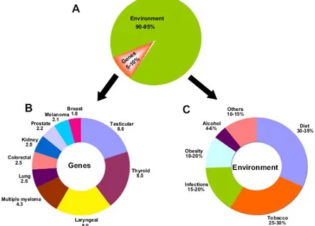
Cancer is caused by both internal factors (such as inherited mutations, hormones, and immune conditions) and environmental/acquired factors (such as tobacco, diet, radiation, and infectious organisms; Fig. 1.1). The link between diet and cancer is revealed by the large variation in rates of specific cancers in various countries and by the observed changes in the incidence of cancer in migrating. For example, Asians have been shown to have a 25 times lower incidence of prostate cancer and a ten times lower incidence of breast cancer than do residents of Western countries, and the rates for these cancers increase substantially after Asians migrate to the West.1
Main cause of cancer insurgence and development.
Diet and Exercise.
The importance of lifestyle factors in the development of cancer was also shown in studies of monozygotic twins. Only 5–10% of all cancers are due to an inherited gene defect. Although all cancers are a result of multiple mutations, these mutations are due to interaction with the environment. These observations indicate that most cancers are not of hereditary origin and that lifestyle factors, such as dietary habits, smoking, alcohol consumption, and infections, have a profound influence on their development. Although the hereditary factors cannot be modified, the lifestyle and environmental factors are potentially modifiable. The lesser hereditary influence of cancer and the modifiable nature of the environmental factors point to the preventability of cancer. The important lifestyle factors that affect the incidence and mortality of cancer include tobacco, alcohol, diet, obesity, infectious agents, environmental pollutants, and
radiation. 1
In the United States, overweight and obesity contribute to 14% to 20% of all cancer-related mortality. Overweight and obesity are clearly associated with increased risk for developing
many cancers, including cancers of the breast in postmenopausal women ,colon, endometrium, adenocarcinoma of the esophagus, and kidney. Evidence is highly suggestive that obesity also increases risk for cancers of the pancreas, gallbladder, thyroid, ovary, and cervix, and for multiple myeloma, Hodgkin lymphoma, and aggressive prostate cancer.
These findings are supported by both epidemiologic studies in humans and other research.
Overweight and obesity are thought to affect risk of these cancers through a variety of mechanisms, some of which are specific to particular cancer types. These mechanisms include effects on fat and sugar metabolism; immune function; levels of several hormones, including insulin and estradiol; factors that regulate cell proliferation and growth, such as insulin-like growth factor-1; and proteins that make hormones more or less available to tissues, such as sex hormone-binding globulin.
Overweight and obesity may increase risk of adenocarcinoma of the esophagus by increasing risk of gastroesophageal reflux disease and Barrett’s esophagus.
Most research on energy imbalance and cancer focuses on increased risks associated with overweight and obesity. Recently, studies exploring intentional weight loss suggest that losing weight may reduce the risk of breast cancer.
Surgery to treat morbid obesity and short-term intentional weight loss have been shown to improve insulin sensitivity and biochemical measures of hormone metabolism, which have been
postulated to contribute to the relationship between obesity and certain cancers. The surgical removal of intra-abdominal fat has also been shown to reduce the metabolic syndrome.
Even though our knowledge about the relationship between weight loss and cancer risk is incomplete, individuals who are overweight or obese should be encouraged and supported in their efforts to reduce weight. 2
Chemicals.
A very large amount of organic and inorganic compounds are deputed or suspected to provoke cancer.
It should be an infinite list.
Among the most prominent we can cite chemicals released from tobacco burning (such as nitrosamine and polycyclic aromatic hydrocarbons), asbestos, benzene, alcoholic beverage. 3
Infection.
Worldwide approximately 18% of cancers are related to infectious diseases.[1] This proportion varies in different regions of the world from a high of 25% in Africa to less than 10% in the developed world. 1
Viruses are the usual infectious agents that cause cancer but bacteria and parasites may also have an effect.
A virus that can cause cancer (called oncovirus) include human papillomavirus (cervical carcinoma), Epstein-Barr virus (B-cell lymphoproliferative disease and nasopharyngeal carcinoma), Kaposi's sarcoma herpesvirus (Kaposi's Sarcoma and primary effusion lymphomas), hepatitis B and hepatitis C viruses (hepatocellular carcinoma), and Human T-cell leukemia virus-1 (T-cell leukemias). Bacterial infection may also increase the risk of cancer, as seen in Helicobacter pylori-induced gastric carcinoma.[23] Parasitic infections strongly associated with cancer include Schistosoma haematobium (squamous cell carcinoma of the bladder) and the liver flukes, Opisthorchis viverrini and Clonorchis sinensis (cholangiocarcinoma). 4
Radiation.
Principal Cellular and Tissue Effects of Radiation can be resumed in: cell killing, mutagenesis, chromosomal aberrations and neoplastic transformations.
Radiation can kill cells by two distinct mechanisms. The first is apoptosis, also called programmed cell death or interphase death.Cells undergoing apoptosis as an immediate consequence of radiation damage usually die in interphase within a few hours of irradiation, irrespective of and without intervening mitosis. The second mechanism for cell killing is radiation-induced reproductive failure. Radiation in sufficient doses can inhibit mitosis; that is, the cell’s ability to divide and proliferate indefinitely. The inhibition of cellular proliferation is the mechanism by which radiation kills most cells.
The major potential consequence of radiation-induced mutations in human populations is heritable genetic effects resulting from mutations induced in germinal cells. 5
Hormones.
Neoplasia of hormone-responsive tissues currently accounts for more than 35% of all newly diagnosed male and more than 40% of all newly diagnosed female cancers in the United States. Given that endogenous hormones apparently affect the risk of these cancers and their overall frequency, concern exists about the effects on cancer risk if the same or closely related hormones are administered for therapeutic purposes.
Oral contraccettive
The relationship of oral contraccettive (OC) use to breast cancer risk has been the topic of many review articles. A recent combined analysis of 54 studies that included over 150,000 women has provided many important answers about the risk of breast cancer among users of combination OC (COC), i.e., OCs which provide an estrogen and progestin in combination in a single pill. This analysis indicates that a modest increased risk of breast cancer is observed among current (relative risk, RR = 1.24) and recent (RR = 1.16) COC users. Age at first COC use modifies the association with recent use. For recent users, the risks are highest for those who began COC use before the age of 20 years. However, total duration of COC use was not associated with increased risk of breast cancer, once recency of use was taken into account. Although the scope of this combined analysis was broad, it still provides little information on COC effects 10 or more years after cessation of use. Moreover, most women who stopped use 10 or more years ago had used COCs for only short periods of time. Women who began use as teenagers are now becoming perimenopausal and postmenopausal. Current studies now underway will be able to examine more complex patterns of COC use, as related to breast cancer risk.
Hormone replacement terapy
Hormone replacement therapy in the form of unopposed estrogen therapy gained widespread popularity in the United States during the 1960s and 1970s. Concomitant with this increasing usage, incidence rates of endometrial cancer in postmenopausal women also increased rapidly, especially on the West Coast, where use of ERT was particularly common. By 1975, the results of epidemiologic case-control studies, demonstrating a strong overall association between ERT and risk of endometrial cancer were being published.Literally dozens of studies have now documented a high relative increase in the risk of endometrial cancer following ERT.Risk is strongly related both to dose and duration of use, but high relative increments in risk follow even moderate doses taken for moderately long periods of time. Women who use ERT for 5 years or longer have approximately a 3.5-fold increase in risk compared with that of women who have never used such therapy. 6
A lot of therapies exist, but even if cancer treatments have lead to prolonged life-span, no one definitive treatment is yet in use.
Bibliography
1 Anand P. et al. Cancer is a preventable disease that requires major lifestyle changes.
Pharmaceutical Research, 2008, 25, 2097-2116.
2 L. H. Kushi et all. American Cancer Society Guidelines on Nutrition and Physical Activity for cancer prevention: reducing the risk of cancer with healthy food choices and physical activity. CA:
A Cancer Journal for Clinicians 2006 56, 254–281.
3 Kuper, H.; Adami, H. O.; Boffetta P.; Tobacco use, cancer causation and public health impact. Journal of internal medicine World Health Organization 2007 251 455–66.
4 Vassilis, S.;Petros R., I.; Eleni G., M.; Peppas, G.; Falagas, M., E.; Chronic bacterial and parasitic infections and cancer: a review The Journal of Infection in Developing Countries 2010 4 267–281.
5 Little, J., B.; "Chapter 14: Ionizing Radiation". In Bast RC, Kufe DW, Pollock RE, et al.. Holland-Frei Cancer Medicine (5th ed.) 2000.
6 Henderson, B., E.; Bernstein, L.; Ross, R., K.; Chapter 13: Hormones and the Etiology of Cancer.
CHAPTER 2
Matrix metalloproteinases (MMPs) are an important family of zinc- and calcium-dependent peptidases involved in the regulation of the cellular behavior by proteolytic processing of the pericellular environment. 1 The founding member of the matrix metalloproteinase (MMP) family, collagenase, was identified in 1962 by Gross and Lapiere, who found that tadpole tails during metamorphosis contained an enzyme that could degrade fibrillar collagen IV. Subsequently, an interstitial collagenase, collagenase-1 or MMP1, was found in diseased skin and synovium. In vitro, MMP1 initiates degradation of native fibrillar collagens, crucial components of vertebrate extracellular matrix, by cleaving the peptide bond between Gly 775–Ile 776 or Gly 775–Lys 776 in native type I, II or III collagen molecules. 2 Based on domain organization and substrate preference, MMPs are grouped into collagenases, gelatinases, stromelysins, matrilysins, membrane-type (MT)-MMPs and others. 3
Collagenases
Collagenases (MMP-1, MMP-8, MMP-13 and MMP-18) cleave interstitial collagens I, II and III into characteristic 3/4 and 1/4 fragments but they can digest other ECM molecules and soluble proteins. Recent studies indicated that MMP-1 activates protease activated receptor (PAR) 1 by cleaving the same Arg-Ser bond cleaved by thrombins, which promotes growth and invasion of breast carcinoma cells. Two other matrixins: MMP-2 and MMP-14 (MT1-MMP), have collagenolytic activity, but they are classified into other subgroups because of their domain compositions.
Gelatinases
Gelatinases (MMP-2 and MMP-9) readily digest gelatin with the help of their three fibronectin type II repeats that binds to gelatin/collagen. They also digest a number of ECM molecules including type IV, V and XI collagens, laminin, aggrecan core protein, etc. MMP-2, but not MMP-9, digests collagens I, II and III in a similar manner to the collagenases. The collagenolytic activity of MMP-2 is much weaker than MMP-1 in solution, but because proMMP-2 is recruited to the cell surface and activated by the membrane-bound MT-MMPs, it may express reasonable collagenolytic activity on or near the cell surface.
Stromelysins
Stromelysins (MMP-3, MMP-10 and MMP-11) have a domain arrangement similar to that of collagenases, but they do not cleave interstitial collagens. MMP-3 and MMP-10 are similar in structure and substrate specificity, but MMP-11 (stromelysin 3) is distantly related. The MMP-11 gene is located on chromosome 22, whereas MMP-3 and MMP-10 are on chromosome 11, along with MMP-1, -7, -8, -12, -20, -26 and -27. MMP-3 and MMP-10 digest a number of ECM molecules and participate in proMMP activation. MMP-11, on the other hand, has very weak activity toward ECM molecules, but cleaves serpins more readily. MMP-11 has a furin recognition motif RX[R/K]R at the C-terminal end of the propeptide and therefore it is activated intracellularly. An intracellular 40-kDa MMP-11 isoform (β-stromelysin 3) is found in cultured cells and placenta.
This transcript, resulting from alternative splicing and promoter usage, lacks the signal peptide and the pro-domain. The function of this isoenzyme is not known.
Matrilysins
Matrilysins (MMP-7 and -26) lack a hemopexin domain. MMP-7 is synthesized by epithelial cells and is secreted apically. Besides ECM components it processes cell surface molecules such as pro-α-defensin, Fas-ligand, pro-tumor necrosis factor α, and E-cadherin. MMP-26 is expressed in normal cells such as those of the endometrium and in some carcinomas. It digests several ECM molecules, and unlike most other MMPs, it is largely stored intracellularly.
MT-MMPs
MT-MMPs in mammals includes four type I transmembrane proteins (MMP-14, -15, -16, and -24) and two glycosylphosphatidylinositol-anchored proteins (MMP-17 and -25). They all have a furin recognition sequence RX[R/K]R at the C-terminus of the propeptide. They are therefore activated intracellularly and active enzymes are likely to be expressed on the cell surface. All MT-MMPs, except MT4-MMP (MMP-17) can activate proMMP-2. MT1-MMP (MMP-14) has collagenolytic activity on collagens I, II, and III. MT1-MMP null mice exhibit skeletal abnormalities during post-natal development, which is attributed to the lack of collagenolytic activity.
Seven MMPs are not grouped in the above categories although MMP-12, MMP-20 and MMP-27 have similar structures and chromosome location as stromelysins.
Metalloelastase, Enamelysin, Epilysin and other MMPs.
Metalloelastase (MMP-12) is expressed primarily in macrophages, but is also found in hypertrophic chondrocytes and osteoclasts. It digests elastin and a number of ECM molecules. It is essential in macrophage migration.
MMP-19 digests many ECM molecules including the components of basement membranes. It is also called RASI (rheumatoid arthritis synovial inflammation) as it is found in the activated
autoantigen in patients with rheumatoid arthritis and systemic lupus erythematosis. It is, however, widely expressed in many organs including proliferating keratinocytes in healing wounds.
Enamelysin (MMP-20) is expressed in newly formed tooth enamel and digests amelogenin.
MMP-21 was originally found in Xenopus and more recently in mice and humans. It is expressed in various fetal and adult tissues and in basal and squamous cell carcinomas. It digests gelatin, but information about the action on ECM components is not known.
MMP-23 is a unique member as it has unique C-terminal cysteine-rich immunoglobulin-like domains instead of a hemopexin domain. The propeptide lacks a cysteine switch. It is proposed to be a type II membrane protein having a transmembrane domain at the N-terminal of the propeptide, but the enzyme is released from the cell as the membrane anchored propeptide is cleaved by a proprotein convertase.
MMP-27 was first found in chicken embryo fibroblasts. Chicken MMP-27 digests gelatin and casein and causes autolysis of the enzyme, but little information is available on the activity of mammalian enzyme.
Epilysin (MMP-28) is expressed in many tissues such as lung, placenta, heart, gastrointestinal tract and testis. The enzyme expressed in basal keratinocytes in skin is considered to function in wound repair. It is also elevated in cartilage from patients with osteoarthritis and rheumatoid arthritis. MMP-21, MMP-23 and MMP-28 have a furin recognition sequence before the catalytic domain and therefore they are likely to be activated intracellularly and secreted as active enzymes. 3
Interest in MMPs increased in the late 1960s and early 1970s following observations that MMPs are up-regulated in diverse human diseases including rheumatoid arthritis and cancer.
However, recent clinical data indicate that the relationship between MMPs and disease is not simple; for example, increased MMP activity can enhance tumour progression or inhibit it.
This complex relationship between MMP expression and cancer has increased the basic and clinical interest in understanding MMP function in vivo, but it has also focused attention on MMPs and pathologies.
The activity of MMPs is controlled at many levels and the regulation of their activity remains a topic of intense research.
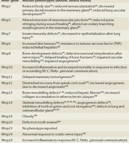
Analysis of MMPs genetic knockouts animals have offers opportunities to identify essential functions of every single MMP and to validate candidate substrates by looking for cleavage products in the control animal and uncleaved proteins in the mutant animal.
The initial characterizations have described surprisingly subtle phenotypes, with all MMP knockout lines surviving to birth (table 2.2). 5
Table 2.2. Relation between MMP-null rats and impaired functions. 5
Main Structure of MMPs
MMPs are members of the metzincin group of proteases, which are named after the zinc ion and the conserved Metionine residue at the active site.
The first structure of an MMP in complex with a synthetic inhibitor (MMP-1 catalytic domain) was reported by Lovejoy and co-workers. 6
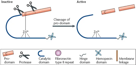
Mammalian MMPs share a conserved domain structure (figure 2.1) that consists of a catalytic domain and an auto-inhibitory pro-domain.
The catalytic activity of MMPs is regulated at four points: gene expression, compartmentalization (i.e., pericellular accumulation of enzyme), proenzyme (or zymogen) activation, and enzyme inactivation and is further controlled by substrate availability and affinity. 7
Most of the matrix metalloproteinases consist of four distinct domains, which are N-terminal pro-domain, catalytic pro-domain, hinge region, and C-terminal hemopexin-like domain.
Figure 2.1. MMP domains and activation mechanism. 5
The N-terminal prodomain.
The MMPs are initially synthesized as inactive zymogens with a pro-peptide domain that must be removed in order to activate the enzyme.
The prodomain essentially consists of three helices that are arranged nearly perpendicular to each other and connected via rather flexible and proteolysis susceptible loops. In case of MMP-1 and MMP-2, the loop between helix H1 and helix H2 contains the region of the first activation cleavage. This first cleavage is supposed to destabilize the threehelix bundle of the MMP prodomains. 8
The catalytic domain
The catalytic power of MMPs is encoded in four essential structural elements:
1. Binding of the main chain of the substrate in the active site cleft.
2. The specificity subsite pockets that define the active site, in particular the S1' subsite. 3. The zinc ion-binding histidine triad and catalytic glutamate.
4. Binding to secondary substrate sites outside the active site.
The catalytic domain of the MMPs contains two zinc atoms. One of these atoms plays a catalytic role and the other a structural role. The thiol group of the cysteine residue within the propeptide
before the metalloproteinase can degrade matrix proteins. 9 Main chain binding of protein or peptide substrates to the active site occurs through formation of an antiparallel β-sheet structure, with hydrogen bonds formed between the substrate and enzyme. A polarized water molecule bound to the active site Zn2+ ion and general base glutamate residue is essential in catalyzing peptide bond cleavage. In MMPs, the Zn2+ ion is coordinated by the three histidines in the HExxHxxGxxH metzincin signature motif. Mutagenesis of any one of these His residues ablates catalytic activity. The nucleophilic attack by the catalytic water molecule in MMPs occurs on the carbonyl of the substrate scissile peptide bond to form a tetrahedral transition-state intermediate in which the negatively charged oxygen is stabilized by coordination with the catalytic Zn2+ ion. 10
The catalytic domain of all MMPs is similar in showing a shallow active-site cleft notched into the front surface and separating the smaller ‘lower subdomain’ from the larger ‘upper subdomain’. This cleft extends horizontally across the molecule and would bind a peptide substrate from left to right. The upper subdomain encompasses a characteristic five-stranded, highly twisted -sheet, flanked by three surface loops on its convex side and by two long regular -helices on its concave side. Four of the five -strands are aligned in a parallel fashion, only the cleft-sided ‘edge strand’ IV runs in opposite direction. 8
The active site is constituted by specificity subsite pockets that define it:
S1, S2, S3, S4, S1’, S2’, S3’ and S4’ and the substrate posses the corresponding portions named P1, P2, P3, P4, P1’, P2’, P3’, P4’ (figure 2.2).
The S1' specificity pocket of MMPs accommodates this side chain and so the size and chemical characteristics of the S1' subsite are important in determining peptide bond preference for cleavage. For example, the small hydrophobic S1' pocket of MMP-1 and MMP-7 restrains substrate preference to small hydrophobic residues at P1'. In contrast, other MMPs such as MMP-2, MMP-3, MMP-8, MMP-9, and MMP-13 have large S1' pockets and can accommodate a more diverse range of amino acids at P1'. 9, 10
The S1’ pocket is relatively deep for most MMP enzymes (e.g., MMP-2, MMP-3, MMP-8, MMP-9, MMP-13, etc.), but for certain MMP enzymes (e.g., MMP-1, MMP-7, and MMP-11) it is partially or completely occluded due to an increase in the size of the side chain of the amino acid at position 193 8 numbering) from leucine to arginine 1), tyrosine 7), glutamine (MMP-11), or one of the amino acid residues that form the pocket. It has also been shown that the mutation of S1’ subunit tyrosine of MMP-7 to leucine changes the substrate specificity to be more like that of the deep pocket enzyme MMP-3. Homology models for MMP-2 and MMP-9 based on the structure of MMP-3 suggest that there may be differences in the shape of the bottom of the S1’ subunit for the deep pocket enzymes. In the case of MMP-2, the S1’ pocket may be a channel with no bottom, whereas that for MMP-9 is said to be a pocket- like subsite. Generally the subunit S2’ is a solvent-exposed cleft and the S3’ subunit is a really ill-defined solvent-solvent-exposed region.
Substrate selectivity
The preferred amino acid in substrate subunit P3 is proline for all the kinds of examined enzymes. Arginine is preferred at P2 for MMP-2 selectivity, whereas Leucine and Methionine are preferred for MMP-7. Phenylalanine is preferred over Leucine and Methionine at P2 for MMP-3. Valine at P1 results in negligible cleavage for all of the enzymes. At the P1 position, Glutamine provides significant cleavage by MMP-7 and MMP-8, and negligible by MMP-1, MMP-2, and MMP-9. At P1’, the presence of a Tyrosine residue results in highly selective cleavage by MMP-8 as well as Leucine and Methionine appear to be preferred for broad-spectrum cleavage; however, on the other hand, the phage-displayed results suggest that Methionine at P1’ gives minimal cleavage with MMP-7. However, the substrate specificity studies suggest that Phenylalanine at P1’ is preferred for cleavage by MMP-3 over the other enzymes, whereas the phage-displayed results indicate that Phenylalanine at P1’ provides negligible cleavage. It has also been shown that 11 and MMP-14 cleave substrates containing unusual amino acids with extremely long side chains at their P1’ position more efficiently than the corresponding substrates with natural phenylalanine or leucine amino acids. There is a very little selectivity by the various substitutions at P2’ position and generally Tryptophan is preferred for efficient cleavage. The same is true for amino acid changes at P3’.
The S3–S1 subsites from a shallow region bordered on one side by the -strand IV that features a hydrophobic proline binding cleft at S3.
It is interesting to note that the selective replacement of catalytic zinc of MMP-3 catalytic domain with other transition metals that are Co2+, Mn2+, Cd2+, and Ni2+ results in the retention of protease activity. However, substitution of the catalytic metal influences the substrate specificity of enzyme, since the active-site geometry is altered and hence affects substrate binding. 9
The hinge region.
The hinge-region in MMPs is a segment of 15–65 amino acids. It is of critical importance for the stability of the enzyme and also for the degradation of complex substrates such as fibrillar collagen by collagenases which require the concerted action of the catalytic and of the hemopexin domain. The hinge region itself contributes to the collagen binding and unwinding and its breakdown.
The structures of human and porcine MMP-1 show an elongated peptide segment which is in close contact with the catalytic and the hemopexin-like domain.
Highly conserved residues for the collagenases in this area are prolines, which are in direct contact with the catalytic and the hemopexin domain thus stabilizing the domain arrangement in MMP-1. In accordance with these structures, mutagenesis experiments showed an 98.5% drop in activity when four prolines in the hinge are replaced by alanines. A single mutation of the central glycine in the center of the hinge region of MMP-1 to aspartate also strongly reduces collagenolysis, which might be explained by reduced flexibility at this site. 8
The C-terminal Hemopexin domain.
This domain has a beautiful common fold. The overall shape is a squat cylinder comprised of four sheets each representing a hemopexin module and each forming a blade of the four-bladed β-propeller structure. Each blade is a twisted four-stranded β-sheet that links to the adjacent blades by small connecting strands on the rim of the domain and from a longer loop at the center. Extensive packing faces on the blade sides between adjacent modules arrange one next to another in a stable configuration. Packing of the wedge-shaped modules also forms a central pore in the domain, in which are bound either one or two Ca2+ ions. The central Ca2+ ions in the MMP-2 hemopexin C
domain are structurally important—their chelation eliminates heparin and fibronectin interactions. Although X-ray crystallography also showed a potential Zn2+ ion binding site on the MMP-2 hemopexin C domain, inductive coupled plasma mass spectrometry analysis did not measure any significant zinc molar content in a recombinant human MMP-2 hemopexin C domain. The hemopexin C domain has homology to the collagen-binding domain of vitronectin (which therefore will also likely have domains with a four-bladed β-propeller structure). 10
Regulation of MMP Activity
As for all secreted proteinases, the catalytic activity of MMPs is regulated at four points: - gene expression
- compartmentalization (i.e., pericellular accumulation of enzyme) - proenzyme (or zymogen) activation, and enzyme inactivation - substrate availability and affinity.
Some members, such as MMP-2, MMP-19, MMP-28, and several MT-MMPs, are expressed in normal tissues, implying a role in homeostasis. However, most MMPs are not expressed in resting tissue yet are induced in repair or remodeling processes and in diseased or inflamed tissues.
The cisteine switch
Harold Van Wart and Henning Birkdahl-Hansen proposed this process as general and required step in the activation of all proMMPs (1990), and they named this mechanism the “cysteine switch”, a term that remains valid and widely accepted. In essence, the thiol-Zn2+ interaction can be broken - and a latent MMP can gain catalytic activity – by three mechanisms:
- direct cleavage of the pro-domain by another proteinase
- reduction of the free thiol by oxidants or by nonphysiologic reagents such as alkylating agents, heavy metal ions, and disulfides,
- allosteric perturbation of zymogen (Figure 2.5).
Thiol reduction and allosteric controls would lead to inter or intramolecular autolytic cleavage of the prodomain.
An important component of the cysteine-switch mechanism is that the prodomain does not need to be removed for a zymogen to acquire activity; only disruption of the zinc-thiol interaction is absolutely required.
Furin is a type 1 membrane serine protease present in the trans-Golgi network.
About one-third of MMPs contain an RXKR or RRKR sequence between the pro and catalytic domains, which serves as a target sequence for proprotein convertases or furins. MMPs with such cleavage site are processed intracellularly before secretion (figure 2.3).
Figure 2.3. Schematic representation of MMP activation. 7
TIMPs: the endogenous inhibitors of MMPs.
TIMPs is an acronym that stand for tissue inhibitors of metalloproteinases. Four TIMP family members exist: TIMP-1, -2, -3 and TIMP-4.
Each of their N- and C-terminal domains contains 6 conserved cysteine residues that form three disulfide loops. The N-terminal region binds to the MMPs’ catalytic domain and inhibits MMP activity, whereas, the terminal region interacts with the the proforms of MMP-2 and MMP-9 C-terminal hemopexin domain to stabilize the pro-enzyme inhibitor complex. TIMP-2 is the only TIMP member that specifically interacts on the cell surface with both MT1-MMP and pro-MMP-2
MMP inhibitor and activator. TIMPs can inhibit all active MMPs, however, not with the same efficacy. TIMP-1 preferentially inhibits MMP-7, MMP-9, MMP-1 and MMP-3, whereas, TIMP-2 inhibit more effectively MMP-2. TIMP-3 can inhibits MMP-2 and MMP-9 among other enzymes (ADAMs), whereas TIMP-4 inhibits MT1-MMP and MMP-2 catalytic activity. TIMPs regulate proteolytic activity and all the MMP-mediated activities. 12
Collagene Hydrolysis: Reaction mechanism
The reaction mechanism for the proteolysis by MMPs has been delineated on the basis of structural information and shown in Figure 2.4. It is proposed that the scissile amide carbonyl coordinates to the active-site zinc (II) ion. This carbonyl is attacked by a water molecule, which is both hydrogen bonded to a conserved glutamic acid (Glu-198 in MMP-8) and coordinated to the zinc(II) ion. The water molecule donates a proton to the Glu residue that transfers it to the nitrogen of the scissile amide, which is followed by the Glu residue shuttling the remaining proton from the water molecule to the nitrogen of the scissile amide with resultant peptide bond cleavage. In this process, the positively charged zinc(II) ion helps to stabilize a negative charge at the carbon of the scissile amide and a conserved alanine (Ala-161 in MMP-8) residue helps stabilize positive charge at the nitrogen of the scissile amide.
H C C O N HC Zn2+ His His His HOH O O Glu O P1 P1' Ala H C C O N HC Zn2+ His His His HO O O Glu O P1 P1' Ala H H H C C O N HC Zn2+ His His His HO O O Glu O P1 P1' Ala H H H C C HN HC Zn2+ His His His O O Glu O P1 P1' Ala H H O O
Figure 2.4. Mechanism of collagen degradation. 9
Despite the huge amount of metalloproteinases that have been discovered, we have choose just three of them: MMP-2, MMP-9 and MMP-13.
MMP-2
MMP-2 (gelatinase A, 72-kDa type IV collagenase) is one of the two described human gelatinases in the MMP family, named for their ability to proteolytically degrade gelatine (denatured collagen). MMP-2 is ubiquitously expressed as a 72-kDa proenzyme and subject to extensive glycosylation. Expression of MMP-2 is constitutive and most proinflammatory stimuli fail to increase the expression level since the gene, in contrast to that of MMP-9, lacks binding sites for pro-inflammatory transcription factors such as activator protein-1.
The MMP-2 proenzyme is activated by forming a complex with TIMP-2 under appropriate stoichiometric conditions, which in turn is a substrate for the membrane bound membrane type-1 MMP (MMP-14) that removes the prodomain of proMMP-2 by proteolytic cleavage (aided by free active MMP-2), yielding the truncated 64-kDa active enzyme. If the concentration of TIMP-2 is too high, both MMP-14 and active MMP-2 will be inhibited and no further activation will ensue. Besides this activation pathway, proMMP-2 can also be activated by thrombin and activated protein C. MMP-2 differs from other MMPs in that the catalytic domain contains cysteine-rich inserts that resemble the collagen-binding regions of the type II repeats in fibronectin. These inserts are required for binding and cleavage of collagen and elastin. MMP-2 is capable of cleaving gelatine, type I, IV and V collagens, elastin and vitronectin.
Through their ability to degrade collagen in the vascular basal membranes, the gelatinases are involved in neovascularisation both under physiological conditions and in pathologies such as tumor metastasis.
The state of activation seems to be of crucial importance for the function of MMP-2 in angiogenesis. Recent research has demonstrated that the fully mature active form induces apoptosis in endothelial cells and inhibits neovascularization, while an intermediate activated complex with MMP-14 enhances cell survival and promotes angiogenesis. MMP-2 can also facilitate migration of cells by direct degradation of the basement membrane thus allowing infiltration of, for instance, neutrophils and lymphocytes, or liberation of chemoattractants. This latter process, named ‘ectodomain shedding’, is an important physiological function of the membrane-bound ADAM proteases, but has also been described for many other members of the metzincin superfamily. MMP-2 has been known to be involved in both promoting and inhibiting inflammation by liberation of pro-inflammatory mediators.
Interestingly, MMP-2 knockout mice exhibit a normal phenotype under physiological conditions, although the animals do show different response patterns upon allergen challenge, which may be attributed to disturbance of the important role of clearing immune cells.
These findings indicate that MMP-2 function may be interchangeable with other metalloproteases, an hypothesis that is supported by the observation that expression of the second gelatinase, MMP-9, is greatly increased in MMP-2 null mice. 13
MMP-9
MMP-9 (gelatinase B, 92-kDa type IV collagenase) was first discovered in neutrophils in 1974. MMP-9 is expressed as a 92-kDa proenzyme, which can be activated to the 83-kDa mature enzyme. The larger size of MMP-9 relative to MMP-2 can be attributed to a heavily O-glycosylated collagen V-like insert that links the metalloprotease domain to the hemopexin- like domain. MMP-9 activation may be mediated by removal of the prodomain by serine proteases or other MMPs, or may be a direct response to oxidative stress that disrupts the cysteine switch (as a matter this particular MMP can be usefull as a marker for acute ischemic stroke). 14 While a considerable overlap exists in the substrates degraded by MMP-2 and -9, MMP-9 is incapable of direct proteolysis of collagen I.
MMP-9 has been described to release the biologically active form of vascular endothelial growth factor (VEGF), which plays an important role in angiogenesis. This process is complemented by the direct proteolytic degradation of vascular basement membrane proteins, indicating that MMP-9 (even more than MMP-2) may play a crucial role in the formation of new blood vessels.
Interestingly, the hemopexin domain of MMP-9 was reported to be an inhibiting factor in angiogenesis as demonstrated by decreased invasion of glioblastoma cells overexpressing the MMP-9 hemopexin domain in a xenograft model.
MMP-9 null mice show decreased fertility, since MMP-9 is crucial during several stages of the female reproductive cycle (implantation of the embryo and remodeling of endometrial tissue that occurs during the menstrual cycle). Absence of MMP-9 also leads to disorders in bone development, specifically delayed bone ossification due to insufficient angiogenesis in growth plates and reduced osteoclast recruitment.
Whereas MMP-2 is primarily inhibited by TIMP-2, MMP-9 is mostly inhibited by TIMP-1 and contrary to MMP-2, which is expressed ubiquitously under physiological conditions, MMP-9 is increased in malignant cell lines and correlates with their metastatic potential.
The role of gelatinases in pathology has been studied extensively, especially in lung diseases and cancer. The amount of both gelatinases in BALF and sputum of patients suffering from chronic
asthma is higher than in healthy individuals and this increase may be responsible for the characteristic tissue remodeling events observed in chronic asthma such as thickening of the basement membrane, smooth muscle tissue hypertrophy and reduced epithelial thickness. Presence and activity of gelatinases is elevated in many other pulmonary diseases, such as cystic fibrosis, bronchiectasis, acute respiratory distress syndrome (ARDS) and infections. 13
MMP-13
MMP-13 (collagenase-3) is the latest human collagenase described in literature. This enzyme exhibits preference toward cleavage of type II collagen, effectively completing the substrate spectrum of the collagenases. Collagenase-3 was first cloned from breast cancer tissue in 1994. MMP-13 expression can be influenced by a wide range of hormones and cytokines, such as parathyroid hormone (indicative of the important role of MMP-13 in bone development), insulin-like growth factors I and II, platelet derived growth factor, basic fibroblast growth factor, transforming growth factor b1 (interestingly both up- and downregulates MMP-13 expression depending on the tissue), interleukin-1 and -6, tumor necrosis factor a and many more. ProMMP-13 can be activated by auto-proteolysis or propeptide removal by various other MMPs like stromelysin-1 (MMP-3), yielding a mature enzyme of 48 kDa, which in turn can be inhibited by TIMP-1, -2 and -3. Active MMP- 13 is a key factor in the activation pathway of several MMPs. Besides the TIMP route of inactivation, MMP-13 can bind to a specific receptor on the surface of osteoblasts and fibroblasts resulting in internalization and degradation of the protease.
MMP-13 plays an important role in bone development and remodeling, as may be anticipated from its capability to cleave type II collagen (a major component of cartilage).
This rather specific function is reflected in a limited expression profile of MMP-13 during development and adulthood, which is restricted to developing skeletal tissue. MMP-13 has, contrary to the other collagenases, a relatively high specific activity toward gelatine, indicating that the proteolytic role of MMP-13 expands past the first step of cleavage of triple-helical collagens. Further identified substrates of MMP-13 include aggrecan and perlecan, transforming growth factor , biglycan, the large protein species of tenascin-C, fibrillin-1 and -2, fibrinogen and two serpins (a2-antichymotrypsin and plasminogen activator inhibitor-2). MMP-13 expression has been regularly referred to in the literature as indicative of various cancerous processes including chondrosarcoma, breast cancer, head and neck tumors and melanoma. In all cases, high expression of MMP-13 seems to be related to
aggressiveness of the tumor, and MMP-13 may even be required for metastasis of certain tumors as demonstrated for malignant melanoma. Regarding the important role of MMP-13 in bone turnover,
it is not surprising that this enzyme has been linked to various bone-related diseases. Since MMP-13 degrades both type II collagen and aggrecan, it has been linked to cartilage destruction in rheumatoid and osteoarthritis. The specialized role of MMP-13 in bone development and disease has made it an attractive target for selective MMP-13 inhibitors as therapeutic compounds. 13
MMPs and cancer
MMPs have a relevant role in the pathophysiology of diseases suchas arthritis, multiple sclerosis, periodontal disease, atherosclerosis, and cardiac injury and remodeling. 15 A number of cancer studies have correlated MMP expression and disease outcome.
MMPs can contribute to tumor growth not only by degradation of the extracellular matrix (ECM) but by the release of sequestered growth factors or the generation of bioactive fragments. For instance, MMP-9 mobilizes VEGF from the ECM and cleaves type IV collagen to generate tumstatin, an angiogenesis inhibitor. MMPs are also important in tumor progression, promoting invasion, immune escape and many other events. However, particularly in carcinomas, MMPs are associated with the supporting ‘host’ cells rather than the tumor cells and this finding has emphasized the emerging theme of the importance of the microenvironment in tumorigenesis. MMP-12, MMP-13 and cathepsin K showed an increase expression in human tumors compared with normal lung. Immunohistochemical analysis also confirmed MMP-12 expression in the stroma of human lung tumor samples.
It is notable that animal studies have shown that MMPs sometimes have ‘protective’ roles in cancer development. Mmp8-null male or ovariectomized female mice develop significantly larger numbers of skin papillomas after a chemical carcinogen treatment and higher grade and more aggressive skin tumors developed in Mmp9-null mice. In a human study of MMP-12 in squamous cell carcinoma it was found that high expression in tumor cells was associated with more aggressive tumors and high expression in tumor macrophages was correlated with lower grade tumors.
It is possible that obtaining a detailed understanding of the potential role of MMPs in the different cell types associated with tumors may be vital for the design of therapeutic anti-MMP strategies.16
MMPs also have complex roles in angiogenesis. It is known that MMPs can promote endothelial cell migration and trigger angiogenic switch. For example, MMP-9 participates in switching angiogenesis by releasing VEGF from ECM. Also, MMP-2 activity was suggested to be necessary for the switch to angiogenic phenotype in an animal model. Furthermore, MMPs increase the bioavailability of the pro-angiogenic growth factors, vascular endothelial growth factor (VEGF), fibroblast growth factor- 2 (FGF-2), and TGF-, which stimulate proliferation and migration of endothelial cells. MT1-MMP regulates VEGF-A expression by promoting VEGFR-2 cell surface localization thereby activating VEGFR-2-Src-Akt-mTOR pathway. MMP-7 degrades soluble VEGF receptor-1 (sVEGFR-1/sFlt-1), an endogenous VEGF inhibitor that sequesters VEGF and blocks its access to VEGF receptors. The degradation of sVEGFR-1 then liberates VEGF from the endogenous trap and allows its access to membrane receptors on endothelial cells. However, MMPs may have adverse effects on angiogenesis. For example, MT1- MMP-mediated endoglin shedding inhibits tumor angiogenesis. The generation of angiostatin and endostatin, two potent angiogenesis inhibitors, also involves MMPs.
MMP-7 and MMP-9 hydrolyze plasminogen to generate angiostatin fragments. Endostatin is the C-terminal proteolytic product of the collagen XVIII a1 chain and MMP-14 can cleave collagen XVIII to generate endostatin-spanning fragment. Hence, we speculate that the effects of MMPs on angiogenesis may be context-dependent. The outcome relies on the availability of specific MMPs and the balance between two opposing effects of individual MMPs on angiogenesis. 4
MMP Inhibitors
As described above, matrix metalloproteinases are involved in the regulation of the cellular turnover and in the spreading of metastasis, and their regulation should be a very usefull weapon for fight cancer.
Both small molecules (synthetic and natural products) and macromolecular endogenous inhibitors have been considered as potential therapies for diseases in which excess MMP activity has been implicated.
A tetracycline derivative, doxycycline (Periostat; CollaGenex Pharmaceuticals Inc., Newtown, PA, USA), is currently the only MMPI approved by the U.S. FDA and is used as an adjunct therapy in adult periodontitis. One of the main reasons for the limitations of hydroxamate-based inhibitors is due to its poor bioavailability and the toxicity arising from its metabolic stability limitations (the
carboxylic acids). On the other hand, most of the inhibitors tested were broad-spectrum MMP inhibitors that make no distinction between these enzymes. 17
Many inhibitors have been developed either by pharmaceutical companies and academies.
Most MMP inhibitors (MMPi) are constituted from a functional groups which are capable to form H bonds with the structure enzyme and one or more residues that can interact through Wan der Waals interactions with the enzyme subsite and a zinc-binding group (ZBG), which is deputate of the catalytic Zn2+ chelation. The most common moiety used for the synthesis of sudden inhibitors are hydroxamates, carboxylates, phosphinates and barbituric. The ZBG however is not always present, especially in the most recent and innovative inhibitors.
MMPsI can be classified in peptidomimetics and not peptidomimetics.
Either peptidomimetics and not peptidomimetics can be classified for their ZBG moiety. Here under will be report the most important and innovative class of inhibitors.
Hydroxamates.
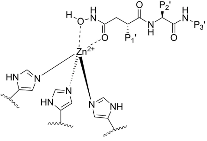
The hydroxamate is the most common and the most powerful moiety for the chelation of the catalytic zinc, it acts as a bidentate ligand and it’s widely used for the developmental of MMPsI.
N HN N HN N NH Zn2+ H N O O N H O H N P3' P2' P1' O H
Figure 2.5. Hydroxamate chelation mechanism. 18
Efficiency of hydroxamates is supported by good in vitro activity data and a very huge amount of good selective nanomolar inhibitors have been synthesised, but unfortunately have result in a fail in clinical trials.
The failure of hydroxamic acid based MMPi at a clinical level may stem, in part, from the lack of selectivity of hydroxamic acids towards the Zn2+ ion, poor pharmacokinetics, and poor oral
Consequently, some efforts have been made to identify alternatives to the hydroxamic acid ZBG. In most of these studies there have been only a comparison between hydroxamic acids and the corrisponding carboxylic acids (usually the synthetic precursor of most hydroxamic acids) and generally a loss in potency has been observed likely due to the change in binding mode and diminished donor ability of the carboxylate ligand.
Castelhano and co-workers reported the selectivity and potency of MMPi with several different ZBGs such ’reverse’ hydroxamates, carboxylates, thiols and phosphinates on a common indolactam/isobutyl backbone moiety.
As a result of these studies, the use of ZBGs other than a hydroxamate, resulted in a 10- to 250-fold loss in potency, but generally no changes in selectivity based on the nature of the
ZBG. 15 Here under are report two of the most important MMP inhibitors that must be considered in this class as broad-spectrum MMPsI:
S S O NH NH O O HOHN Batimastat N H O H N NHOH O OH O Marimas tat
Scheme 2.6. Molecular structure of Batimastat and Marimastat.
Unfortunately these two MMPsI failed during clinical trials due to muscolo-skeletal toxicity.
Pirones
A 2008 paper 15 report an interesting MMP inhibitory activity of some pirone based molecules. Docking representation show the chelating effect of pyrone ring over zinc ion (figure 2.7).
Figure 2.7. Pirone based scaffold in MMP-3 active site. 11
X-ray structures of thiomaltol and maltol tris(pyrazolyl)borate zinc(II) complexes reveal that the hydroxythiopyrone and hydroxypyrone ligands coordinate to tris(pyrazolyl) borate zinc(II) in different geometries (Fig. 3),12 suggesting that substituents at the same positions of thiopyrone and pyrone rings may interact with MMP active sites in different fashions. These findings promote us to investigate 5- and 6-substituted hydroxypyrones and hydroxythiopyrones. 11
Figure 2.8. Geometries of maltol- and thiomaltol-tris(pyrazolyl)borate zinc(II) complexes. 11
Barbiturates.
(barbiturates) are inhibitors of the MMPs with good gelatinase selectivity. This kind of MMP inhibitory with barbiturate-based scaffold are without the sedative effects of the classical agents, and impart a better in vivo stability respect to other zinc binding group (e.g., hydroxamates).
The ionised barbiturate ring chelates the zinc atom in a tridentate way directing the 5-substituents into the S1 and S2 substrate binding pockets. The class exhibit broadly similar SAR to non-barbiturate MMP inhibitor families. The 5-aromatic substituents (biphenyl or phenyloxyphenyl) bind in the deep hydrophobic S1 substrate pocket. The second 5-substitutent is directed in this arrangement into the S2 pocket towards solvent. Several elegant studies have demonstrate that C5- piperazine or piperidine substitution is favourable for high potency and moreover that the heterocycle imparts flexibility in the design of a range of barbiturate- based pharmacological, clinical and biochemical tools.
The structural basis for barbiturate interactions with the MMPs was revealed in several X-ray studies. 19 HN N O O N N O HO N N (a)
Figure 2.9. (a) Barbiturate inhibitor RO-206-0222 (b) Interaction between inhibitor and active site region of MMP-9. Ionic bonds to the catalytic zinc and intermolecular hydrogen bonds are shown as broken lines (distances in Å). 20
MMP inhibitors without a zinc-binding group
High-throughput screening of chemical libraries has led to the discovery of unusual MMP inhibitors, especially for MMP-13. Among these, MMP inhibitors with no zinc-binding group that do not engage in direct interactions with the zinc active site ion has emerged. These compounds without the zinc tether can be tailored to target the depth of the MMP S1’ cavity keeping a reduced molecular weight, in contrast to inhibitors containing a zinc-binding group. The first highly potent and selective inhibitor of MMP-13 reported was compound 1 (Ki = 8 nM towards MMP-13 and no activity detected against MMP-1, -2, -3, -7, -8, -9, -10, -11, -12, -14 and -16 when tested at 100 M). The particular binding mode of 1 in MMP-13 active site is illustrated by comparing the X-ray structure of MMP-13 in interaction with compounds 1 and 2 (for chemical structures see Scheme 3 and 2 respectively). Compound 8 binds in the bottom part of the S1’ cavity of MMP-13 and extends into an additional cavity termed the “S1’ side pocket” as depicted in figure 2.10.
HN N N HN F F O O 1 N H N O O -O O O F 2
Figure 2.10. Superimposition and comparison of cartoon representations of two crystal structures of MMP-13 in complex with a non-chelating inhibitor 1 (pink stick, PDB code: 1XUD) and with a pyrimidinetrione inhibitor 2 (yellow stick, PDB code: 1YOU) illustrating the different S1’ loop conformation and orientation of Lys249 in the two complexes. 21
An excellent paper published from Gao et all. of Boehringer Ingelheim Pharmaceuticals 22, report a library of compounds born from the same scaffold. Some of them show a very good activity joined to an exceptional selectivity for MMP-13. Here is report the main structure of sudden inhibitors.
O HN Ar O HN O NH O R'
Figure 2.11. Main scaffold of selective MMP-13 inhibitors without zinc-binding group SAR studies of non-zinc-chelating MMP-13 inhibitors: Improving selectivity and metabolic stability. 22
Bibliography
1. Diaz, N; Sua, D.; Valdes, H.; From the X-ray Compact Structure to the Elongated Form of the Full-Length MMP-2 Enzyme in Solution: A Molecular Dynamics Study J. Am. Chem. Soc. 2008,
130, 14070–14071.
2. Page-McCaw, A.; Ewald, A. J.; Werb, Z. Matrix metalloproteinases and the regulation of tissue remodelling. Nature review, Molecular cell biology 2007, 8, 221-233.
3. Huxley-Jones, J.; Clarke, T.K.; Beck, C.; Toubaris, G.; Robertson, D.L.; Boot-Handford, R.P. The evolution of the vertebrate metzincins; insights from Ciona intestinalis and Danio rerio. Bio
4. Hua, H.; Li, M.; Luo, T.; Yin, Y.; Jiang, Y.; Matrix metalloproteinases in tumorigenesis: an evolving paradigm. Cell. Mol. Life Sci. 2011 68 3853–3868
5. Andrew, J.; Ewald, Werb, Z.; Page-McCaw, A.; Matrix metalloproteinases and the regulation of tissue remodelling. Nature reviews, Molecular cell biology, 2007, 8, 221-233.
6. Lovejoy B.; Cleasby A.; Hassell A.M.; Longley K.; Luther M.A.; Weigl D.; McGeehan G.; McElroy A.B.; Drewry D.; Lambert M.H.; Structure of the catalytic domain of fibroblast collagenase complexed with an inhibitor. Science. 1994, 263, 375-7
7. Ra, H.J.; Parks, W.C.; Control of matrix metalloproteinase catalytic activity Matrix Biology,
2007, 26, 587-596.
8. Maskos, K.; Crystal structures of MMPs in complex with physiological and pharmacological inhibitors Biochimie 87 (2005) 249–263
9. Whittaker, M.; Floyd, C.,D.; Brown, P.; Gearing, A., J. H.; Design and Therapeutic Application of Matrix Metalloproteinase Inhibitors. Chem. Rev. 1999, 99, 2735-2776.
10. Overall, C. M. Molecular determinants of metalloproteinase substrate specificity: matrix metalloproteinase substrate binding domains, modules, and exosites. Mol. Biotechnol. 2002, 22, 51– 86.
11. Yan, Y.-L.; Miller, M.T.; Cao, Y.; Cohen, S.M.; Synthesis of hydroxypyrone- and
hydroxythiopyrone-based matrix metalloproteinase inhibitors: Developing a structure–activity relationship. Bioorganic & Medicinal Chemistry Letters 2009 19 1970–1976
12. Bourboulia, D.; Stetler-Stevensona W. G.; Matrix MetalloProteinases (MMPs) and Tissue Inhibitors of Metallo Proteinases (TIMPs): positive and negative regulators in tumor cell adhesion. Semin Cancer Biol. 2010, 20, 161–168.
13. Klein, T.; Bischoff, R.; Physiology and pathophysiology of matrix metalloproteases Amino
14. Ramos-Fernandez, M.; Bellolio, M. F.; Stead, L. G.; Matrix Metalloproteinase-9 as a Marker for Acute Ischemic Stroke: A Systematic Review. Journal of Stroke and Cerebrovascular Diseases
2011 20 47-54.
15. Agrawal, A.; Romero-Perez, D.; Jacobsen, J., A.; Villarreal, F. J.; Cohen, S. M.; Zinc-binding groups modulate selective inhibition of MMPs. ChemMedChem 2008, 3, 812 – 820.
16. Murphy, G.; Nagase, H.; Progress in matrix metalloproteinase research. Mol. Aspects Med,
2008, 29, 290-308.
17. Marques, S. M.; Tuccinardi, T.; Nuti E.; Santamaria, S.; Andre, V.; Rossello, A.; Martinelli, A.; Santos, A.; Novel 1-Hydroxypiperazine-2,6-diones as New Leads in the Inhibition of Metalloproteinases. J. Med. Chem. 2011, 54, 8289−8298.
18. Jacobsen, F.,E.;, Lewis J., A.; Cohen S., M.; The design of inhibitors for medicinally relevant metalloproteins. ChemMedChem 2007, 2, 152 – 171.
19. Wang, J.; Medina, C.; Radomski, M., W.; Gilmer, J. F.; N-Substituted homopiperazine barbiturates as gelatinase inhibitors. Bioorg. Med. Chem. 2011 19 4985–4999.
20. Tochowicz A., Maskos K., Huber R., Oltenfreiter R., Dive V., Yiotakis A., Zanda M., Bode W, Goettig P. Crystal Structures of MMP-9 Complexes with Five Inhibitors: Contribution of the Flexible Arg424 Side-chain to Selectivity J. Mol. Biol. (2007) 371, 989–1006.
21. Devel, L.; Czarny, B.; Beau, F.; Georgiadis, D.; Stura, E.; Dive, V. Third generation of matrix metalloprotease inhibitors: Gain in selectivity by targeting the depth of the S10 cavity. Biochimie
2010, 921, 501-1508.
22. Gao, D., A.; Xiong, Z.; Heim-Riether, A.; Amodeo, L.; August, E.,M.; Cao, X.; Ciccarelli, L.; Collins, B., K.; Harrington, K.; Haverty, K.; Hill-Drzewi, M.; Li, X.; Liang, S.; Margarit, S., M.; Moss, N.; Bioorganic & Medicinal Chemistry Letters. 2010, 20, 5039–5043.
CHAPTER 3
Developmental of Matrix
Metalloproteinases Inhibitors
Benzo[d]isothiazol-3(2H)-one-1,1-dioxide Derivatives
I started my research program designing and synthesizing novel MMP-inhibitor (MMPI) candidates bearing a benzo[d]sothiazole heterocyclic core, developed as constrained analogues of the known sulphonamido-based non peptidic MMPIs of the literature.
Carrying out a throughout literature survey, I realized that the presence of a sulphonamide moiety in the non peptidic MMPIs scaffold often plays a pivotal role in allowing the compound to interact suitably with the protein binding site. As evidenced in the following Table (Table 1), a number of potent and effective MMPIs exploit this group as the key fragment of their templates.
Table 3.1. MMP-Inhibitors characterized by a sulphonamide moiety
Inhibitory Activity (IC50, nm)
Inhibitor name MMP-2 MMP-9 MMP-13 O O2S N HOHN O N CGS 27023A1 20 9 --O2S N O HOHN O Compound 13 12,46 12.4 O2S N O HOHN O Compound 22 590 2.44 S O O O O CF3 N O O HO H O ABT-5183 0.61 0.32
--It has been pointed out in many studies that a co-occurrent presence of a sulphonamide group and a zinc binding group, on the same heterocyclic scaffold, may promote a strong and effective coordination of the catalytic zinc ion within the MMP active site.
Literature examples clearly demonstrate that the sulphonamide group of the inhibitor may be involved in strong hydrogen bonds with amino acid residues from the active site cleft of the enzyme, thus stabilizing considerably the enzyme-inhibitor adduct.
The sulphonamide group does not participate in hydrogen bonds, but may enhance the participation of the two oxygen atoms, by donating them a major share of the lone pairs of electrons, to form coordinating bonds. This lead to the development of partial negative charges on the oxygen atoms, to the extent they attract lone pairs from the sulphur atom, turning out able to form hydrogen bonds. The strength of this latter clearly depends on the partial negative charges developed on the oxygen atoms.
Regarding the nitrogen atom, adjacent to the sulphur one, it has a free lone pair of electrons. Accordingly, it can be assumed to participate in some change-transfer phenomenons with the enzyme, acting either as a donor or as an acceptor of charges, depending upon the positive or negative charge.
A positive one implies that the nitrogen is acting as an acceptor, so that its ability to donate electrons decreases, decreasing the strength of charge transfer phenomenon.
A negative charge, on the other hand, means that the nitrogen would act as a donor so that it becomes more capable of donating electron, resulting in stronger charge-transfer phenomenon. The sulphonamide moiety of the non peptidic MMPIs may not only be exploited to anchor the backbone of the protein through hydrogen bonds, but also used to direct properly the hydrophobic substituent of the MMPI to the S1’ pocket of the enzyme, thus enabling it to plunge in deeply. This is clearly emphasized by Ohtani and co-workers, who developed a number of sulphonamido and carboxamido derivatives as effective inhibitors of type IV collagenases, MMP-2 and MMP-9.7 As schematically represented in Figure 3.1, the C-N-S-C fragment, representing the sulphonamide portion of the 2-(biphenyl-4-ylsulfonamido)-N-hydroxy-3-phenylpropanamide, 3, adopts exclusively a gauche conformation, thus allowing the biphenyl substituent of the compound to direct properly toward the S1’ pocket.
On the contrary, when the same fragment is replaced by the C-N-(C=O)-C portion, as in the N-(1-(hydroxyamino)-1-oxo-3-phenylpropan-2-yl)biphenyl-4-carboxamide, 4, the torsion adopts a trans conformation, thus directing the biphenyl substituent to the shallow S2’ and S3’ subsites. Such a change heavily affects the inhibitory efficacy of the resulting compound. Actually, while compound
3 proves to inhibit potently both MMP-2 and MMP-9, showing IC50 values in the low micromolar range, derivative 4 turns out rather ineffective (Figure 1).
HO O H N N H S O O HO O H N N H O MMP-2 0.017 µM MMP-9 0.030 µM MMP-2 > 10 MMP-9 > 50 µµMM 3 4
Figure 3.1. Schematic interaction of the sulphonamido and carboxamido inhibitors 3 and 4.
Tetrahedral geometry at the sulphur atom, as opposed to planarity of the carboxamide linkage, provides ideal geometry to link S1’ directed hydrophobic substituent with ZBG.
Elongation of the sulphonamide substituent reduces the inhibitory activity against enzymes with shallower S1’ pocket, mainly MMP-1 and MMP-7, while retains efficacy against enzymes with open S1’ pockets, such as MMP-2, -8 and -13, and consequently enhances the selectivity.
Therefore, the sulphonamide substituent can be profitably manipulated somehow to alter the selectivity pattern of the resulting compounds.4, 5
Intrigued by the biological versatility of the sulphonamide portion, I focused my attention on the benzo[d]sothiazole heterocyclic core, in which this key fragment is constrained into a five membered skeleton. Aiming to disclose novel hits, to develop as MMPI leads, I explored the versatility of this scaffold designing and synthesizing a number of derivatives, summarized in Figure 3.2. R2 S N O O O R1 R1: H, (CH2)nCOOH
R2: H, NHCOCH3, NHCOC6H5, NHCOC6H4-p-X n: 1-3
Figure 3.2. General formula of benzo[d]isothiazole MMPIs
At first, to investigate a possible role of the sulphonimido moiety as distinctive ZBG, only alkyl- and aryl-amido substituents were inserted in the position 6 of the heterocyclic scaffold. Later, to
S H N O O HN O HO Zn2 + S1 ' Po c ke t S2 ', S3 ' Cle ft NH HN O HO Zn2 + S1 ' Po c ke t S2 ', S3 ' Cle ft O
residues of different length were inserted in the position 2 of the core. Finally, both the substitution patterns were explored.
The novel benzisothiazole derivatives were prepared from the key intermediate 5, 6-nitro-1,2-benzisothiazol-1,1-dioxide-3(2H)-one, which were synthesized following a previously reported procedure6 (Scheme 3.3). O2N CH3 O2N CH3 O2N S Cl O O O2N CH3 S NH2 O O S NH O O O ClSO3H NH4OH CrO3 5 6 7 8
Scheme 3.3. Synthetic Pathway of 6-nitro-1,2-benzisothiazol-1,1-dioxide-3(2H)-one.
Reaction of the commercially available 4-nitrotoluene, 6, with chlorosolfonic acid afforded the corresponding sulphonil chloride, 7. Treatment of 7 with concentrated ammonium hydroxide led to the sulphonamide 8. Cyclization of 8, by means of oxidation of benzylic carbon to carboxylic acid with chromium trioxide, afforded the target 6-nitro-1,2-benzisothiazol-1,1-dioxide-3(2H)-one, 5. To obtain MMPI hits 10 a-c, bearing different substituents in the position 6 of the nucleus, the key intermediate 5 was reduced to the corresponding amino derivative, 9, at room temperature and atmospheric pressure, via a catalytic hydrogenation performed in the presence of Palladium/Carbon as the catalyst. Compound 9 gave the target hits, 10 a-c, by reaction with the suitable acyl chloride, in the presence of Et3N and DMAP (Scheme 3.4).
NH O Pd/C, H2 EtOH NH O R Cl O PhMe/Et3N/DMAP NH O S S S O O O O O O O2N H2N N H 5 9 10 a-c R O a: R = CH3 b: R = C6H5 c: R = C6H4-p-OCH4
Scheme 3.4. Synthetic Pathway to obtain MMPI hits 10 a-c.
Scheme 3.5 describes the synthetic pattern employed to obtain inhibitors bearing a carboxylic group in the position 2 of the core.
NH O 1) EtONa/EtOH N O S S O O O O COOEt n 11 12 a-c 2) Cl(CH2)nCOOEt N O S O O COOH n 13 a-c 1) NaOH 2) HCl a: n = 1 b: n = 2 c: n = 3 Scheme 3.5. Synthetic Pathway to obtain MMPI hits 13 a-c.
Reaction started with the commercially available 1,2-benzisothiazol-1,1-dioxide-3(2H)-one, 11, which was first converted to the corresponding sodium salt, through treatment with sodium ethoxide, than alkylated with the suitable alo-esters, with a number of carbon atoms ranging from two to four, to give compounds 12 a-c. Finally, the hydrolysis of the ester group, to achieve the target acids 13 a-c, was performed with sodium hydroxide in methanol/water, followed by acidification with diluted hydrochloric acid.
Compounds substituted in both positions 2 and 6 of the benzisothiazole scaffold were synthesized as depicted in Scheme 3.6.
6-Nitro-1,2-benzisothiazol-1,1-dioxide-3(2H)-one, 5, was converted to the corresponding sodium salt, then alkylated with ethyl bromoacetate to obtain the ester 14. Catalytic hydrogenation of the nitro group afforded the amino derivative 15, which gave the 6-substituted esters, 16 a,b, by reaction with the suitable acyl chloride in the presence of triethyl amine and DMAP. Hydrolysis of the ester group, realized with sodium hydroxide in methanol/water, followed by acidification with diluted hydrochloric acid, gave the target MMPI hits 17 a,b.
NH O 1) EtONa/EtOH N O S S O O O O COOEt 5 14 2) ClCH2COOEt H2, Pd/C R Cl O PhMe/Et3N/DMAP N O S 1) NaOH 2) HCl O O HN R O COOEt O2N O2N EtOH N O S O O COOEt 15 H2N 16 a,b N O S O O HN R O COOEt 17 a,b
All the novel hits were tested for their inhibitory activity and selectivity against specific MMPs, namely MMP-2, MMP-9 and MMP13. Assays were performed by the research group of Professor Armando Rossello, serving at our Faculty, exploiting a fluorescent method. Biological results are reported in Table 2 and expressed as IC50.
All the synthesized compounds showed IC50 values in the high micromolar range, thus turning out as novel and exploitable MMPIs hits.
Products 10 a-c, characterized by the presence of the sulphonimido moiety as distinctive ZBG, showed the best inhibitory activity. On the contrary, the insertion of an additional carboxylic moiety, as in 13 a-c, determined a reduction of efficacy, more marked for the propionic and the butyric residues.
Table 3.2. Inhibitory activity of MMPI hits 10 a-c, 11, 17 a,b and 13 a-c.
N O S O O R1 R IC50 (M) N R R1 MMP-2 MMP-9 MMP-13 11 H H 338 290 511 10a* H NHCOCH3 120 69 85 10b* H NHCOC6H5 190 125