Please cite this article in press as: Paba, P., et al., Performance evaluation of the COBAS/TaqMan HIV-1 v2.0 in HIV-1 positive patients with low viral load: A comparative study. J. Virol. Methods (2011), doi:10.1016/j.jviromet.2011.03.014
ARTICLE IN PRESS
G ModelVIRMET 11493 1–4
Journal of Virological Methods xxx (2011) xxx–xxx
1
Contents lists available atScienceDirect
Journal of Virological Methods
j o u r n a l h o m e p a g e :w w w . e l s e v i e r . c o m / l o c a t e / j v i r o m e tShort communication
1
Performance evaluation of the COBAS/TaqMan HIV-1 v2.0 in HIV-1 positive
patients with low viral load: A comparative study
2 3
Pierpaolo Paba
a, Lavinia Fabeni
a, Massimo Ciccozzi
b, Carlo Federico Perno
a, Marco Ciotti
a,∗ 4aLaboratory of Molecular Virology, Foundation Polyclinic Tor Vergata, Viale Oxford, 81-00133 Rome, Italy
5
bDepartment of Infectious, Parasitic and Immunomediated Diseases, National Institute of Health, Viale Regina Elena, 299-00161 Rome, Italy
6 7 8 Article history: 9 Received 7 December 2010 10
Received in revised form 4 March 2011
11 Accepted 9 March 2011 12 Available online xxx 13 Keywords: 14 HIV-1 15 Real-time PCR 16
Low viral load
17
a b s t r a c t
HIV-1 viral load determination is a crucial step for monitoring the efficacy of highly active antiretroviral therapy (HAART) and predicts disease progression. Real-time PCR based assays are available for moni-toring the viral load. They differ in sensitivity, genomic target region and dynamic range. In this study, the performance of the Roche Cobas Taqman HIV-1 v2.0 was evaluated on plasma samples from HIV-1 positive patients in parallel with the Abbott RealTime HIV-1 assay in a routine diagnostic setting. Overall, there was a good agreement between the two assays. However, some samples detected by the Abbott RealTime HIV-1 assay but below the limit of quantitation of the assay were found negative result when tested with the Roche Cobas Taqman HIV-1 v2.0. It is conceivable that signal anomalies or background noise may affect the lower-end precision of the Abbott RealTime HIV-1 assay. Based on these results, it is concluded that it is not recommended to switch platform during longitudinal viral load monitoring of HIV-1 positive patients.
© 2011 Published by Elsevier B.V.
1. Introduction
18
Quantitation of HIV-RNA is a critical step to monitor highly
19
active antiretroviral therapy (HAART) and predict disease
progres-20
sion (Mellors et al., 1997). Viral load levels below the limit of
21
quantitation usually reflect adherence to treatment and efficacy of
22
HAART (Gross et al., 2001; Bagchi et al., 2007). On the contrary,
23
quantifiable HIV-1 RNA may suggest poor treatment adherence
24
or virological failure. In elite controllers, individuals who control
25
spontaneously viral replication without antiretroviral drugs, low
26
levels of viremia (down to 2 copies/ml) have been detected by
27
ultrasensitive methods and were related to higher HIV-1-specific
28
antibody responses and low levels of CD4+ (Pereyra et al., 2009).
29
Understanding the meaning of very low viral load may be relevant
30
for patient management (Di Mascio et al., 2004).
31
Currently, several commercial assays are available for
quantita-32
tion of plasma HIV-1 RNA. They differ in sensitivity, dynamic range,
33
target region, and amplification method (Peter and Sevall, 2004).
34
Real-time PCR is the method used most widely and it offers a series
35
of advantages over the conventional molecular methods: such as
36
(i) increased analytical sensitivity, (ii) faster results, (iii) reduced
37
risk of contamination, and (iv) wider dynamic range.
38
∗ Corresponding author. Tel.: +39 06 20902087; fax: +39 06 20902078. E-mail address:[email protected](M. Ciotti).
The extensive genetic variability of HIV-1 strains circulating 39
world-wide has an important impact on the management of this 40
infection, from the identification of infected persons to viral load 41
determination and monitoring of treatment. There is currently no 42
assay able to quantify the whole spectrum of circulating HIV-1 43
strains. Differences in primers/probe design, target region, technol- 44
ogy used may be responsible for underestimation of the viral load 45
or failure of detection with direct implications for clinical manage- 46
ment and detection of treatment failure (Peeters et al., 2010). 47
Amplification of multiple targets of HIV-1 genome may improve 48
the accuracy of viral load determination. In this study, plasma 49
samples from HIV-1 positive patients were tested by the Abbott 50
RealTime HIV-1 assay in comparison with the Roche Cobas Taq- 51
man HIV-1 v2.0 (Roche Diagnostics, Branchburg, NJ, USA), which 52
uses a multiplex real-time PCR approach to amplify the gag and 53
LTR regions within the HIV-1 genome. The goal of the study was to 54
verify whether the Roche multiplex real-time approach improves 55
the accuracy of viral load determination especially in samples with 56
low or not quantitated viremia (<40 copies/ml) as measured by the 57
Abbott RealTime HIV-1 assay. 58
2. Materials and methods 59
2.1. Study group 60
The study was carried out on 109 plasma samples from 59 61
HIV-1 positive patients (25 women and 34 men) admitted at the 62
0166-0934/$ – see front matter © 2011 Published by Elsevier B.V. doi:10.1016/j.jviromet.2011.03.014
Please cite this article in press as: Paba, P., et al., Performance evaluation of the COBAS/TaqMan HIV-1 v2.0 in HIV-1 positive patients with low viral load: A comparative study. J. Virol. Methods (2011), doi:10.1016/j.jviromet.2011.03.014
ARTICLE IN PRESS
G ModelVIRMET 11493 1–4
2 P. Paba et al. / Journal of Virological Methods xxx (2011) xxx–xxx
Table 1
Range of HIV-1 viral load in the three groups of patients examined.
Abbott RealTimeHIV-1 (copies/ml) Cobas Taqman HIV-1 v2.0 (copies/ml) N. viral subtypes N. samples Viral load range N. samples Viral load range
Group I 28 40–100 3 TND 2, B; 1, F 4 <20 detected 2, NA; 2, B 6 20–40 2, CRF02 AG; 2, F; 1, B; 1, G 2, 6 40–100 NA; 3, B; 1, G 9 >100a 2, G; 6, B; 1, CRF01 AE Group II 12 >100 0 TND 0 <40 3 40–100 2, B; 1, CRF02 AG 9 >100 2, NA; 6, B; 1, C
Group III 69 <40 17 TND 14, B; 2, NA; 1, G
22 <20 detected 15, B; 2, G; 1, NA; 1, C; 1, F; 2, CRF02 AG
14 20–40 10, B; 1, F; 1, CRF02 AG, 2, NA;
11 40–100 10, B; 1, G
5 >100b 5, B
TND: target not detected; NA: not available.
aViral load >100 copies/ml detected by Cobas Taqman HIV-1 v2.0 in group I. bViral load >100 copies/ml detected by Cobas Taqman HIV-1 v2.0 in group III.
Infectious Diseases ward of the Polyclinic Tor Vergata. Of these
63
patients, four were infected by the recombinant form CRF02 AG,
64
one by CRF01 AE, five by subtype G, four by subtype F, one by
65
subtype C, and 37 by subtype B. No information on the viral subtype
66
was available for 7 patients. CD4+ count was also performed as
67
part of the routine investigation.
68
2.2. HIV-1 RNA extraction and amplification
69
Two aliquots of 1 ml each of plasma were collected from each
70
patient and tested independently by the Roche and Abbott systems.
71
In the case of Roche assay, the COBAS Ampliprep instrument was
72
used for automated specimen processing and the Cobas TaqMan48
73
for the automated amplification and detection (Roche Molecular
74
System, Inc., Branchburg, NJ, USA). Samples tested by the Abbott
75
assay were run on the m2000 system, a platform capable of
76
automated RNA extraction and PCR set-up, followed by
amplifi-77
cation/detection (Abbott Molecular Inc., Des Plaines, IL, USA).
78
The Cobas Ampliprep/Cobas Taqman HIV-1 v2.0 can
quan-79
titate HIV-1 RNA over the range of 20–10,000,000 copies/ml,
80
while the Abbott HIV-1 RealTime test quantitates over a range of
81
40–10,000,000 copies/ml.
82
When the samples were discordant qualitatively
(posi-83
tive/negative), an “in-house” nested PCR targeting a 220 bp
84
fragment of the V3 region within the gp120 gene was performed.
85
The sensitivity of the method is 10 copies/ml (data not shown).
86
Primers sequences are available upon request. Viral RNA was
87
reverse transcribed using the SuperScript®One-Step RT-PCR Sys-88
tem (Invitrogen, Milan, Italy) according to the following thermal
89
profile: 1 cycle at 50◦C, 30 min, then 1 cycle at 94◦C, 2 min, 40
90
cycles at 95◦C, 30 s, 51◦C, 30 s, 72◦C, 50 s; with a final extension
91
step at 72◦C, for 10 min. In the second round PCR the conditions
92
were: 1 cycle at 93◦C, 12 min; 40 cycles at 95◦, 30 s, 52◦C, 30 s,
93
72◦C, 40 s; followed by 1 cycle at 72◦C, 10 min. Precautions were
94
taken to avoid contamination and controls were included in each
95
PCR run. The amplicons were checked on a 2% agarose gel under
96
UV light.
97
2.3. Statistical analysis
98
Linear regression and correlation analysis were employed to
99
determine assays relationship. The method of Bland–Altman was
100
applied to assess the agreement between the two assays.
101
3. Results 102
Of the 109 plasma samples tested by the Abbott HIV-1 RealTime 103
assay, 28 had a viral load between 40 and 100 copies/ml (group I), 104
12 >100 copies/ml (group II), while 69 were positive but below the 105
limit of quantitation of the assay (<40 copies/ml, group III). When 106
the same samples of the group I were tested by Cobas TaqMan HIV- 107
1 v.2.0, 3 were negative (target not detected), 4 were <20 copies/ml 108
(detected), 6 between 20 and 40 copies/ml, 6 between 40 and 100 109
copies/ml, and 9 above 100 copies/ml. In the group II, 3 samples 110
had a viremia between 40 and 100 copies/ml, and 9 >100 copies/ml. 111
Finally, in the group III, where all 69 samples tested by the Abbott 112
assay were <40 copies/ml, 17 were undetected, 22 were positive 113
but <20 copies/ml, 14 had a viremia between 20 and 40 copies/ml, 114
11 between 40 and 100, and 5 >100 copies/ml. 115
In the group I, three patients had a subtype G, two a subtype 116
F, two a CRF02 AG recombinant form, one CFR02 AE recombinant 117
form, two an unknown genotype, and 22 were infected by subtype 118
B. 119
In the group II, one patient had subtype C, one subtype 120
CRF02 AG, two an unknown genotype and eight a subtype B. 121
Group III, three patients were infected with the recombinant form 122
CFR02 AG, two with subtype F, two with subtype G, one with 123
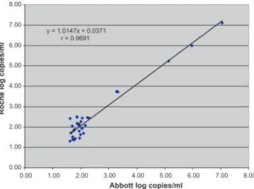
y = 1.0147x + 0.0371 r = 0.9691 0.00 1.00 2.00 3.00 4.00 5.00 6.00 7.00 8.00 8.00 7.00 6.00 5.00 4.00 3.00 2.00 1.00 0.00
Abbott log copies/ml
Roche log copies/ml
Fig. 1. Correlation of viral load results obtained using Abbott HIV-1 RealTime PCR
Please cite this article in press as: Paba, P., et al., Performance evaluation of the COBAS/TaqMan HIV-1 v2.0 in HIV-1 positive patients with low viral load: A comparative study. J. Virol. Methods (2011), doi:10.1016/j.jviromet.2011.03.014
ARTICLE IN PRESS
G ModelVIRMET 11493 1–4
P. Paba et al. / Journal of Virological Methods xxx (2011) xxx–xxx 3
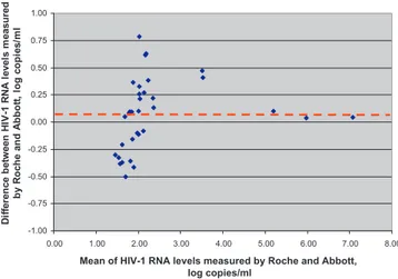
-1.00 -0.75 -0.50 -0.25 0.00 0.25 0.50 0.75 1.00 8.00 7.00 6.00 5.00 4.00 3.00 2.00 1.00 0.00
Mean of HIV-1 RNA levels measured by Roche and Abbott, log copies/ml D if fe re nc e be tw e e n H IV -1 R N A l e v e ls m e a s ur e d by R o c h e a n d A bbot t, l o g c o pi e s /m l
Fig. 2. Bland–Altman analysis of the samples tested by Abbott HIV-1 RealTime PCR and Cobas TaqMan HIV-1 v2.0.
subtype C, and 29 with subtype B. Based on the viral sub-types
124
distribution within the three groups of patients examined,
discor-125
dant results do not seem to be related to different viral subtypes
126
(Table 1).
127
Linear regression analysis carried out on the 33 samples
quan-128
tified by both systems showed a good correlation (r = 0.9691)
129
between the two assays as shown inFig. 1, although this result
130
could be biased by the low number of samples with high viremia
131
tested. Bland–Altman analysis showed a good agreement between
132
the two assays (Fig. 2).
133
Of the 17 discordant samples, negative by the Roche assay but
134
detected (<40 copies/ml) by the Abbott test, extra plasma of 9
sam-135
ples was available for further testing. The nested PCR targeting the
136
V3 region of the gp120 gene was negative on all samples tested,
137
confirming the result observed with the Roche assay.
138
4. Conclusions
139
Accurate determination of HIV-1 viral load is crucial for
eval-140
uating the response to treatment and the adherence to therapy.
141
Achieving low levels of viremia during antiretroviral treatment
pre-142
dicts a sustained virological response. For this reason, samples with
143
low viral load were selected and tested in parallel by two
commer-144
cial kits: the Cobas TaqMan HIV-1 v.2.0 and the Abbott RealTime
145
HIV-1 assay. Both assays are based on real-time PCR with a wide
146
dynamic range and high sensitivity. Overall, a good agreement was
147
found between the two test systems when considering the
sam-148
ples quantified within the dynamic range of both tests, although
149
this result is partly biased by the low number of samples with high
150
viremia tested. Bland–Altman analysis showed a good agreement
151
between the two methods. Of the samples tested, it is worth
not-152
ing that 30 samples not quantitated by the Abbott RealTime HIV-1
153
assay were quantitated by the Roche Cobas TaqMan HIV-1 v.2.0
154
(Table 1). The multiplex PCR approach of the Roche assay with the
155
amplification of two viral regions (LTR and gag gene) instead of
156
the amplification of one viral region (pol gene) as with Abbott may
157
explain this difference. The apparent under-quantitation of HIV-1
158
RNA levels observed in 14 samples (9 group I and 5 group II) with
159
the Abbott assay could also be attributed to the issue of primer
160
and probe binding polymorphisms as a result of genetic diversity
161
within different viral subtypes. The amplification of two viral
tar-162
gets in one reaction could increase the accuracy of the viral load
163
determination and the spectrum of quantifiable HIV-1 isolates.
164
Recent studies addressing the performance of the Roche and
165
Abbott HIV-1 real-time assays have detected an increasing number
166
of positive samples with viral load below the limit of quantitation 167
of both assays (Wirden et al., 2009; Sloma et al., 2009). Similar 168
results were reported in studies comparing the Cobas TaqMan HIV- 169
1 v.2.0 assay with the Cobas Amplicor assay (Lima et al., 2009; 170 Gatanaga et al., 2009) where several samples were detected but 171
not quantified by both assays. The clinical meaning of such findings 172
is still unclear. Some studies showed that these episodic low-level 173
viremias do not necessarily reflect the appearance of drug-resistant 174
strains or virological failure (Manavi, 2008; Smit et al., 2009), 175
while others hypothesized that these viremic blips are linked to 176
altered specimen-processing procedures (Rebeiro et al., 2008). Sim- 177
ilar results were obtained in this study where several samples were 178
below the limit of quantitation by both assays irrespective of the 179
viral subtype tested. 180
Finally, 17 samples positive by the Abbott assay but below the 181
limit of quantitation of the assay gave a negative result when tested 182
by Roche assay. This negative results were confirmed by an “in- 183
house nested” PCR when plasma was available for further testing. 184
Since all patients examined in this study were HIV-1 positive, arte- 185
facts due to cross-hybridization of primers/probe can be excluded. 186
It is conceivable that in some circumstances signal anomalies or 187
background noise may affect the lower-end precision of the Abbott 188
RealTime HIV-1 assay (Shain and Clemens, 2008). 189
In the light of these findings, virological follow-up should be 190
performed using the same assay and viremic blips should be inter- 191
preted with caution before changing therapy. A strict follow-up 192
may indicate whether there is a virological relapse or blip with no 193
impact on the ongoing antiviral therapy. Amplification of multiple 194
viral targets by real-time PCR represents an important step forward 195
for a virus with high genetic variability such as HIV-1. It should 196
improve the reliability and accuracy of the virological follow-up. 197
References 198
Bagchi, S., Kempf, M.C., Westfall, A.O., Maherya, A., Willing, J., Saag, M.S., 2007. Can 199
routine clinical markers be used longitudinally to monitor antiretroviral therapy 200
success in resource-limited settings? Clin. Infect. Dis. 44, 135–138. 201
Di Mascio, M., Markowitz, M., Louie, M., Hurley, A., Hogan, C., Simon, V., Follmann, D., 202
Ho, D.D., Perelson, A.S., 2004. Dynamics of intermittent viremia during highly 203
active antiretroviral therapy in patients who initiate therapy during chronic 204
versus acute and early human immunodeficiency virus type 1 infection. J. Virol. 205
78, 10566–10573. 206
Gatanaga, H., Tsukada, K., Honda, H., Tanuma, J., Yazaki, H., Watanabe, T., Honda, 207
M., Teruya, K., Kikuchi, Y., Oka, S., 2009. Detection of HIV TYPE 1 LOAD BY THE 208
Roche Cobas TaqMan assay in patients with viral loads previously undetectable 209
by Roche Cobas Amplicor Monitor. Clin. Infect. Dis. 48, 260–262. 210
Gross, R., Bilker, W.B., Friedman, H.M., Strom, B.L., 2001. Effect of adherence to newly 211
antiretroviral therapy on plasma viral load. AIDS 15, 2109–2117. 212
Lima, V., Harrigan, R., Montaner, J.S., 2009. Increased reporting of detectable plasma 213
HIV-1 RNA levels at critical threshold of 50 copies per millilitre with the Taqman 214
assay in comparison to the Amplicor assay. J. Acquir. Immune Defic. Syndr. 51, 215
3–6. 216
Manavi, K., 2008. The significance of low-level plasma HIV viral load on Cobas Taq- 217
Man HIV-1 assays for patients with undetectable plasma viral load on COBAS 218
Amplicor monitor version 1.5. HIV Clin. Trials 9, 283–286. 219
Mellors, J.W., Mu ˜noz, A., Giorgi, J.V., Margolick, J.B., Tassoni, C.J., Gupta, P., Kingsley, 220
L.A., Todd, J.A., Saah, A.J., Detels, R., Phair, J.P., Rinaldo Jr., C.R., 1997. Plasma viral 221
load and cd4+ lymphocytes markers of HIV-1 infection. Ann. Intern. Med. 126, 222
946–954. 223
Peeters, M., Aghokeng, A.F., Delaporte, E.E., 2010. Genetic diversity among human 224
immunodeficiency virus-1 non-B subtypes in viral load and drug resistance 225
assays. Clin. Microbiol. Infect. 16, 1525–1531. 226
Pereyra, F., Palmer, S., Miura, T., Block, L.B., Wiegand, A., Rothchild, A.C., Baker, B., 227
Rosenberg, R., Cutrel, E., Seaman, M.S., Coffin, J.M., Walker, B.D., 2009. Persistent 228
low-level viremia in HIV-1 elite controllers and relationship to immunologic 229
parameters. J. Infect. Dis. 200, 984–990. 230
Peter, J.B., Sevall, J.S., 2004. Molecular-based methods for quantifying HIV viral load. 231
AIDS Patients Care STDS 18, 75–79. 232
Rebeiro, P.F., Kheshti, A., Bebawy, S.S., Stinnette, S.E., Erden, H., Tang, Y., Sterling, T.R., 233
Raffanti, S.P., D’Aquila, R.T., 2008. Increased detectability of plasma HIV-1 RNA 234
after introduction of a new assay and altered specimen-processing procedures. 235
Clin. Infect. Dis. 47, 1354–1357. 236
Shain, E.B., Clemens, J.M., 2008. A new method for robust quantitative and qualitative 237
Please cite this article in press as: Paba, P., et al., Performance evaluation of the COBAS/TaqMan HIV-1 v2.0 in HIV-1 positive patients with low viral load: A comparative study. J. Virol. Methods (2011), doi:10.1016/j.jviromet.2011.03.014
ARTICLE IN PRESS
G ModelVIRMET 11493 1–4
4 P. Paba et al. / Journal of Virological Methods xxx (2011) xxx–xxx
Sloma, C.R., Germer, J.J., Gerards, T.M., Madrekar, J.N., Mitchell, P.S., Yaho, J.D., 2009. 239
Comparison of the Abbott realtime human immunodeficiency virus type 1
(HIV-240
1) assay to the Cobas Ampliprep/Cobas TaqMan HIV-1 test: workflow, reliability,
241
and direct costs. J. Clin. Microbiol. 47, 889–895.
242
Smit, E., Bhattachrya, S., Osman, H., Taylor, S., 2009. Increased frequency of HIV-1
243
viral load blip rate observed after switching from Roche Cobas Amplcor to Cobas
244
Taqman assay. J. Acquir. Immune Defic. Syndr. 51, 364–365.
Wirden, M., Tubiana, R., Marguet, F., Leroy, I., Simon, A., Bonmarchand, M., Ait- 245
Arkoub, Z., Murphy, R., Marcelin, A.G., Katlama, C., Calvez, V., 2009. Impact of 246
discrepancies between the Abbott realtime and cobas TaqMan assays for quan- 247
tification of human immunodeficiency virus type 1 group M non-B subtypes. J. 248