Durante il Dottorato di ricerca mi sono occupata dello studio della regolazione dell‘espressione genica in Neisseria menigitidis. In particolare la mia attenzione si è focalizzata sulla regolazione mediata da piccoli RNA non codificanti (sRNAs). Ho studiato il fenotipo di un mutante knock-out del gene dell‘RNA chaperone hfq e ho identificato e caratterizzato 2 nuovi sRNAs in meningococco e i loro rispettivi circuiti di regolazione. Ho inoltre identificato altri possibili nuovi sRNAs attraverso l‘analisi del trascrittoma di N. menignitidis tramite la tecnologia ―Illumina sequencing‖. Infine, ho anche caratterizzato il circuito di regolazione di un regolatore trascrizionale della famiglia AraC.
Nel periodo di Dottorato sono stata co-autrice dei seguenti lavori scientifici:
Metruccio M.M., Fantappiè L., Serruto D., Muzzi A., Roncarati D., Donati C., Scarlato V., Delany I. ― The Hfq-dependent small non-coding (s) RNA NrrF directly mediates Fur-dependent positive regulation of succinate dehydrogenase in Neisseria meningitidis.” J. Bacteriol. 2009 Feb;
191(4):1330-42. Epub 2008 Dec 5.
Fantappiè L., Metruccio M.M., Seib K., Oriente F., Cartocci E., Ferlicca F., Giuliani M., Scarlato V., Delany I. ― The RNA chaperone Hfq is involved in the stress response and virulence in Neisseria meningitidis and is a pleiotropic regulator of protein expression‖. Infect. Immun. 2009 May; 77(5):1842-53. Epub 2009 Feb17
Fantappiè L., Oriente F., Muzzi A., Serruto D., Scarlato V., Delany I. ―A novel Hfq-dependent sRNA that is under Fnr control and is synthetized in oxygen limitation in Neisseria meningitidis”. Mol Microbiol. 2011 Feb 21.
doi: 10.1111/j.1365-2958.2011.07592.x. [Epub ahead of print]
Fantappiè L., Scarlato V., Delany I. ―Identification ot the target of an iron-responsive AraC like protein from meningococcus that is in a regulatory cascade with Fur‖. (Submitted to Microbiology, 2011)
Table of Contents
Riassunto ... 7 Abstract ... 9 Introduction ... 12 1.1 Meningococcal disease ... 12 1.2 The pathogen ... 131.3 Colonization and invasion ... 14
1.4 Virulence factors ... 16
1.5 Adaption to the host environment ... 19
1.6 Small regulatory RNAs ... 21
1.7 The pleiotropic regulator Hfq ... 26
1.8 How to identify new sRNAs ... 31
Results ... 37
I Characterization of a hfq knock out in Neisseria meningitidis ... 37
1.9 The hfq locus of N. meningitidis ... 37
1.10 Expression of the Hfq protein and generation of an Hfq null mutant ... 39
1.11 Complementation of the Hfq mutant. ... 42
1.12 Hfq plays a major role in stress tolerance in N. meningitidis ... 44
1.13 Hfq contributes to the survival in ex vivo and in vivo models . 45 1.14 Identification of proteins differentially expressed in hfq mutant ... 49
1.15 Global analysis of gene expression in the Hfq null mutant ... 55
II Identification of AniS, a novel sRNA synthesied under oxygen limitation ... 59
1.16 NMB1205 is a small transcript deregulated in the Hfq mutant 59 1.17 The synthesis of the novel sRNA is regulated by FNR. ... 63
1.18 AniS negatively regulates NMB1468 and NMB0214 genes ... 66
1.19 Validation of the direct targeting of NMB1468 by AniS ... 71
1.20 AniS is a small RNA bound by Hfq ... 73
III Molecular mechanism of action of the sRNA NrrF ... 76
1.21 A bioinformatic approach identified a Fur-regulated sRNA in . the N. meningitidis MC58 genome ... 76
1.22 A base pairing mechanism mediated by Hfq is the molecular mechanism of action of NrrF ... 77
1.23 Fumarate hydratase and superoxide dismutase are regulated by at least a novel Hfq-dependent sRNA. ... 83
IV Global investigation of sRNAs ... 85
1.24 Solexa RNA sequence revealed 19 putative novel sRNAs in meningococcus ... 86
Discussion... 92
1.25 Characterization of a hfq knock out in Neisseria meningitidis 92 1.26 Identification of AniS, a novel sRNA synthesied under oxygen limitation ... 96
1.27 Molecular mechanism of action of the sRNA NrrF ... 101
1.28 Concluding remarks ... 104
Materials and methods ... 105
1.29 Bacterial strains and culture conditions ... 105
1.31 Construction of plasmids and recombinant strains ... 106
1.32 Expression and purification of the Hfq protein ... 109
1.33 In vitro cross-linking ... 110
1.34 Generation of anti-Hfq antiserum ... 111
1.35 Western blot analysis ... 111
1.36 Fractionation of proteins of N. meningitidis ... 112
1.37 Separation of total proteins by 2D gel electrophoresis ... 113
1.38 In-gel protein digestion and MALDI-TOF mass spectrometry analysis. ... 113
1.39 In vitro antimicrobial stress assays ... 114
1.40 Ex vivo human serum assay and ex vivo whole-blood model of meningococcal bacteremia. ... 115
1.41 In vivo animal model. ... 116
1.42 RNA extraction ... 116
1.43 Microarray procedures: design, cDNA labelling, hybridization, and data analysis. ... 117
1.44 Primer extension, S1 nuclease mapping, Northern blot and real time PCR. ... 119
1.45 cDNA library construction and Illumina sequencing ... 121
1.46 Read mapping and visualization ... 122
1.47 DNase I footprinting ... 123
1.48 Generation of in vitro transcripts ... 123
1.49 Electrophoretic mobility shift assays of in vitro transcription products. ... 125
Tables ... 127
RIASSUNTO
I piccoli RNA batterici (sRNAs) sono regolatori post-trascrizionali coinvolti nelle risposte a diversi stress. Questi piccoli trascritti non codificanti sono sintetizzati in risposta a un certo segnale e controllano l‘espressione genica dei loro reguloni modulando la traduzione o la stabilità di mRNA targets, spesso insieme all‘ RNA chaperone Hfq. In questa tesi si è caratterizzato in Neisseira meningitidis un mutante knock out per il gene
hfq, dimostrando che ha un fenotipo pleiotropico. Questo suggerisce un
importante ruolo della proteina Hfq nell‘adattamento agli stress e nella virulenza del batterio, nonchè la presenza di sRNAs dipendenti da Hfq. Inoltre, un‘analisi globale dell‘espressione genica, per identificare trascritti regolati nel mutante di Hfq, ha rivelato l‘esistenza di uno sRNA, impropriamente annotato come una open reading frame (ORF), che abbiamo chiamato AniS. Si è dimostrato che la sintesi di questo nuovo sRNA è indotta in condizioni di anaerobiosi, grazie all‘azione del regolatore trascrizionle FNR che ne attiva la trascrizione. Abbiamo inoltre identificato 2 possibili targets di AniS attraverso analisi dell‘espressione genica globale.
In questa tesi si è anche effettuata una dettagliata analisi molecolare dell‘azione dello sRNA NrrF, il primo ad essere stato identificato in N.
meningitidis. Si dimostra che NrrF regola la succinato deidrogenasi
formando un duplex con una zona di complementareità all‘interno della regione sdhDA del trascritto della succinato deidrogenasi. Hfq aumenta
l‘efficenza del legame tra i 2 RNA e questo probabilmente determina una rapida degradazione del trascritto in vivo. Inoltre,allo scopo di identificare altri possibili sRNAs in N. menigitidis abbiamo sequenziato il trascrittoma del batterio in condizioni standard di crescita in vitro e in condizioni di carenza di ferro. Questa analisi ha rivelato geni che sono attivamente trascritti nelle 2 condizioni. La nostra attenzione si è rivolta in particolare alle regioni non codificanti attivamente trascritte nel genoma. Questo ha permesso di identificare 19 possibili nuovi sRNAs e regioni 5‘ e 3‘ UTRs (regioni non tradotte). Ulteriori studi saranno focalizzati sull‘identificazione dei circuiti regolatori di questi sRNAs e sui loro targets.
ABSTRACT
Bacterial small regulatory RNAs (sRNAs) are posttranscriptional regulators involved in stress responses. These short non-coding transcripts are synthesised in response to a signal, and control gene expression of their regulons by modulating the translation or stability of the target mRNAs, often in concert with the RNA chaperone Hfq. Characterization of a Hfq knock out mutant in Neisseria meningitidis revealed that it has a pleiotropic phenotype, suggesting a major role for Hfq in adaptation to stresses and virulence and the presence of Hfq-dependent sRNA activity. Furthermore, global gene expression analysis of regulated transcripts in the Hfq mutant revealed the presence of a regulated sRNA, incorrectly annotated as an open reading frame, which we renamed AniS. We demonstrated that the synthesis of this novel sRNA is anaerobically induced through activation of its promoter by the FNR global regulator and through global gene expression analyses we identified at least two predicted mRNA targets of AniS.
In this thesis we also performed a detailed molecular analysis of the action of the sRNA NrrF, the first one identified in N. meningitidis. We demonstrated that NrrF regulates succinate dehydrogenase by forming a duplex with a region of complementarity within the sdhDA region of the succinate dehydrogenase transcript, and Hfq enhances the binding of this sRNA to the identified target in the sdhCDABmRNA; this is likely to result in rapid turnover of the transcriptin vivo.
In addition, in order to globally investigate other possible sRNAs of N.
both standard in vitro and iron-depleted conditions. This analysis revealed genes that were actively transcribed under the two conditions. We focused our attention on the transcribed non-coding regions of the genome and, along with 5‘ and 3‘ untranslated regions, 19 novel candidate sRNAs were identified. Further studies will be focused on the identification of the regulatory networks of these sRNAs, and their targets.
INTRODUCTION
1.1Meningococcal disease
N. meningitidis is a strictly human pathogen responsible for meningitis and
sepsis, two devastating diseases that can kill children and young adults within hours, despite the availability of effective antibiotics.
Studies performed in Europe (Caugant et al., 2007) have demonstrated that carriage rates are very low in the first few years of life, but sharply rise during adolescence, peaking at 10–35% in 20–24-year olds, before decreasing to less than 10% in older age groups (Caugant et al, 2007; Claus
et al., 2005). Compared with the carriage rate, meningococcal disease is
rare, and disease rates vary in different geographic regions of the world (Hill et al., 2010). What changes the colonization state of the organism into a disease state is not entirely clear. It appears that a combination of bacterial virulence factors and host susceptibility, including age, prior viral infection, smoking (Cartwright & Ala'Aldeen, 1997) and genetic polymorphisms (reviewed in Emonts et al., 2003) may ultimately lead to meningococcal disease. The human nasopharyngeal mucosa, in fact, is the only natural reservoir of N. meningitidis, however meningococcus can invade the pharyngeal mucosal epithelium and, in the absence of bactericidal serum activity, disseminate into the bloodstream, causing septicaemia. In a subset of cases, the bacteria can also cross the blood-brain barrier and infect the cerebrospinal fluid, causing meningitis.
In general, mortality occurs in up to 10% of patients with invasive meningococcal disease (Stephens, 2007). Mortality rates are dependent on the type and severity of invasive disease, and are greatest for fulminant
septicaemia (up to 55%) followed by meningitis with associated septicaemia (up to 25%), and lowest for meningitis without sepsis (generally <5%) (Brandtzaeg & van Deuren, 2005). However, patients who survive invasive meningococcal disease often live with a number of physical and mental sequelae, including amputation of limbs and digits, scarring of skin, speech impairment and seizures (Borg et al., 2009).
1.2 The pathogen
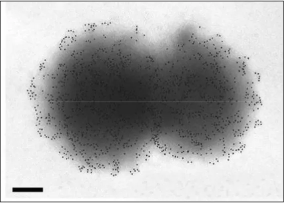
N. meningitidis is a Gram-negative diplococcus (Fig. 1). It is aerobic,
non-motile, non-sporulating, usually encapsulated and piliated. Traditionally different N. meningitidis strains are classified into serogroups according to the immunological reactivity of their capsule polysaccharides. With this method 13 different serogroups have been identified, but only A, B, C, Y, X and W135 commonly cause invasive infections. Meningococci are further classified into serotype and serosubtype, based on antigenic differences in their major outer membrane proteins (OMPs), PorA and PorB. The serological classification system, however, is limited due to high frequency of phase and antigenic variation of outer-membrane structures, which has led to the development of DNA-based approaches to characterize meningococcal strains. The most important of these methods is multilocus sequence typing (MLST), which characterizes isolates on the basis of the nucleotide sequences of internal fragments of seven housekeeping genes defining their sequence type (ST) (Maiden et al., 1998). Menigococci can in this way be classified into lineages, termed clonal complexes (cc). A clonal complex is a group of STs that share at least four of the seven loci in common with a central ancestral genotype. Despite huge diversity in
meningococcal population, only a minority of these clonal complexes are associated with invasive disease, known as hyperinvasive lineages (Maiden, 2008). Why hyperinvasive meningococcal lineages are more pathogenic than others remain still unknown.
Figure 1: ImmunoGold labelling and transmission electron microscopy of N. meningitidis. Analysis of the strain was performed with antisera raised against NadA adhesin (Scale bars: 200 nm.) (Pizza et al., 2000)
1.3 Colonization and invasion
Colonization of the upper respiratory mucosal surfaces by N. meningitidis is the first step in the establishment of a human carrier state and invasive meningococcal disease (Stephens, 2009). Initial contact with nasopharyngeal epithelial cells is mediated by Type IV pili. Then, meningococci proceed to proliferate on the surface of human non-ciliated epithelial cells, forming small microcolonies at the site of initial attachment
(Stephens, 2009). After the initial colonization, there is a loss or down regulation of the capsule, which sterically masks the outer membrane proteins. This event is thought to occur both via cell contact induced repression (Deghmane et al., 2002), and by selection of low or no-capsule expressing bacteria due to phase variation (Hammerschmidt et al., 1996). Close adherence of meningococci to the host epithelial cells is mediated by a variety of possible redundant adhesins, previously masked by the capsule. This results in the appearance of cortical plaques and the recruitment of factors leading to the formation and extension of epithelial cell pseudopodia that internalize the bacteria (Stephens, 2009). Once internalized in the epithelial cells meningococcus can evade the host immune response, find more available nutrients and can also cross the epithelium and enter the bloodstream (Stephens, 2009). Intracellular survival is determined by factors including IgA1 protease, which degrades lysosome-associated membrane proteins, and upregulation of expression of capsule, which is anti-opsonic and anti-phagocitic and therefore aids survival in blood (Stephens, 2009; (Virji, 2009)). (Fig 2). The next steps of meningococcal invasion of the bloodstream and the passage across the brain vascular endothelium, which results in infection of the meninges and the cerebrospinal fluid, are still poorly understood.
Virji, 2009 Figure 2: Stages in the pathogenesis of N. meningitidis. N. meningitidis may be acquired through the inhalation of respiratory droplets. The organism establishes intimate contact with non-ciliated mucosal epithelial cells of the upper respiratory tract, where it may enter the cells briefly before migrating back to the apical surfaces of the cells for transmission to a new host. Asymptomatic carriage is common in healthy adults in which bacteria that enter the body by crossing the epithelial barrier are eliminated. Besides transcytosis, N. meningitidis can cross the epithelium either directly following damage to the monolayer integrity or through phagocytes in a ‗Trojan horse‘ manner. In susceptible individuals, once inside the blood, N. meningitidis may survive, multiply rapidly and disseminate throughout the body and the brain. Meningococcal passage across the brain vascular endothelium (or the epithelium of the choroid plexus) may then occur, resulting in infection of the meninges and the cerebrospinal fluid (Nassif, 1999).
1.4 Virulence factors
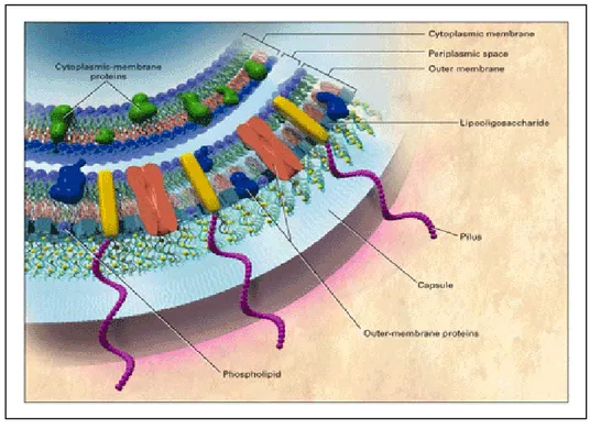
The major virulence factor (Fig 3) of meningococcus is the polysaccharide capsule that protects the bacterium during airborne transmission between hosts (Romero & Outschoorn, 1997), confers protection from effectors of the innate immunity (Vogel et al., 1997), allows survival in blood, as mentioned before, and may shield bacterial surface from the host immune
effectors mechanisms (Virji, 2009). The second principle virulence factor, mainly involved in the interface between the host and the bacterium, are the pili, long surface proteins that protrude from the capsule.
Figure 3: Virulence factors in meningococcal cell membranes (from Rosenstein et al., 2001)
Together with the outer membrane adhesins, pili facilitate adhesion to host tissues having a crucial role in the initial establishment of encapsulated bacteria on mucosal surfaces, helping the penetration of the negatively charged barrier at the host-pathogen interface (Heckels et al., 1976). In addition to adhesion, pili, are involved in several other functions, for example facilitating uptake of foreign DNA from the extracellular environment, increasing transformation frequency of bacteria and maintaining the genetic diversity that supports the success of Neisseria in the human host (Helaine et al., 2007). The pilus of meningococcus is expressed from the pilE locus, but homologous recombination between the
pilE gene and a number of non-expressed ‗silent‘ pilS genes results in a
change in the pilE sequence. These variants differ in their transformability, adherence and immunogenicity (Virji et al., 1992). Additionally, N.
meninigitidis spp. possess host specific iron acquisition systems and
numerous immune evasion mechanisms such as factor H binding protein that is able to down-regulate complement deposition (Virji, 2009). Close adhesion and invasion is mainly mediated by an array of proteins such as opacity proteins (Opc, Opa) and other adhesins. These proteins are believed to be responsible for the host specificity as well as for tissues within the host (Virji, 2009). Numerous additional apparently minor adhesins (several of which were identified by homology searching of the available genomes) are generally expressed at low levels during in vitro growth but may be important in in vivo infections. Their expression can in fact be regulated at different levels in the different environment the bacterium encounter during its infection. It has been reported, for example that in conditions that mimic the host infection such as iron (Delany et al., 2006) and oxygen limitation (Bartolini et al., 2006) or interaction with epithelial cells (Dietrich et al., 2003) and blood (Echenique et al, in press) the transcriptome of N.
meningitidis is considerably altered and, as a result, some virulence factors
may be over-expressed. Furthermore, several adhesins are subject to antigenic variation and/or phase variation, which allow bacteria to generate a broad and variable repertoire of surface structure that facilitates evasion of immune effectors mechanisms and adaptation to different niche (Virji, 2009).
1.5 Adaptation to the host environment
N. meningitdis, during its infection, is subjected to constant selective
pressures and its ability to adapt rapidly to environmental changes is essential for its survival (Hill et al., 2010). Phase and antigenic variation of a number of surface components permits immune evasion during infection, but since the bacterium can infect diverse sites within the human host, which represent a unique niche with respect to nutrients, environmental factors and competing microorganisms, it has to rapidly change its metabolism and cellular composition to adapt to different environments. Much of this adaptation is carried out at the transcriptional level. Different transcriptional regulators, activated by different stresses encountered during infection, regulate the transcription of many genes important for survival and virulence. However, only few transcriptional regulators are found in the pathogenic Neisseria (menigitidis and gonorrhoeae): 35 putative regulators in N. meningitidis MC58 strain, compared to Escherichia coli, which harbours more than 200 transcriptional regulators. This reveals a striking limitation for transcriptional regulation, which is possibly related to the restricted ecological niche of the Neisseriacae (Schielke et al., 2010). The mechanisms of adaptation of N. meningitidis to two different conditions it encounters during infection are described below.
Iron limitation
It has been very well established that bacterial pathogenesis and survival are dependent on the ability to acquire iron within the host (Andrews et al., 2003; DeVoe, 1982). Cell growth and multiplication, in fact, require essential nutrients such as iron, which is limiting in the human host being sequestrated by human iron proteins. Although N. meningitidis does not
produce siderophores for iron acquisition, it possesses outer membrane receptors that have been postulated to scavenge the iron-loaded siderophores secreted by other bacteria colonizing the nasopharyngeal tract (Carson et al., 1999). Once inside the host, the organism must compete for iron with host iron proteins, and meningococcus possesses receptors for transferrin, lactoferrin, and hemoglobin (Perkins-Balding et al., 2004). However, iron overload results in toxicity for the bacterium; therefore, iron uptake is tightly regulated and in menigococcus, as in many bacteria, this regulation is mediated by the ferric uptake regulator (Fur) protein (Delany
et al., 2003; Escolar et al., 1999). Fur senses internal iron concentration and
binds to and represses iron uptake genes using ferrous iron as a co-repressor (Delany et al, 2003). Fur has been also reported to act positively in the expression of certain genes, both with a direct mechanism (binding upstream promoter sequences (Delany et al., 2004) and an indirect mechanism which involves a posttranscriptional regulation mediated by a Fur repressed small regulatory RNA named NrrF (Mellin et al., 2007; Metruccio et al., 2009). The regulatory circuit of NrrF is a part of this thesis work.
Oxygen limitation
During its infection N. meningitidis encounters numerous complex extracellular and intracellular environments, since it moves from the upper respiratory tract, to mucous membranes, blood and CSF (Archibald & Duong, 1986), being exposed to highly divergent partial pressures of oxygen (high in the upper respiratory tract and low in the mucus membranes and in the blood (Archibald & Duong, 1986). It has been shown that although N. meningitidis fails to grow under strictly anaerobic
conditions, under oxygen limitation the bacterium expresses a denitrification pathway system that supplement growth (Anjum et al., 2002; Rock & Moir, 2005). Pathogenic Neisseria use FNR as a global transcription factor, to control these responses under oxygen limitation, in particular to induce the denitrification and sugar fermentation pathways (Bartolini et al, 2006) as an alternative to aerobic respiration. FNR is only active as a dimer containing [4Fe-4S] cluster. The cluster dissociates in the presence of oxygen, destabilizing the dimer, with loss of FNR activity (Kiley & Beinert, 2003). Only a total of 9 transcriptional units have been identified as being responsive to the FNR regulator in N. meningitidis (Bartolini et al, 2006). Interestingly factor H binding protein (fHBP,) which enables the bacterium to evade complement-mediated killing by binding factor H (Madico et al., 2006; Schneider et al., 2006), and which is a component of the MenB vaccine currently in development, has been shown to be positively regulated by oxygen limitation through a FNR dedicated promoter (Oriente et al., 2010). This result, together with the observation that a knock-out of FNR in N. menigitidis is attenuated in the mouse and infant rat animal models (Bartolini et al, 2006), indicate the importance of these responses for the pathogenesis and the survival of meningococcus in the human host. The identification and analysis of a novel sRNA induced under oxygen limitation by FNR is a part of this thesis work.
1.6 Small regulatory RNAs
Small regulatory RNAs (sRNAs) are crucial regulatory elements in bacterial stress responses and virulence (reviewed in: (Gottesman, 2004), (Waters & Storz, 2009), (Papenfort & Vogel, 2010). These regulators
mostly function as coordinators of adaptation processes in response to environmental changes, integrating environmental signals and controlling target gene expression, primarily at posttranscriptional levels (Wassarman, 2002), Gottesman, 2004). Regulatory RNAs can modulate transcription, translation, mRNA stability and DNA maintenance or silencing (Waters & Storz, 2009). They can act using diverse mechanisms, including changes in RNA conformation, protein binding, base pairing with others RNAs and interaction with DNA. There are different classes of sRNAs classified according to their mechanism of action in bacterial cells. One class of sRNA comprises riboswitches. They are sequences at the 5‘ end of mRNAs they regulate that can adopt different conformations in response to environmental changes, (Grundy & Henkin, 2006) regulating in this way the expression of the coding sequence. Another class of sRNAs can bind to proteins, including global regulators, and antagonize their function (Pichon & Felden, 2007). A recently discovered group of RNA regulators, named CRISPR (clustered regulatory interspaced short palindromic repeats) provide resistance to bacteriophage (Sorek et al., 2008) and prevent plasmid conjugation (Marraffini & Sontheimer, 2008) . CRISPR RNAs contain short regions of homology to bacteriophage and plasmid sequences and can interfere with bacteriophage infection and plasmid conjugation, most likely by targeting the homologous foreign DNA. But the largest and most studied group of sRNA, which is also analyzed in this thesis, acts through base pairing with mRNAs, modulating their translation and stability. This class of regulatory RNAs has two distinct broad classes: the cis enconed sRNAs (Fig 4A), that have a perfect complementarity with their targets and the
of partial complementarity and may have many distinct mRNA targets. The binding of sRNAs to target mRNAs is very specific: a single base substitution in the sRNA or target mRNA can be sufficient to disrupt duplex formation. Although sRNAs binding is very specific, each sRNAs can act on more than one target mRNA and each target mRNA can be regulated by multiple sRNAs ( reviewed in (Repoila & Darfeuille, 2009). RNA base-pairing interactions are usually in the 5‘ UTR of the target mRNA and have been shown to alter mRNA structure ultimately leading to changes in translation efficiency and, as a consequence, mRNA stability (Wassarman, 2002, Waters & Storz, 2009). It has recently been shown that sRNAs can also interact with coding regions, regulating their targets not by translational control, but by accelerating RNase E-dependent mRNA targets decay (Pfeiffer et al., 2009). The majority of the regulation by the known trans-encoded sRNAs is negative (Aiba, 2007; Gottesman, 2004); base pairing with the target mRNA usually leads to repression of protein levels through translational inhibition, mRNA degradation or both (Sharma et al., 2007; Vecerek et al., 2007; Morita et al., 2006). However sRNAs can also activate expression of their target mRNAs. Base pairing of the sRNA disrupt an inhibitory secondary structure, which sequesters the ribosome binding site (Hammer & Bassler, 2007; Urban & Vogel, 2008; Prevost et
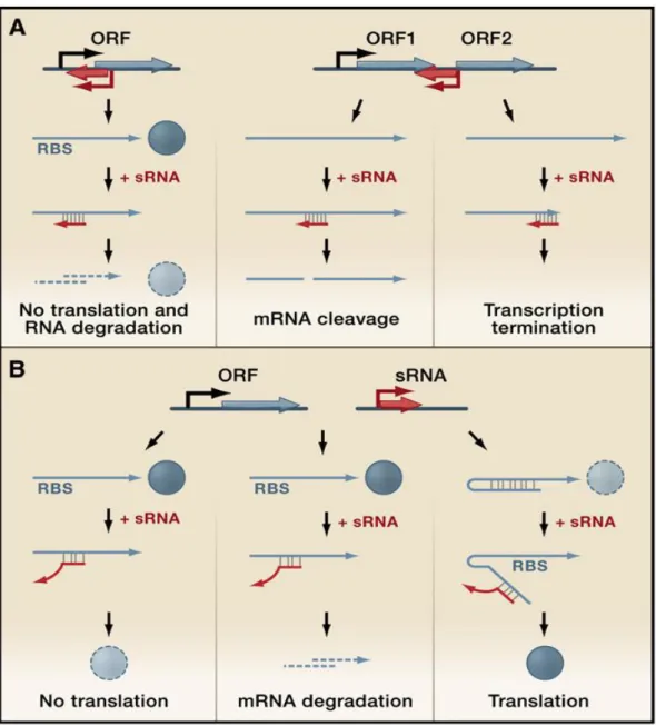
Figure 4 Gene regulation mediated by regulatory RNAs which act by base-pairing (Waters & Storz, 2009)
(A) Two possible configurations of cis-encoded antisense sRNAs (red) and their target RNAs (blue), which share extensive complementarity. (Left panel). A sRNA encoded opposite to the 5‘UTR of its target mRNA. Base pairing inhibits ribosome binding and often leads to target mRNA degradation. (Right panels) A sRNA encoded opposite to the sequence separating two genes in an operon. Base pairing of the sRNA can target RNases to the region and cause mRNA cleavage, with various regulatory effects, or the sRNA can cause transcriptional termination, leading to reduced levels of downstream genes.
(B) Genes encoding trans-encoded antisense sRNAs (red) are located separately from the genes encoding their target RNAs (blue) and only have limited complementarity. Trans-encoded sRNA can act negatively by base pairing with the 5‘UTR and blocking ribosome binding (left panel) and/or targeting the sRNA-mRNA duplex for degradation by RNases (middle panel). Trans-encoded sRNA can act positively by preventing the formation of an inhibitory structure, which sequesters the ribosome-binding site (RBS) (right panel).
However two recent papers suggest that in Clostridium and Streptococcus the VR-RNA and FasX sRNAs, exert positive regulation of virulence genes primarily at the level of mRNA stabilization (Obana et al., 2010; Ramirez-Pena et al., 2010) and reviewed in (Podkaminski & Vogel, 2010). Those sRNAs for which a function has been elucidated are involved in many different cellular processes including, adaptation and resistance to stresses (Repoila et al., 2003), metabolism and homeostasis (Bejerano-Sagie & Xavier, 2007; Masse et al., 2007), control of the expression of outer membrane proteins (Vogel & Papenfort, 2006), virulence and pathogenesis (Johansson & Cossart, 2003; Romby et al., 2006; Toledo-Arana et al., 2007). The trans-acting antisense class of sRNAs often require the RNA chaperone Hfq as a cofactor, which facilitates the interaction between sRNAs and target mRNAs (Valentin-Hansen et al., 2004; Aiba, 2007; Chao & Vogel, 2010) (Fig 5). The binding of Hfq can affect the structure of sRNA and target mRNAs (Moller et al., 2002; Zhang et al., 2002; (Geissmann & Touati, 2004) and Hfq has been demonstrated to accelerate strand exchange to facilitate dynamic RNA-RNA interactions (Arluison et
al., 2007); furthermore Hfq protects many sRNAs from degradation, most
likely by binding to RNase E cleavage sites within these sRNAs (Moll et
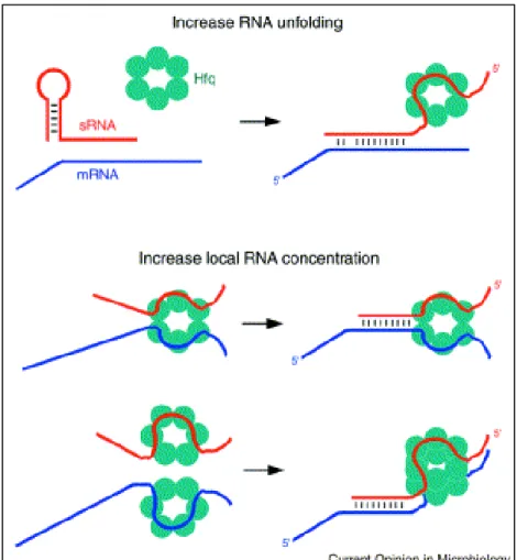
Figure 5: Mechanisms by which Hfq might facilitate sRNA-mRNA basepairing. (Storz et al., 2004)
Hfq (aqua ring) may promote RNA unfolding or may increase the local concentrations of the sRNA (red) and its mRNA target (blue).
1.7 The pleiotropic regulator Hfq
Hfq is a hexameric RNA binding protein which was originally identified in
E. coli as a host factor required for the replication of Qbacteriophage (Kajitani & Ishihama, 1991). It shares structural and functional homology with the Sm proteins in eukaryotes, which have central roles in RNA metabolism (Pannone & Wolin, 2000). It has more recently been described as a pleiotropic regulator that modulates the stability or translation of an
increasing number of mRNAs (Valentin-Hansen et al, 2004; Aiba, 2007; (Brennan & Link, 2007) and facilitates pairing of sRNAs with their target mRNAs (Moller et al., 2002; Kawamoto et al., 2006); Zhang et al., 2002). Available structural data, the latest showing E. coli Hfq bound to polyriboadenilate RNA (Link et al., 2009), suggest that Hfq acts as an RNA chaperone by binding sRNAs and mRNAs (Fig 6).
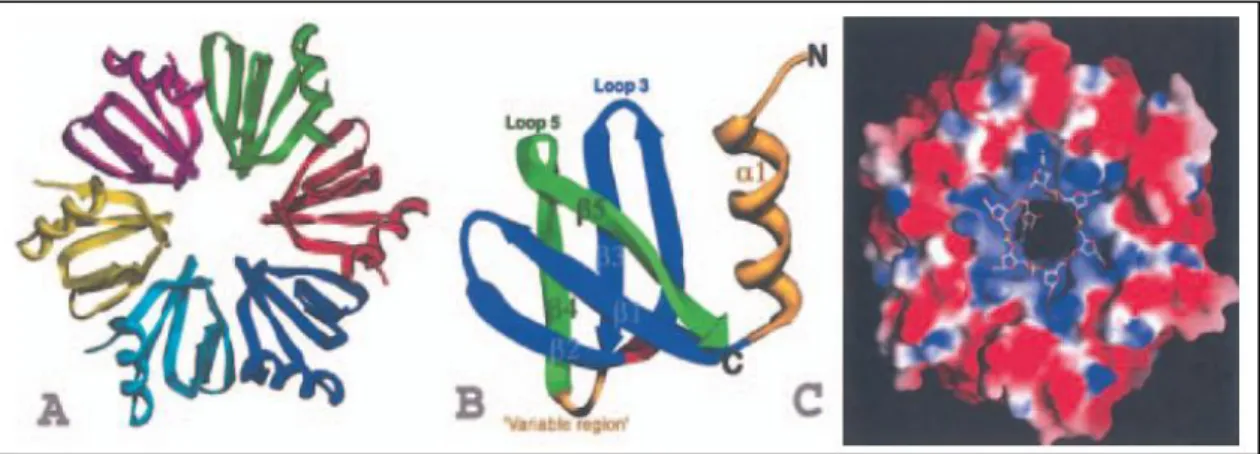
Figure 6. Structures of the S. aureus Hfq and an Hfq–RNA complex. (Valentin-Hansen et al, 2004)
(A) Structure of the Hfq hexamer with each subunit coloured differently.
(B) Ribbon diagram of an Hfq subunit. The Sm1 motif is coloured blue and the Sm2 motif is green. Regions outside the two motifs, i.e. the N-terminal -helix and the variable region, are colored yellow.
(C) Electrostatic surface representation of the RNA binding site of the Hfq hexamer. Blue is electropositive and red is electronegative. The RNA is shown as a stick model, with oxygen, nitrogen, carbon and phosphorus atoms colored red, blue, white and yellow respectively.
It has been shown that Hfq binds with greater avidity to RNA sites containing a short-single stranded stretch of uridines and adenosines (Brennan & Link, 2007; Brescia et al., 2003; Mikulecky et al., 2004; Sun &
Wartell, 2006). The Hfq protein is conserved in a wide range of bacteria and varies in length from 70 to 100 amino acids (Valentin-Hansen et al, 2004) (Fig 7). In all cases, the Sm motif is located in the N-terminal region of the molecule. The C-terminal domain seems not to play a significant role in the major functions of Hfq. In fact, a C-terminal truncated form of the E.
coli Hfq lacking the last 27 amino acids can replace all tested functions of
the intact E. coli Hfq (102 amino acid residues) (Sonnleitner et al., 2002). To date, Hfq candidates have been identified in about half the completed or nearly completed bacterial genomes. Two distinct copies are found in few species, including Bacillus anthracis and Ralstonia sp., and tandem Hfq sequences are present within a single 193-residue protein from
Novosphingobium aromaticivorans (Sun et al., 2002). Hfq can also
modulate the decay of some mRNAs (e.g. rpsO, which encodes for ribosomal protein S15) by binding to their poly(A) tails, stimulating poly(A) adenylation by poly(A) polymerase I (PAP I) and protecting this message from polynucleotide phosphorylase (PNP), RNase II and RNase E, enzymes involved in mRNA degradation (Hajnsdorf & Regnier, 2000); (Mohanty et al., 2004; Folichon et al., 2005, Folichon et al., 2003). It has been shown that Hfq can also protect many sRNAs from degradation, most likely by binding to RNase E cleavage sites within these sRNAs (Moll et al., 2003). However, recently it has been demonstrated that in Rhodobacter
sphaeroides the sRNA RSs0019 is stabilized in the absence of Hfq
(Berghoff et al., 2009) and also in this thesis we provide evidences for a sRNA being more stable in hfq of meningococcus. Based on the literature the contribution of Hfq to sRNA stability is controversial. While it has been shown that the binding of this chaperone, in the absence of basepairing,
stabilizes sRNAs protecting them form RNAseE cleaveage (Moll et al., 2003, Masse et al., 2003); it has also been shown that Hfq-mediated pairing of sRNAs to their targets promotes coupled degradation of the interacting RNAs (Masse et al., 2003). So it could be possible that binding of Hfq to a particular sRNA can accelerate its turnover.
Considering the role of Hfq as a pleiotropic regulator in bacteria it is not surprisingly that the inactivation of the hfq gene results in sensitivity to a wide number of environmental stresses and alters the synthesis of many proteins, including outer membrane porins (Muffler et al., 1997; Sittka et
al., 2007; Tsui et al., 1994; Valentin-Hansen et al., 2007). Hfq also
influences the fitness and virulence of many pathogenic bacteria. Mutants lacking Hfq are often sensitive to host defence mechanisms and highly attenuated in animal models (Ding et al., 2004; Kulesus et al., 2008; McNealy et al., 2005; Sittka et al, 2007; Pichon & Felden, 2005; Sonnleitner et al., 2003). Interestingly, in N. menigitidis by signature-tagged mutagenesis hfq was identified as one of 73 genomic loci that attenuated N. menigitidis virulence in rats (Sun et al., 2000). In this thesis we show that in N. meningitidis Hfq plays a major role in adaptation to stresses and virulence and by a transcriptome study we identified 152 Hfq-regulated genes which suggest the presence of Hfq-dependent sRNAs circuits in this bacterium. Indeed the meningococcal Hfq protein was shown to interact specifically with many sRNAs when expressed in the heterologous system Salmonella typhimurium (Sittka et al., 2009) suggesting that the meningococcal protein has a conserved major function as a sRNA-binding protein. To date, virulence phenotypes of hfq mutants are most dramatic in negative pathogens. The role of Hfq in
Gram-positive species is still under investigation. It has been reported, in fact, that
hfq strains of L. moncytogens and S. aureus have wild type sRNA profiles
(Christiansen et al., 2004; Bohn et al., 2007), but the conservation of key aminoacids (Valentin-Hansen et al, 2004) and the proven role in antisense regulation (Nielsen et al., 2010) suggest the functional conservation of Hfq in such species. We can speculate that transcriptional regulation by sRNAs can occur by both Hfq-dependent and independent mechanisms. It is not still understood if Hfq-independent trans-acting sRNAs can act on their own (depending on the free energy for sRNA-mRNA pairing interaction (Jousselin et al., 2009), or if they depend on other factors that substitute the Hfq protein.
Figure 7: The Hfq family (Sauter et al., 2003)
The organisms corresponding to the sequences are indicated on the left. Conserved polar, basic and acidic residues appear in green, pink and violet, respectively, Gly and Pro in yellow, and a star indicates those involved in RNA binding in S.aureus (Schumacher et al., 2002). Blue boxes are conserved patches of hydrophobic residues. The 2D structure prediction from Jpred is indicated at the bottom as well as the 2D features seen in 3D structures.
1.8 How to identify new sRNAs
It is very well established that sRNAs play critical regulatory roles in many bacterial processes (reviewed in (Zhou & Xie, 2011; Beisel & Storz, 2010; Liu & Camilli, 2010; Toledo-Arana et al., 2007; Repoila et al., 2003). However, unlike regulatory proteins, non-coding regulatory sRNAs have not been readily identified and annotated in the bacterial genome sequences that are available in the databases, and in many systems, including
Neisseria spp., their existence is still largely unexplored. For this reason
experimental strategies have become increasingly important for sRNAs discovery. The first sRNAs were discovered about four decades ago fortuitously using genetic screens, or through radiolabeling of total RNA and subsequent isolation from gels (Wassarman et al., 1999). But it was only very recently that many new sRNAs have been identified and characterized in a wide range of bacterial species. This was mainly possible thanks to the increased availability of novel technologies such as computational predictions of sRNAs, tiling arrays and high-throughput cDNA sequencing (RNA-seq) that are used to study sRNAs at the genome-wide level (Sharma & Vogel, 2009). The last two techniques not only allow the identification of novel sRNAs, but also the analysis of the whole transcriptome of bacteria in different growth conditions. The high density (tiling) arrays carry up to hundreds of thousands of DNA oligonucleotides systematically covering the sense and antisense strand of a genome, including the intergenic regions (IGRs) from which most known sRNAs are expressed. An important issue in this kind of technique, as well as the RNA-seq, is choosing physiological conditions in which sRNAs are
expressed. Genomic tiling arrays have been successfully used to study the transcriptome of Listeria monocytogenes (Toledo-Arana et al., 2009),
Bacillus subtilis (Rasmussen et al., 2009) and Mycoplasma pneumoniae
(Guell et al., 2009), as well as specific genomic features in E. coli (Selinger
et al., 2000) and Caulobacter crescentus (McGrath et al., 2007). However,
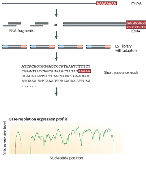
the array-based approach requires hundreds of thousands probes and is limited by background noise and cross hybridization, and therefore requires extensive normalization (Sorek & Cossart, 2010). In contrast to microarray methods, RNA-seq directly determine the cDNA sequence. This technique uses recently developed deep-sequencing technologies. In general, a population of RNA is converted to a library of cDNA fragments with adaptors attached to one or both ends. Each molecule, with or without amplification, is then sequenced in a high-throughput manner to obtain short sequences. The reads are typically 30–400 bp, depending on the DNAsequencing technology used. To generate a transcriptome map, these reads are computationally mapped to the reference genome (Fig 8).
Figure 8: A typical RNA-seq experiment. (Wang et al., 2009)
Briefly, long RNAs are first converted into a library of cDNA fragments through either RNA fragmentation or DNA fragmentation. Sequencing adaptors (blue) are subsequently added to each cDNA fragment and a short sequence is obtained from each cDNA using high-throughput sequencing technology. The resulting sequence reads are aligned with the reference genome or transcriptome and used to generate a base-resolution expression profile. In principle, any high-throughput sequencing technology can be used for RNA-Seq (Wang et al, 2009), and the Illumina, Applied Biosystems SOLiD and Roche 454 Life Science systems have already been applied for this purpose in bacteria (Passalacqua et al., 2009; Perkins et al., 2009; Sittka
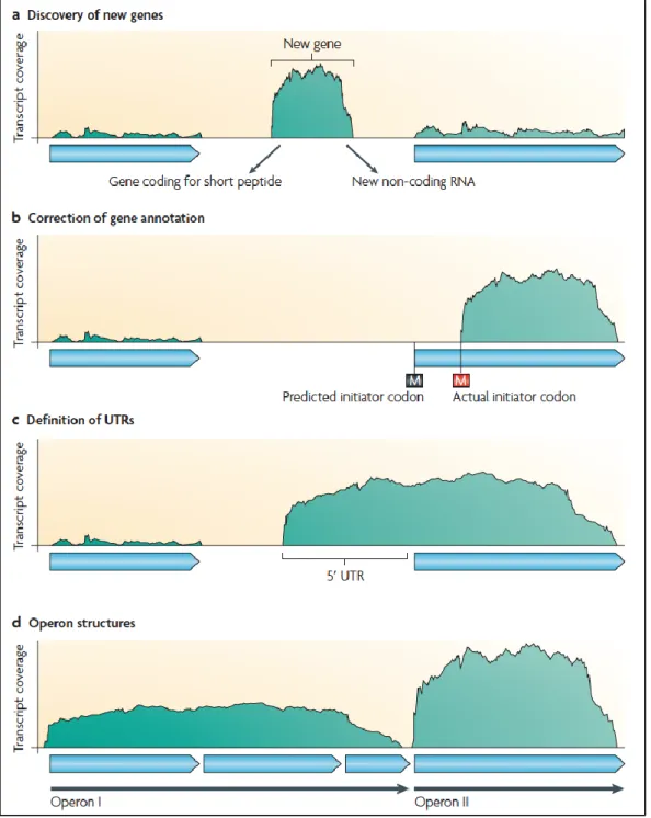
advantage of RNA-Seq is that it has very low, if any, background signal because DNA sequences can been unambiguously mapped to unique regions of the genome. A transcriptome analysis can be used to improve genome annotation by enabling: the discovery of new genes, the correction of gene annotation; the detection of UTRs and transcription start sites and the determination of operon relationship (Sorek and Cossart, 2009 and Fig 9). Furthermore, whole-transcriptome analysis now allows the global interrogation of sRNA abundance and antisense RNAs in any species primarily by detecting expression from non-protein-coding regions. In this way, for example, 13 sRNAs that were induced during niche switching in the opportunistic pathogen B. cenocepacia were recently discovered (Yoder-Himes et al, 2009). Similarly, Perkins et al. detected 55 intergenic regions that are likely to encode new sRNAs in Salmonella Typhi Ty2 (Perkins et
al, 2009), and the number of known sRNAs in L. monocytogenes has been
more than doubled to 50 sRNAs by a tiling arraybased study (Toledo-Arana
et al., 2009). Interestingly, sRNAs can also be enriched by
co-immuoprecipitation with the Hfq protein (Zhang et al., 2003; Stikka et al, 2008) and subsequently sequenced. In conclusion, with whole-transcriptome analysis it is now possible to study the involvement of elements such as sRNAs, riboswitches and cis-antisense regulators in the physiology and pathogenicity of any prokaryote.
Figure 9: Different applications of RNA-seq (Sorek & Cossart, 2010)
The x axis represents a schematic genomic region and the y axis represents transcript coverage as derived from RNA-seq. The light blue arrows depict annotated genes. Transcriptomic information can be used to improve genome annotation by enabling: the discovery of new genes or sRNAs (a); the correction of gene structure annotations (b); the black M depicts the predicted first methionine and the red M is the first methionine in the corrected annotation); the detection of UTRs and transcription start sites (c); and the determination of operon relationships (d).
RESULTS
I.
Characterization of a hfq knock out mutant in Neisseria
meningitidis
1.9 The hfq locus of N. meningitidis
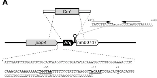
In N. meingitidis, the hfq gene (NMB0748) is predicted to encodea protein protein of 97 amino acids and shows 65% identity with the Hfq protein from E. coli. In the MC58 genome hfq is flanked upstream by a 147-bp
intergenic region possibly containing its promoter and is followed downstream by a putativetranscriptional terminator (Fig. 10A). This is in contrast toE. coli and other similar enterobacteria, in which hfq appearsto be located in a cluster of genes which form an operon, usually transcriptionally linked to the upstream gene miaA (Tsui et al., 1994). We mapped the promoter of the hfq gene of N. meningitidis inthe intergenic region directly upstream by performing S1 nuclease protection analysis. Total RNA was isolated from the MC58 strainduring in vitro growth and then hybridized with a hfq-specific probe and digested with S1 nuclease. Figure 10B shows an RNA-protectedband which corresponds to the 5' end of the hfq mRNA, whichmaps the transcriptional initiation site (+1) to 49 nucleotides upstream of the ATG start site of the gene. Analysis of the nucleotide sequence upstream revealed –10 (TACAAT) and –35 (TGGATA) hexamers, suggesting that the hfq gene is transcribed from a
70
promoter. The intensity of this band doesnot vary significantly in RNA prepared from various time pointsin the growth curve, suggesting that the
hfq promoter is transcribed constitutively throughout the various growth phases.
Figure 10: The hfq locus of N. meningitidis. (A) Diagram of the hfq locus in the genome of N. meningitidis strain MC58, showing the strategy used to construct the hfq null mutant by allelic replacement with a chloramphenicol cassette. The genes flanking the hfq gene in meningococcus are an upstream gene encoding a putative D-alanine-endopeptidase and a downstream gene encoding a conserved hypothetical protein with homology to RNA methylases. These genes are different from genes in other negative and Gram-positive bacteria in which a similar genetic organization has often been observed. In the sequence of the intergenic region upstream of the hfq gene underlined bold type indicates -10 and -35 hexamers and the transcriptional start site at position +1, as shown in panel B.
The sequence of the putative rho-independent transcriptional terminator and its position with respect to the initiation of transcription at position +1 are also shown. (B) Mapping of the hfq promoter by an S1 nuclease protection assay. The DNA probe was radioactively labeled at one end, hybridized to 15 g of total meningococcal RNA (lanes T1, T2, T3, T5, and T7), and digested with S1 nuclease for mapping of the 5‘ end of the hfq transcript. Total RNA was prepared after 1, 2, 3, 5, and 7 h of growth in GC broth (lanes T1, T2, T3, T5, and T7, respectively). Two control samples with E. coli tRNA instead of total RNA were processed in parallel with (lane +) and without (lane -) addition of S1 nuclease. The position of the RNA-specific S1 nuclease-protected band corresponding to the 5‘ end of the hfq transcript is indicated. Lane G+A contained a G-A sequence reaction mixture for the DNA probe used as a size marker. The nucleotide sequence of the coding strand upstream of the transcriptional start site is shown on the left.
1.10 Expression of the meningococcal Hfq protein and
generation of an Hfq null mutant
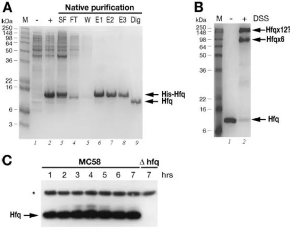
In order to investigate the expression of the Hfq protein in theMC58 strain, we purified the Hfq protein and raised antibodiesto Hfq in mice. We cloned the hfq gene into an expression vector,expressed it as a recombinant His-tagged protein in E. coli, and then purified it by Ni-NTA affinity chromatography. Figure11A shows the results of an SDS-PAGE analysis of various fractionsobtained for the expression and purification steps. The Hfq protein was highly expressed in the soluble fraction (Fig. 11A,lane 3) and gave rise to a highly pure recombinant His-Hfq proteinpreparation (lanes 6 to 8), and the N-terminal His tag was cleaved and removed after purification, giving rise to an untagged Hfqprotein (lane 9). Using in vitro cross-linking, we investigated whether this protein oligomerized into its functional hexamericform. As Fig. 11B shows, after treatment for 1 h with the DSScross-linker, two high-molecular-weight oligomers were visibleon the gel. One of the major cross-linked forms migrated ata molecular weight
of approximately 70,000, which is consistent with the hexameric form of the protein, and the other more slowlymigrating form may represent two cross-linked hexamers, suggestingthat the recombinant Hfq protein forms hexamers in solution.This protein preparation was used to raise antibodies againstmeningococcal Hfq in mice, and the resulting antiserum was tested in an immunoblot analysis with total protein samples taken from time course cultures of the MC58 strain and an Hfq null mutantstrain.
Figure 11 : (A) Expression and purification of the recombinant Hfq protein. SDS-PAGE was performed with protein extracts from uninduced (lane -) and IPTG-induced (lane +) cultures of E. coli containing the pET15bHfq expression plasmid, the cleared soluble fraction before (lane SF) and after (lane FT) binding to the Ni-NTA resin, the wash fraction (lane W), elution fractions (lanes E1 to E3), and untagged Hfq protein after thrombin digestion (lane Dig). (B) In vitro cross-linking of Hfq reveals hexameric forms in solution. For in vitro cross-linking, the untagged protein was not treated (lane -) or treated (lane +) with the cross-linking reagent DSS. The relative positions of the molecular weight markers (lane M) are indicated on the left. The positions of the oligomeric forms of the Hfq protein are indicated on the right. (C) Western blot showing the expression of the Hfq protein in
wild type strain MC58 or the hfq mutant over time. Total protein samples were taken at the time points shown in Fig. 12A. Five micrograms of total protein was loaded for each time point. Anti-Hfq antiserum recognizes a band at approximately 11 kDa in the wild type strain but not in the Hfq null mutant. Antiserum against NMB2091 was also used to stain the blot as a loading control for total protein, as indicated by the asterisk.
The null mutant of MC58 was generated by replacing the hfq genewith a chloramphenicol cassette, as indicated in Fig. 10A. Thismutant, when it is grown on solid medium, has an obvious growthphenotype in that it forms small pinpoint colonies after overnight incubation. Furthermore, after approximately 2 to 3 days ofgrowth on solid medium, the centers of the colonies have a concavemorphology, and, where there is confluent growth on the plate,the culture appears to be more flat and acquires a silver sheen. We grew the wild type and null mutant in liquid GC medium and took samples every hour over the time course. Consistent with the growth phenotype on plates, the null mutant exhibited asignificantly altered growth curve compared with the wild type in liquid medium (Fig. 12A). In particular, it had an increased lag phase and did not reach a cell density equivalent to thatof the wild type. Western blot analysis of the expression of Hfq throughout the time course (Fig. 11C) revealed that the anti-Hfq antiserum recognizes a protein band migrating to a positioncorresponding to approximately 11 kDa in the wild type, which is absent from the null mutant strain. Furthermore, the proteinis expressed during all phases of the growth curve, which is consistent with results shown in Fig. 10B, and no significant differences in the level of expression were detected, except possibly for slight accumulation of the protein in late log and stationary phases.
1.11 Complementation of the Hfq mutant.
In order to investigate downstream polar effects due to thehfq mutation, we
performed RT-PCR analysis of the NMB0747 gene using RNA prepared from the wild type and the Hfq knockout mutant.The results showed that there was a 10-fold reduction in RNAlevels for NMB0747, indicating that readthrough of the putativerho-independent terminator occurs and that it is likely that hfq is coexpressed with the downstream gene. In order to
determineif the phenotypes of the mutant were directly related to thelack of expression of the Hfq protein or possible polar effects, we generated a complemented mutant strain expressing a single copy of the hfq gene in
trans and related this expression to possible restoration of the wild type phenotype. In this strain, hfq_C, the expression of the hfq gene was inducibleby addition of IPTG, as its transcription is under the controlof the Ptac promoter and the LacI repressor. This strain was grown in liquid cultures in the presence of increasing amountsof IPTG, along with the wild type and mutant strains, and the growth curves are shown in Fig. 12A. Samples were collected atmid-log phase, and total protein extracts of each strain wereused to monitor Hfq expression by Western blotting (Fig. 12B). The generation time of the mutant was significantly less (78± 10 min) than that of the wild type (48 ± 7 min), while the generation times of the complemented mutant were shorterin cultures with IPTG-induced increases in Hfq expression. Itis worth noting that the levels of Hfq expression in the complementingstrain at the time of maximal induction were approximately two-to threefold less than the wild type levels, and this may explain the incomplete restoration of the growth phenotype. These resultsindicate that
the growth defect of the hfq mutant can be restoredby expression of Hfq in a dose-dependent manner.
Figure 12 : (A) Growth curves for the wild type, the hfq mutant, and the hfq_C complemented mutant in GC medium supplemented with different concentrations of IPTG (in M, as indicated after the mutant designation). Three independent sets of cultures were grown on different days, and the symbols indicate the average of the three cultures for each strain; the error bars show the standard deviations. The calculated generation times of the cultures were as follows: MC58, 48 ±7 min; hfq, 78 ±10 min; and hfq_C with 0, 1, 10, 100, and 1,000 _M IPTG, 80 ± 5, 80 ± 5, 70 ± 17, 55 ± 8, and 51 ± 10 min, respectively. (B) Western blotting of cell lysates of the MC58 wild type strain, the hfq mutant, and the complemented mutant, showing Hfq expression. IPTG was added at the final concentrations indicated to the growth medium for induction of Hfq in the complemented mutant. Cells were grown in GC broth to an OD600 of 0.5, and 10 g of total protein was added to each lane.
1.12 Hfq plays a major role in stress tolerance in N.
meningitidis.
As Hfq is reported to be involved in stress tolerance in many pathogens (Christiansen et al., 2004; Robertson & Roop, 1999; Schiano et al., 2010; Meibom et al., 2009) and a large number of proteins are differentially expressed in the hfq mutant, we tested the ability of the meningococcal
hfq mutant to resist several environmental stresses. A series of growth assays and killing assays were performed with the wild type, the hfq mutant, and the hfq_C complemented mutant strain in the presence of different antimicrobial compounds. The hfq mutant was significantly more sensitive than the wild typestrain to three membrane-perturbing detergents (the nonionic detergents Triton X-100 and Tween, as well as the anionic detergentSDS), and the results of a representative viability assay areshown in Fig. 13A. Likewise, the lack of expression of the hfq gene resulted in increased sensitivity of the null mutant strainto killing by oxidative stress, as investigated using paraquat (which generates superoxide anion radicals inside the cell),xanthine-xanthine oxidase (which generates superoxide and H2O2outside the cell), and H2O2 assays (Fig. 14B). We also challengedthe strains with 4.5 M NaCl in an osmotic stress assay. Again,the hfq mutant was significantly more sensitive than the strainsexpressing the Hfq protein (Fig. 13C). Finally, we tested whetherthe meningococcal mutant was more sensitive to antimicrobial peptides, such as polymyxin B or LL-37. Antimicrobial peptidesare an important part of the innate immune response, which canattack the outer membrane, forming holes and thereby causing misfolding of the periplasmic proteins and envelope stress. The results
shown in Fig. 13C indicate that Hfq contributes considerablyto resistance to antimicrobial peptides in meningococcus. In all assays performed, the complemented mutant exhibited resistancepatterns very similar to those of the wild type (Fig. 13A to C),confirming that inactivation of Hfq results in considerable sensitivity of the mutant strain to these antimicrobial compounds. The wild type, the mutant, and the complemented mutant all behavedsimilarly when they were incubated for the duration of the assayin GC broth, indicating that the differences in survival arenot due to intrinsic growth defects (data not shown). Based on all these analyses, the Hfq protein plays an important rolein the resistance of meningococcus to a wide range of stresses, many of which may be particularly physiologically relevant tothe infectious cycle.
1.13 Hfq contributes to the survival of meningococcus in ex
vivo and in vivo models
To examine how the strains responded to conditions comparableto those in the host, their survival in ex vivo whole-blood and serum assays was assessed. The human blood assay was used to assess both cellular and humoral mechanisms of killing (including the action of complement, antibody-mediated serum bactericidalactivity, and opsonophagocytosis, as well as killing by neutrophils, macrophages and antimicrobial peptides), while the serum assay was used to assess killing of N. meningitidis mediated by thehumoral immune response. The Hfq mutant was less able to survivein human blood and exhibited serum sensitivity (Fig. 14A and B). Once the serum was heat inactivated, the sensitivity of thehfq mutant was
lost, and the number of CFU did not decreaseover the time course of the assay, which implies that the hfq mutant is sensitive to a heat-labile component of the complement pathway. For in vivo infection studies, an isogenic hfq mutant of theadapted 2996 strain was generated for use in the infant ratmodel. Groups of six mice were inoculated intraperitoneallywith either a high (105 CFU) or low (103 CFU) dose of the wildtype or the hfq mutant strain. After 18 h, the animals werebled, and bacteria were counted by colony plating. Compared to the counts for the wild type strain, the bacterial counts for the hfq mutant were significantly decreased (Fig. 14C). Althoughwe cannot exclude the possibility that the growth phenotype may contribute to the reduced survival in the rat, these resultssuggest that Hfq plays a role in the pathogenesis of meningococcus in ex vivo human models and in the in vivo infant rat model.
Figure 13: Assays of the viability of wild type strain MC58, the hfq mutant, and the
hfq_C complemented mutant. Cells were grown to an OD600 of 0.5 in GC broth (1 mM IPTG was added for expression of Hfq in the complemented mutant), diluted, and incubated with the antimicrobial compounds indicated. Samples were taken at 15, 30, and/or 60 min, and viable cell counts were determined by plating. The final concentrations of compounds used were as follows: Tween, 0.05%; SDS, 0.01%; Triton X-100, 0.1%; hydrogen peroxide, 5 mM; xanthine oxidase, 4.3 mM xanthine and 300 mU xanthine oxidase; paraquat, 5 mM; NaCl, 4.5 M; LL-37, 2 M; and polymyxin B, 10 g/ml.
Figure 14: Survival of strain MC58, the hfq mutant, and the hfq_C complemented mutant in ex vivo whole blood (A) and serum (B) assays and an in vivo infant rat model (C). There was a reduced level of bacteremia with the hfq mutant in an infant rat infection model. Groups of animals were inoculated with two initial doses (approximately 103 and 105 CFU; specific levels of the initial inoculum were determined in each case and are indicated on the x axis), and bacterial counts for the wild type (diamonds) and the hfq mutant (triangles) recovered from the blood of each animal were determined after an 18-h period. The P values for comparisons of the levels of the wild type and the hfq mutant recovered with initial doses of 103 and 105 CFU are 0.0007 and 0.0057, respectively
1.14 Identification of proteins differentially expressed in the
hfq mutant
To obtain a preliminary picture of global changes in the expressionprofiles of proteins altered in the hfq mutant, we comparedthe whole-cell protein patterns in SDS-PAGE gels for culturesamples obtained at mid-log phase (Fig. 15). The lack of Hfqin the mutant results in global changes in protein expression,and many protein bands are more abundant or less abundant in the Hfq-deficient strain. Furthermore, the expression profileof the mutant is restored to a profile similar to that of thewild type after restoration of Hfq expression in the hfq_C strainby IPTG addition. In order to identify some of the proteins differentially expresseddue to deletion of the Hfq protein, total protein from logarithmically grown cultures of wild type and hfq mutant cultures were separated by one-dimensional (1D) or 2D electrophoresis. Furthermore,cell cultures were also fractionated to obtain secreted, cytoplasmic, and envelope fractions and separated by 1D SDS-PAGE. Representativegels for triplicate samples are shown in Fig. 15, and the most altered protein spots and bands that were identified by mass spectrometry are shown in Fig. 15B and C, respectively. Thesespots and bands were excised and digested with trypsin, and the resulting peptides were analyzed by MALDI-TOF mass spectrometry.It is worth noting that a greater number of high-molecular-weightproteins appeared to be released into the supernatants of thehfq mutant cultures than into the supernatant of
the wild typestrain cultures, although many of these proteins could not be identified. Using mass spectrometry analysis following 1D and 2D separation, we were able to identify 27 proteins that showed altered
accumulation in the hfq mutant, and results of theseanalyses are shown in Table 1. Twenty of the proteins appear to be upregulated in the hfq mutant, and seven proteins aredownregulated. The functions of the proteins whose abundance is altered can be subdivided into various groups, including energy metabolism, amino acid biosynthesis, oxidative stress responses, and outer membrane proteins (OMPs), many of which are associated with pathogenicity.
Figure 15 : Identification of differentially abundant proteins in the hfq knockout mutant. (A) SDS-PAGE of cell lysates of the MC58 wild type strain, the hfq mutant, and the complemented mutant, showing pleiotropic effects of Hfq on protein expression in the hfq mutant. The arrows on the right indicate the positions of derepressed protein bands, and the
filled circles indicate downregulated protein bands in the hfq null mutant. (B) Total proteins of logarithmically grown wild type strain MC58 and the hfq mutant were separated by 2D gel electrophoresis. Proteins were first focused on a nonlinear pH 3 to 10 gradient and then separated on a 9 to 16.5% SDS-polyacrylamide gel. Cells were grown to an OD600 of 0.5 in GC broth. (C) 1D SDS-PAGE analysis of total proteins (Total) or cytoplasmic (Cyto) or envelope (Env1 and Env2) fractions of cells of the wild type (lane +) and the Hfq mutant (lane -) grown to log phase in GC broth. Both 1D and 2D gels were stained with Coomassie brilliant blue R-250. In addition, wild type and Hfq mutant cultures were grown to an OD600 of 0.2 in Mueller-Hinton medium containing 0.25% glucose, and 22 ml of cell-free supernatant was precipitated and loaded (Supr)