letters to the editor
609
Nefrologia 2013;33(4):601-22
disease. A subset of Grover´s disease. J Cutan Pathol 1996;23(2):151-64. 5. Frampton JE, Plosker GL. Icodextrin: a
review of its use in peritoneal dialysis. Drugs 2003;63(19):2079-105.
Elías Jatem1, Irene Agraz1, M. Eugenia Semidei2, Berta Ferrer2, Rosa Ramos1, Joan Fort1
1Servicio de Nefrología. Hospital Universitari
Vall d’Hebron. Barcelona. (Spain).
2Servicio de Anatomía Patológica. Hospital
Universitari Vall d’Hebron. Barcelona. (Spain).
Correspondence: Elías Jatem
Servicio de Nefrología. Hospital Universitari Vall d’Hebron, Idumea. 08035 Barcelona. (Spain).
[email protected] [email protected]
Acute renal failure
induced by acute
interstitial nephritis
secondary to cocaine
Nefrologia 2013;33(4):609-11
doi:10.3265/Nefrología.pre2013.Feb.11809 To the Editor:Cocaine has been used by 2.6% of the Spanish population aged between 15 and 64 at some point in their life, mak-ing it one of the most consumed illegal drugs after cannabis.1Cocaine use is as-sociated with multiple complications: neurological, cardiovascular, psychi-atric, pulmonary, gastrointestinal and nephrological.
Renal complications associated with cocaine use have received little atten-tion, despite the existence of several mechanisms, in addition to secondary high blood pressure, that can cause acute renal failure (ARF) or worsen a pre-existing case of chronic renal failure.2
Drug-induced acute interstitial nephri-tis (DIAIN) represents a high percent-age of acute renal failure in clinical
atitis A, B, and C and mycoplasma did not detect active infection. The ultra-sound showed normal-sized, diffusely echogenic kidneys with appropriate ar-terial and venous flow.
The electrocardiogram was normal. The chest x-ray showed a cardiothoracic ra-tio <0.5 and lung fields without infil-trates.
After admission, urinary output re-mained at 50 to 75ml/h and creatinine remained unchanged. The patient un-derwent a renal biopsy.
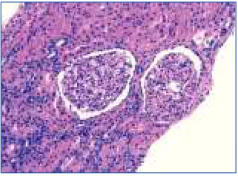
Histological findings are as follows: optical microscopy showed a total of 13 glomeruli, all normal, without sclerosis, proliferation or necrotic lesions (Figure 1). Basement membranes and the glomerular mesangium were normal. The interstitium displayed moderate mononuclear inflammatory infiltrate with abundant eosinophils (Figure 2), with presence of focal tubulitis and at-rophy (Figure 2). The arterioles did not display remarkable lesions and immune deposits were not shown in the im-munofluorescence.
The findings were compatible with the pathological diagnosis of acute tubu-lointerstitial nephritis (ATN).
This fact, along with the clinical char-acteristics and recent use of cocaine led us to define this case as cocaine-in-duced AIN.
The patient obviously suspended drug use and was treated with oral pred-nisone (initial dose 1mg/kg/day), which was progressively decreased and dis-continued after 12 weeks.
In the subsequent follow-up, his pro-gression was good with a gradual im-provement in renal function until com-plete recovery in the month in which treatment started.
DISCUSSION
We report the case of a patient with ARF, with acute tubulointerstitial lesion associated with DIAIN, in which no re-practice. Some studies indicate that
DI-AIN is the lesion responsible for renal failure in about 15% of biopsies with ARF. Furthermore, in many cases of DIAIN, no biopsy is performed and di-agnosis is based on clinical data and re-cent administration of a new drug which, as described below, is some-times not very easy to identify.3-5
CASE REPORT
28-year-old male, admitted with pain at the dimples of Venus, fatigue and nau-sea, with preserved diuresis.
The patient had used intranasal cocaine (1g) five days before admission. He de-nied having taken non-steroidal anti-in-flammatory drugs or other medication. The physical examination showed a good general condition, with slightly high blood pressure of 147/97mmHg and without fever, rash or arthralgia. Cardiovascular and respiratory exami-nations were normal. The abdomen was soft, depressible and painless and the liver was palpable 1cm below the costal margin and there was slight pain on bi-lateral palpation of lower back. The initial blood test showed an unre-markable complete blood count (with-out eosinophilia), normal liver func-tion and albumin within the normal range, serum creatinine: 160µmol/l, urea: 7.5mmol/l, potassium: 3.9mmol/l, sodium: 139mmol/l chlo-ride 101mmol/l. Total creatine phos-phokinase was normal (3.3µkat/l) with normal MB fraction. Urine sediment showed 2 leukocytes and 3 erythro-cytes per high power field and no dys-morphic erythrocytes or eosinophils. Urine biochemistry: sodium: 46mmol/l, potassium: 33mmol/l and chloride: 63mmol/l, protein ratio: cre-atinine 5g/mol, negative urine culture. Protein electrophoresis, immunoglobu-lins, complement, levels of angiotensin converting enzyme and antinuclear an-tibody titres were normal. Serology for human immunodeficiency virus, Ep-stein-Barr virus, cytomegalovirus,
letters to the editor
610 Nefrologia 2013;33(4):601-22
The pathogenesis of AIN involves an idiosyncratic allergic reaction to drug exposure. It often involves a type IV hypersensitivity T cell response. Mole-cular mimicry or direct binding of the drug to the tubular basement membrane are the main mechanisms involved,9 and this maybe the underlying process in our case.
Early recognition of DIAIN is crucial because patients may ultimately devel-op chronic kidney disease.
The key element of treatment is the in-terruption of the causative agent. How-ever, as DIAIN is an inflammatory al-lergic process, it is necessary to consider the use of immunosuppressive agents, including corticosteroids.10 In corticoresistant AIN, there are re-ports of cases that suggest the benefit of cyclophosphamide and cyclosporine, as well as potential beneficial effects of mycophenolate mofetil.11
Growing evidence based on different studies suggests that steroids lead to a quicker and more complete recovery of renal function.4
As a consequence of interstitial infiltra-tion typical of AIN, a rapid progression towards interstitial fibrosis can occur in a few weeks. Based on these data, we used corticosteroids at the time of diag-lated agent was identified, except for
cocaine. Currently people are starting to become aware of cocaine-induced ARF in adults; in fact, the two most common causes are rhabdomyolysis and malignant high blood pressure in-duced by intense arterial vasocon-striction.
There are few reported cases of co-caine-related DIAIN.2,6 The mecha-nism remains unclear, and it remains to be demonstrated whether this enti-ty is related to cocaine per se or natu-ral impurities, adulterants or dilu-ents.7 In fact, in the case of crack, contamination is highly likely. In this case the patient may have been sensitised to cocaine or its additives by previous consumption. Hypersen-sitivity to the drug is the most likely cause in our patient.7,8
Our patient did not have any “classic” symptoms of ARF, such as fever, rash, or eosinophilia, but recent studies suggest that AIN is a heterogeneous disorder and, therefore, these “clas-sic” symptoms are only seen in fewer than 30% of cases.8Eosinophiluria is usually interpreted as a feature of DI-AIN; however, it has very low sensi-tivity (67%). The eosinophiluria specificity for the diagnosis of AIN is 87%, but it may be present in other diseases that may also present with acute renal failure.6
nosis to prevent potential progression to irreversible interstitial fibrosis. The re-sult was positive and displayed rapid normalisation of renal function. DIAIN should be recognised as a po-tential cause of acute renal failure in cocaine users and the history of poten-tial use should be carefully investigat-ed in patients with AIN with no obvi-ous cause.
Conflicts of interest
The authors declare that they have no conflicts of interest related to the con-tents of this article.
1. Encuesta domiciliaria sobre alcohol y drogas en España (EDA-DES), 2007-2008. Delegación del Gobierno para el Plan Nacional sobre Drogas. Madrid: Ministerio de Sanidad y Consumo; 2008. 2. Wojciechowski D, Kallakury B, Nouri P. A
case of cocaine-induced acute interstitial nephritis. Am J Kidney Dis 2008;52(4):792-5.
3. Haas M, Spargo BH, Wit EJ, Meehan SM. Etiologies and outcome of acute renal insufficiency in older adults: a renal biopsy study of 259 cases. Am J Kidney Dis 2000;35:433-47.
4. Perazella MA, Markowitz GS. Drug-induced acute interstitial nephritis. Nat Rev Nephrol 2010;6(8):461-70.
5. Praga M, González E. Acute interstitial nephritis. Kidney Int 2010;77(11):956-61.
6. Gitman MD, Singhal PC. Cocaine-induced renal disease. Expert Opin Drug Saf 2004;3(5):441-8.
7. Baker RJ, Pusey CD. The changing prolife of acute tubulointerstitial nephritis. Nephrol Dial Transplant 2004;19(1):8-11. 8. Bomback AS, Markowitz GS. Increased prevalence of acute interstitial nephritis: more disease or simply more detection? Nephrol Dial Transplant 2013;28(1):16-8. 9. Decelle L, Cosyns JP, Georges B, Jaoul M, Lefebvre C. Acute interstitial nephritis after cocaine sniffing. Clin Nephrol 2007;67(2):105-8.
10. González E, Gutiérrez E, Galeano C, Chevia C, de Sequera P, Bernis C, et al. Early steroid treatment improves the recovery of renal function in patients with drug-induced acute interstitial
Figure 1. Renal biopsy.
Renal biopsy displaying normal glomeruli with marked inflammatory interstitial infiltrate (haematoxylin-eosin; original magnification x 200).
Figure 2. Renal biopsy.
Mononuclear inflammatory interstitial infiltrate, infiltrating the epithelium of some tubules, along with oedema and abundant focal eosinophils
(haematoxylin-eosin; original magnification x 200).
letters to the editor
611
Nefrologia 2013;33(4):601-22
and no data for anaemia. He was trans-ferred to Nephrology where a radi-ograph and ultrasound (Figure 2) were performed with normal results. The pa-tient began fluid therapy. He showed progressive worsening of renal function (creatinine of 10.49mg/dl on the fifth day) and oliguria; he did not display acute haemodialysis criteria. Renal function gradually improved until crea-tinine levels of 2.23mg/dl were record-ed on the eighth day, in the polyuric phase; haemoglobin at admission was 14.9g/dl and at discharge it was 12g/dl, with creatinine of 1.52mg/dl. The diag-nosis was acute renal failure, acute tu-bular necrosis in remission and haemo-globinuria. After 10 days in the outpatient service, he had normal renal function and sediment.
DISCUSSION
Haemoglobinuria secondary to extreme exercise was observed in 1881 as a re-sult of microtrauma due to strenuous marching of soldiers, and was known as “march haemoglobinuria”. In 1964, Davidson1 demonstrated that it volved transient extracorpuscular in-travascular haemolysis due to erythro-cyte microtraumas on their passage through the capillaries with the in-travascular passage of haemoglobin fol-lowing erythrocyte lysis;2this process saturates haptoglobin, which causes free haemoglobin to be filtered by the glomeruli, with subsequent haemoglo-binuria; filtered haemoglobin dimers are absorbed by tubular cells and bro-ken down; iron is stored as haemosiderin and it is excreted in chronic forms, presenting haemosider-inuria. This symptom occurs, in most cases, without anaemia expression.3 In-travascular haemolysis is associated with the reduction of serum haptoglo-bin, sometimes to undetectable levels. However, it is not a specific indicator of intravascular haemolysis, as it may be low or absent with rapid onset extravas-cular haemolysis; moreover, it is an acute phase reactant. Accordingly, it may be normal in the presence of in-flammation or infection.4When haemo-globin is broken down in tubules, haem
nephritis. Kidney Int 2008;73(8):940-6. 11. Preddie DC, Markowitz GS, Radhakrishnan
J, Nickolas TL, D’Agati VD, Schwimmer JA, et al. Mycophenolate Mofetil for the Treatment of Interstitial Nephritis. Clin J Am Soc Nephrol 2006;1:718-22.
Rosana Gelpi1, Omar Taco1, Montse Gomà2, Joan Torras1, Rafael Poveda1, Teresa Álvarez 2, Josep M. Grinyó1, Xavier Fulladosa1 1 Servicio de Nefrología. Hospital Universitari
de Bellvitge. IDIBELL. Barcelona. (Spain).
2Servicio de Anatomía Patológica. Hospital
Universitari de Bellvitge. IDIBELL. Barcelona. (Spain).
Correspondence: Rosana Gelpi
Servicio de Nefrología. Hospital Universitari de Bellvitge. IDIBELL. C/ Feixa Llarga s/n. 08907 L'Hospitalet. Barcelona. (Spain). [email protected]
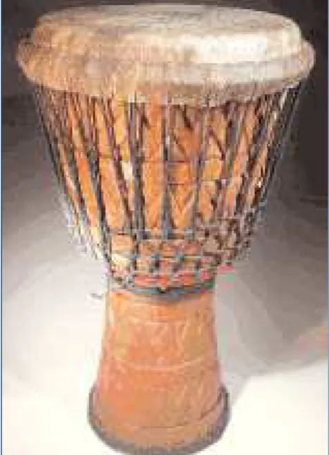
Energetic beating of
the dyembe (African
drum) as a cause of
acute renal failure
Nefrologia 2013;33(4):611-2
doi:10.3265/Nefrologia.pre2012.Dec.11844
To the Editor:
A 29-year-old male came to the Emer-gency department with abdominal pain with a 48 hour progression and diar-rhoea without pathological characteris-tics. He informed us that he had been energetically playing African drums (dyembe, Figure 1) in the last 3 days, in sessions lasting 9 hours each, with little fluid intake, hyporexia and subsequent reddish urine. He always experiences these symptoms whenever he plays the drum. Physical exam was unremark-able: athletic body, constants within normal range. He had very positive dip-stick results for blood, with urine sedi-ment negative for erythrocytes and pro-teinuria of 150mg/dl, creatinine of 4.45mg/dl, urea 80mg/dl, creatine phosphokinase (CPK) of 255U/l, lac-tate dehydrogenase of 509U/l, compen-sated metabolic acidosis, normal ions
pigments are released. These can cause renal damage due to tubular obstruc-tion, direct lesion or vasoconstriction at a spinal level, with predisposing factors such as volume depletion, acidosis and ischaemia.5
The shear force required to produce the aforementioned erythrocyte lysis is 3000dyn/cm; in vivo it occurs with peak tangential forces6of 6000dyn/cm. Many cases have been published relat-ed to physical activity in the lower limbs, and only 3 cases in the upper limbs: in 1974 in the United States, there was a case of a young man who had positive pigmenturia with myoglo-bin and haemoglomyoglo-bin after a percussion session;7 in 2006, in Uruguay, a case was described of 26 individuals after playing drums on a national holiday, and in 2011, a Caucasian man had haemoglobinuria after playing drums, which led to the term “percussion haemoglobinuria”.8
We present our clinical experience of haemoglobinuria secondary to exces-sive percussion of African dyembe drums. We rule out haematuria as the cause of the brownish-red urine, be-cause the urine sediment was negative
Figure 1. African dyembe drum