RESEARCH ARTICLE
SUBJECT COLLECTION: MODEL SYSTEMS IN DRUG DISCOVERY
A calixpyrrole derivative acts as an antagonist to GPER,
a G-protein coupled receptor: mechanisms and models
Rosamaria Lappano
1,*, Camillo Rosano
2,*, Assunta Pisano
1, Maria Francesca Santolla
1,
Ernestina Marianna De Francesco
1, Paola De Marco
1, Vincenza Dolce
1, Marco Ponassi
2, Lamberto Felli
2,
Grazia Cafeo
3, Franz Heinrich Kohnke
3, Sergio Abonante
4and Marcello Maggiolini
1,‡ABSTRACT
Estrogens regulate numerous pathophysiological processes, mainly
by binding to and activating estrogen receptor (ER)
α and ERβ.
Increasing amounts of evidence have recently demonstrated that
G-protein coupled receptor 30 (GPR30; also known as GPER) is also
involved in diverse biological responses to estrogens both in normal
and cancer cells. The classical ER and GPER share several features,
including the ability to bind to identical compounds; nevertheless,
some ligands exhibit opposed activity through these receptors. It is
worth noting that, owing to the availability of selective agonists and
antagonists of GPER for research, certain differential roles elicited by
GPER compared with ER have been identified. Here, we provide
evidence on the molecular mechanisms through which a calixpyrrole
derivative acts as a GPER antagonist in different model systems,
such as breast tumor cells and cancer-associated fibroblasts (CAFs)
obtained from breast cancer patients. Our data might open new
perspectives toward the development of a further class of selective
GPER ligands in order to better dissect the role exerted by this
receptor in different pathophysiological conditions. Moreover,
calixpyrrole derivatives could be considered in future anticancer
strategies targeting GPER in cancer cells.
KEY WORDS: Breast cancer, Calixpyrroles, Cancer-associated fibroblasts, Estrogen, GPR30/GPER, Signal transduction
INTRODUCTION
Breast cancer is the most frequent malignancy in women, and
mortality of affected individuals is mainly caused by the
development of metastatic processes (Siegel et al., 2012), which
is driven at least in part by the tumor microenvironment (Schedin
and Borges, 2009). Fibroblasts play an essential role in wound
healing, regulation of epithelial differentiation and inflammation,
and are the predominant cell type in breast tumor stroma (Tomasek
et al., 2002). Cancer cells produce secreted factors that activate
fibroblasts into proliferative cells, namely cancer-associated
fibroblasts (CAFs), which in turn promote the survival and
growth of cancer cells (Giannoni et al., 2010; Kalluri and
Zeisberg, 2006; Martinez-Outschoorn et al., 2010; Orimo et al.,
2005; Polyak and Kalluri, 2010; Tejada et al., 2006). For instance,
CAFs elicit an active role in the initiation, progression, metastasis
and recurrence of breast tumors (Aboussekhra, 2011).
Estrogens are a group of steroid compounds involved in numerous
pathophysiological processes, including in the development of
hormone-sensitive tumors (Ascenzi et al., 2006; Yager and
Davidson, 2006). In particular, previous studies have supported a
reliable association between estrogens and an increased risk of
breast cancer (Henderson and Fegelson, 2000; Yue et al., 2013). The
mitogenic action of estrogens is mainly mediated by estrogen
receptor (ER)
α and ERβ, which are ligand-activated transcription
factors (O
’Malley, 2005; Zhou et al., 2014). In addition, several
studies have revealed that a member of the G-protein coupled
receptor family, named GPR30 (also known as GPER), is also able
to mediate estrogen signaling in diverse types of normal and
malignant cells, including breast cancer cells and CAFs derived
from breast tumor patients (Madeo and Maggiolini, 2010;
Maggiolini and Picard, 2010). Ligand-activated GPER triggers the
rapid activation of transduction pathways such as epidermal growth
factor receptor (EGFR) and mitogen-activated protein kinases
(MAPKs), leading to a specific gene signature and the migration and
proliferation of cancer cells and CAFs (Albanito et al., 2007;
Lappano et al., 2014; Pandey et al., 2009; Prossnitz and Maggiolini,
2009; Santolla et al., 2012). Of note, GPER expression has been
associated with negative clinical features and poor survival rates in
patients with hormone-sensitive tumors (Filardo et al., 2006; Smith
et al., 2009, 2007; Sjöström et al., 2014), suggesting that GPER
might be involved in the stimulatory action exerted by estrogens in
these malignancies. Considering that GPER and ER bind
promiscuously to many compounds, including endogenous and
environmental estrogens as well as antiestrogens (Lappano et al.,
2012a; Prossnitz and Barton, 2011), an ongoing major challenge in
dissecting the transduction network mediated by GPER is the
discovery of novel agents able to act selectively through this receptor,
although certain ligands have been identified in our and other
previous studies (Bologa et al., 2006; Dennis et al., 2009, 2011;
Lappano et al., 2012b; Maggiolini et al., 2015; Sinicropi et al., 2015).
Calixpyrroles are macrocyclic compounds made up of pyrrole
units linked by quaternary carbon atoms at their 2,5-positions (Gale
et al., 2001). Larger calix[
n]pyrroles (n>4) and hybrid calixpyrroles
in which one or more pyrrole units are replaced by a benzo or other
heterocyclic unit(s) are also known (Cafeo et al., 2002, 2007).
Calixpyrroles have gained considerable interest owing to their
ability to bind anions (Gale et al., 1998, 2001), to act as ditopic
(ion-pair) receptors (Custelcean et al., 2005) and to host neutral
molecules (Allen et al., 1996) that accept NH hydrogen bonds
(Gale, 2011). A
meso-p-nitroaniline-calix[4]pyrrole derivative
trans-coordinated to a platinum(II) [Pt(II)] has been synthesized
Received 3 April 2015; Accepted 7 July 2015
1
Department of Pharmacy and Health and Nutritional Sciences, University of Calabria, Rende 87036, Italy.2U.O.S. Biopolymers and Proteomics, IST-National Institute for Cancer Research, Genova 16132, Italy.3Department of Chemical Sciences, University of Messina, Messina 98166, Italy.4Breast Cancer Unit, Regional Hospital, Cosenza 87100, Italy.
*These authors contributed equally to this work
‡Author for correspondence ([email protected])
This is an Open Access article distributed under the terms of the Creative Commons Attribution License (http://creativecommons.org/licenses/by/3.0), which permits unrestricted use, distribution and reproduction in any medium provided that the original work is properly attributed.
Disease
Models
&
and for the first time characterized both by structural and
in vitro
analysis as a drug delivery system for
trans-Pt (Cafeo et al., 2013).
In order to verify whether similar moieties could be used in
medicinal chemistry as protein ligands, we designed and
evaluated
‘in silico’ diverse calixpyrrole derivatives as suitable
GPER ligands. In accordance with the results obtained in
computational studies, we ascertained the molecular mechanisms
involved in the biological responses to a calix[4]pyrrole derivative
[meso-octamethylcalix[4]pyrrole (C4PY)] (Fig. 1), which had the
ability to act as a GPER antagonist in breast cancer cells and CAFs
used as model systems. Hence, our data suggest that C4PY might be
a useful agent toward a better understanding of the role played by
GPER in cancer cells as well as in important components of the
tumor microenvironment.
RESULTS
Molecular modeling and binding assays show that C4PY
binds to GPER
We previously identified novel ligands of GPER through a
molecular modeling approach in which it was discovered that the
ligand binding pocket of GPER consists of a deep cleft in the protein
core, contoured by both hydrophobic and hydrophilic amino acids
belonging to transmembrane helices (TM) III, TM V, TM VI and
TM VII (Lappano et al., 2010, 2012a,b; Rosano et al., 2012). In
particular, the three-dimensional model of GPER was successfully
tested as a protein target, and docking simulations run
in silico
demonstrated a good affinity of the agonist moiety G-1 for the
receptor (Lappano et al., 2010), in accordance with previous data
(Bologa et al., 2006). Taking into account the aforementioned
findings, we assessed that, among diverse calixpyrroles derivatives,
the C4PY binding modes (which describes the orientations of the
ligand and receptor, and the conformation of each when they are
bound) to GPER are mainly characterized by a network of
hydrophobic interactions formed between the macrocycle rings
and the protein core residues. This structural characteristic, the
dimensions and the conformation adopted meant that C4PY
displayed a full interaction with the receptor binding cleft by
forming a hydrogen bond with Glu115, different hydrophobic
contacts with residues Leu119, Thr201, Phe206, Phe208, Arg299,
His302, Pro303 and His307, and then involving amino acids
belonging to TM II, EL (extracellular loop) 2 and TM VII (Fig. 2).
Table 1 recapitulates the interaction of diverse ligands with the
GPER protein residues for a better appraisal of their binding modes.
In order to confirm the actual ability of C4PY to bind to GPER, we
performed competition assays in ER-negative but GPER-positive
SkBr3 breast cancer cells using radiolabeled 17
β-estradiol (E2) as a
tracer (Lappano et al., 2010). In line with the results obtained in
docking simulations, C4PY showed the same capability as E2 and
G-1 to displace [3H]E2 (Fig. 3A). In our previous study, nicotinic
acid induced stimulatory effects in breast cancer cells and CAFs by
binding to GPER and activating the GPER-mediated signaling
(Santolla et al., 2014). In order to provide additional evidence on the
ligand properties of C4PY to GPER, we performed competition
assays using [5,6-3H] nicotinic acid in SkBr3 cells that do not
express the nicotinic acid receptors (GPR109A and GPR109B)
(Santolla et al., 2014). It is worthy of noting that C4PY displaced the
radiolabeled tracer in a dose-dependent manner, as do nicotinic acid
and G-1 (Fig. 3B). Collectively, these results demonstrate that
C4PY might be considered as a novel ligand of GPER.
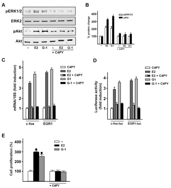
C4PY acts as a GPER antagonist
The evaluation of GPCR-mediated signaling includes the early
response of the MAPK cascade, which has been used in order to
ascertain the potential agonist/antagonist activity of novel drug
candidates (May and Hill, 2008). Because ERK phosphorylation
indicates the binding of ligand to GPER (Filardo et al., 2000;
Maggiolini and Picard, 2010), we aimed to assess the action
triggered by C4PY. In SkBr3 cells, C4PY (ranging from 1 nM to
10 µM) did not trigger ERK phosphorylation (data not shown),
although it was able to prevent the ERK activation by E2 and G-1
(Fig. 4A,B). Likewise, C4PY inhibited the phosphorylation of Akt
induced by both E2 and G-1 (Fig. 4A,B). Considering that the
GPER-MAPK-PI3K transduction pathway regulates a number of
target genes (Maggiolini et al., 2004; Pandey et al., 2009; Sukhatme
Fig. 1. Chemical structure of meso-octamethylcalix[4]pyrrole (C4PY). TRANSLATIONAL IMPACT
Clinical issue
Biological responses to estrogens are mainly mediated by estrogen receptor (ER)α and ERβ, which function as ligand-activated transcription factors. In addition, the G-protein coupled receptor (GPR30/GPER) mediates estrogenic signaling in normal and malignant tissues, including breast cancer cells and cancer-associated fibroblasts (CAFs). Several ER ligands, such as estrogens and ER antagonists, have demonstrated the ability to bind to GPER, eliciting promiscuous and, in certain cases, opposite actions than those elicited via ER binding.
Results
In this study, the authors designed and evaluated ‘in silico’ diverse calixpyrrole derivatives as potential GPER ligands. In accordance with the results obtained in computational studies, the authors established the molecular mechanisms through which a calixpyrrole derivative, named C4PY, might act as a GPER antagonist in breast tumor cells and CAFs that were obtained from individuals with breast cancer. In particular, they showed that C4PY elicits an inhibitory action on GPER-activated signaling, including the repression of both ERK and Akt phosphorylation, gene transcription, cell proliferation and migration in breast cancer cells and in CAFs. Notably, C4PY is selective for GPER and does not interfere with ER-dependent responses upon estrogen exposure.
Implications and future directions
The identification and functional characterization of this novel compound acting as a selective GPER antagonist might represent a valuable tool to further dissect the pharmacology of this receptor and to better differentiate the specific functions elicited by different ER types. In addition, the inhibitory action of C4PY might open new avenues toward innovative pharmacological approaches to target the GPER-mediated stimulatory effects in breast carcinomas. Moreover, this study underlines the fact that strategies against the stimulatory effects exerted by estrogens in ER-negative cancer cells and in key components of the tumor microenvironment (such as CAFs) could be considered as an intriguing opportunity to target breast malignancies.
RESEARCH ARTICLE Disease Models & Mechanisms (2015) 8, 1237-1246 doi:10.1242/dmm.021071
Disease
Models
&
et al., 1988; Vivacqua et al., 2012), we then assessed whether the
E2- and G-1-induced expression of
fos and EGR1 (early growth
response protein 1) is repressed by C4PY in SkBr3 cells (Fig. 4C).
Further
corroborating
these
findings,
C4PY
inhibited
the
transactivation of
fos and EGR1 promoter constructs triggered by
E2 and G-1 (Fig. 4D). Biologically, we ascertained that the
antagonistic action exerted by C4PY through GPER prevents the
proliferation of SkBr3 cells that is induced by E2 and G-1 (Fig. 4E).
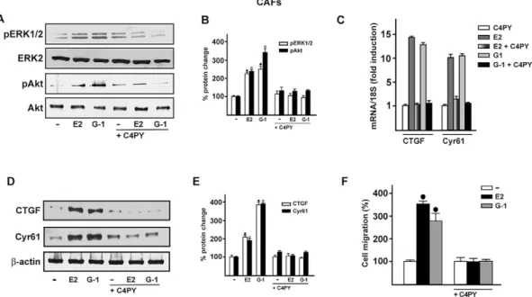
C4PY exerts inhibitory effects through GPER in CAFs
Increasing amounts of evidence demonstrate that CAFs actively
contribute to the growth, expansion and dissemination of breast
cancer cells (Al-Ansari et al., 2012; Lebret et al., 2007; Cheng and
Weiner, 2003; Gao et al., 2010). Therefore, we investigated whether
C4PY elicits an inhibitory action through GPER in CAFs derived
from breast cancer patients, because these cells express GPER and
lack ER (De Francesco et al., 2014; Madeo and Maggiolini, 2010;
Pupo et al., 2013). In accordance with the results obtained in SkBr3
cells, C4PY prevented also in CAFs the rapid ERK and Akt
activation induced upon exposure to E2 and G-1 (Fig. 5A,B). Next,
we aimed to evaluate the potential of C4PY to alter the expression of
two GPER target genes,
CTGF and Cyr61 (Pandey et al., 2009),
which have been implicated in cell migration (Chen et al., 2007).
Notably, the upregulation of CTGF and Cyr61 induced by E2 and
Fig. 2. Ligand binding modes to GPER. (A) C4PY in the protein binding cleft is drawn in green. The protein surface is colored according to its electrostatic potential (blue positive, red negative). The same ligand binding mode is schematically reported in panel B, where the interacting amino acids are indicated as dark gray sticks. (C,D) The agonists GPER-L1 and GPER-L2 are drawn in light green (C) and purple (D) sticks, respectively. Binding mode of G-1 (cyan) is shown in panel E and the full-antagonist MIBE (orange) in panel F.
Disease
Models
&
G-1 in CAFs at both the mRNA and protein levels was abolished in
the presence of C4PY (Fig. 5C-E). As a biological counterpart, the
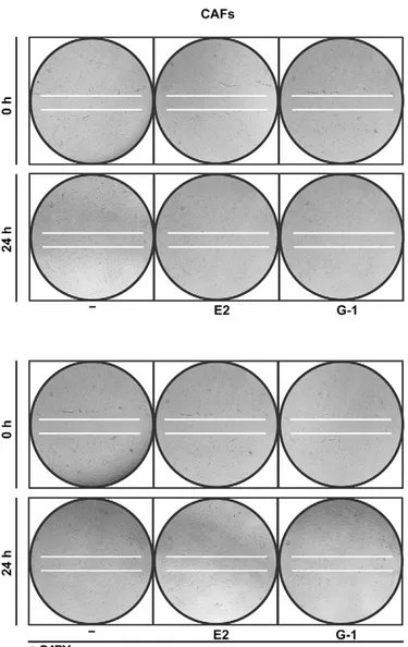
migration of CAFs promoted by both E2 and G-1 was abolished by
C4PY (Fig. 5F and Fig. 6), indicating that this compound is able to
interfere with relevant responses mediated by GPER also in CAFs
that play a stimulatory role within the tumor microenvironment
toward cancer progression (Bhowmick et al., 2004).
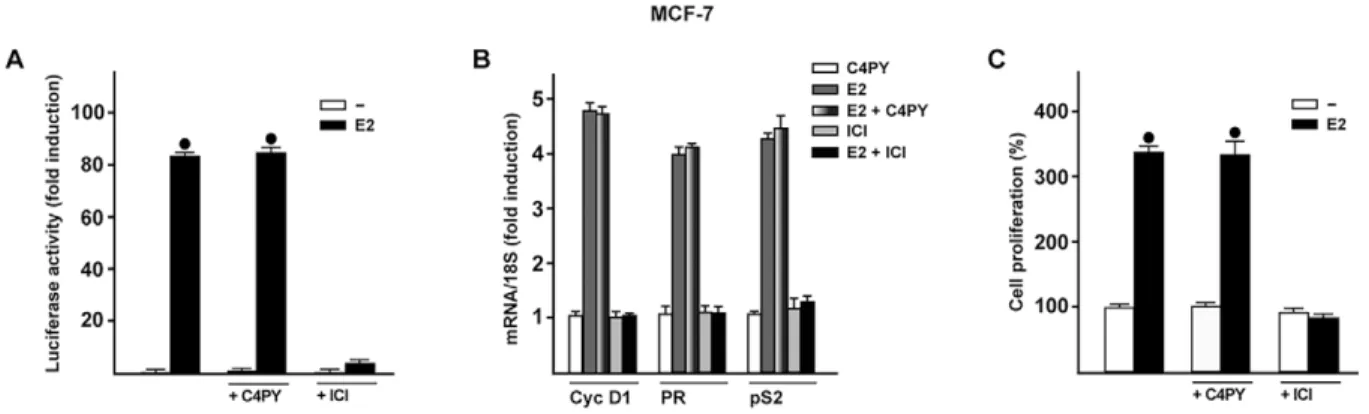
C4PY does not interfere with ER-mediated signaling
In order to verify whether C4PY might also regulate biological
responses mediated by the classical ER, we transiently transfected
an ER reporter gene in MCF-7 breast cancer cells. C4PY neither
displayed the ability to transactivate ER (data not shown) nor to
abrogate the luciferase activity induced by E2 as observed using the
ER antagonist ICI (Fig. 7A). In addition, C4PY did not prevent the
E2-dependent upregulation of ER target genes such as cyclin D1,
progesterone receptor (PR) and pS2, nor the proliferation of MCF-7
cells as shown by ICI (Fig. 7B,C). Together, these data provide
evidence that C4PY acts as a selective GPER antagonist in breast
cancer cells and CAFs.
DISCUSSION
In this study, we have identified a novel antagonist ligand of GPER,
namely C4PY, which exhibits an inhibitory action on
GPER-activated signaling, including the repression of both ERK and Akt
phosphorylation, gene transcription, and cell proliferation and
migration in breast cancer cells and in CAFs. Of note, C4PY acts
selectively through GPER: it does not interfere with the responses
triggered by the ER-dependent transduction pathway upon estrogen
exposure.
GPCRs constitute a large class of receptors of great biological
importance owing to their central role in signal transmission. For
instance, abnormal expression, regulation and function of numerous
GPCRs have been associated with cancer initiation, progression,
invasion and metastasis (Lappano and Maggiolini, 2011, 2012;
O
’Hayre et al., 2014). Therefore, the pharmacological manipulation
Table 1. GPER residues involved in macrocycle binding
Transmembrane helices (TM)
and extracellular loop (EL) Residues
Selective antagonist: C4PY Full antagonist: MIBE Agonist: G-1 Agonist: GPER-L1 Agonist: GPER-L2
TM I Ser62 – – Hyd Hyd –
TM II Glu115 HB HB Halogen
bond
– –
Val116 Hyd – – Hyd Hyd
Leu119 Hyd Hyd – Hyd Hyd
Ser134 – Hyd Hyd Hyd Hyd
Leu137 – Hyd – Hyd Hyd
Gln138 – HB Hyd Hyd –
Met141 – – Hyd Hyd Hyd
EL 2 Thr201 Hyd Hyd – Hyd Hyd
Glu203 – Hyd – – –
Phe206 Hyd Hyd Hyd Hyd Hyd
Phe208 Hyd Hyd Hyd Hyd Hyd
TM VI Trp272 – – – Hyd –
Glu275 – – HB Hyd –
Ile279 – – Hyd Hyd –
TM VII Arg299 Hyd Hyd – Hyd –
His302 Hyd Hyd – Hyd Hyd
Pro303 Hyd Hyd – Hyd Hyd
Gly306 Hyd – – Hyd
His307 Hyd – Hyd Hyd Hyd
Met309 – – – Hyd –
Ser310 – – – HB –
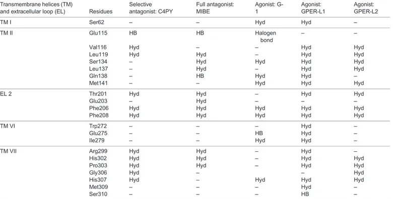
HB, hydrogen bond; Hyd, hydrophobic interaction.
Fig. 3. C4PY is a ligand of GPER. (A) C4PY competes with [3H]E2 for binding to GPER in SkBr3 cells. Competition curves of increasing concentration of unlabeled E2, G-1 and C4PY expressed as a percentage of maximum specific [3H] E2 binding. Each data point represents the mean±s.d. of triplicate samples of three separate experiments. (B) C4PY competes with [5,6-3H] nicotinic acid (NA) for binding to GPER in SkBr3 cells. Competition curves of increasing concentration of unlabeled NA, G-1 and C4PY expressed as a percentage of maximum specific [5,6-3H] NA binding. Each data point represents the mean±s.d. of three separate experiments performed in triplicate.
RESEARCH ARTICLE Disease Models & Mechanisms (2015) 8, 1237-1246 doi:10.1242/dmm.021071
Disease
Models
&
of these receptors is very attractive for the development of novel
ligands that might become part of innovative strategies targeting
tumor development and metastasis. In particular, hormones have been
extensively studied among the mitogens that act through GPCRs
toward the stimulation of cancer cell growth (Dorsam and Gutkind,
2007). In this regard, it has been demonstrated that the aberrant
activity of GPCRs might contribute to the progression of
dependent tumors and their switch into aggressive
hormone-insensitive tumors (Dorsam and Gutkind, 2007). In line with these
observations, several experimental studies have proved that GPER
mediates numerous signaling events in response to estrogens in
different types of cancer cells (Prossnitz and Maggiolini, 2009). As it
concerns breast malignancy, the role exerted by GPER should be
carefully considered owing to its ability to bind not only estrogens but
also ER antagonists such as 4-hydroxytamoxifen (OHT) and ICI
182,780, which elicit stimulatory effects particularly in ER-negative
cancer cells (Filardo et al., 2000; Lappano et al., 2014; Pandey et al.,
2009; Revankar et al., 2005). In addition, GPER signaling is activated
by many ER ligands, including natural estrogens and environmental
contaminants (Albanito et al., 2015; Maggiolini et al., 2004; Pupo
et al., 2012; Thomas and Dong, 2006). It is worthy of noting that we
recently identified a compound, named MIBE, that exhibits the
peculiar feature of acting as an antagonist ligand of both GPER and
ER in breast cancer cells (Lappano et al., 2012a). Overall, the
discovery of selective agonist/antagonist ligands of GPER has widely
aided research toward the evaluation of the specific activities
Fig. 4. C4PY exerts inhibitory effects through GPER in SkBr3 breast cancer cells. (A) ERK1/2 and Akt activation in SkBr3 cells treated for 15 min with 100 nM E2 or 1 µM G-1 is prevented in the presence of 1 µM C4PY. (B) Densitometric analysis of the blots normalized to ERK2 and Akt, respectively. Each data point represents the mean±s.d. of three independent experiments. (C) The mRNA expression of fos and EGR1 induced in SkBr3 cells by 1 h treatment with 100 nM E2 and 1 µM G-1 is inhibited in the presence of 1 µM C4PY, as evaluated by real-time PCR. Results obtained from experiments performed in triplicate were normalized for 18S expression and shown as fold change of RNA expression compared to cells treated with vehicle. Each data point represents the mean±s.d. of three independent experiments performed in triplicate. (D) The transactivation of fos and EGR1 luciferase reporter genes transfected in SkBr3 cells induced by 100 nM E2 and 1 µM G-1 is inhibited by 1 µM C4PY. Luciferase activity was normalized to the internal transfection control Renilla luciferase; values are presented as fold change (mean±s.d.) of vehicle control and represent three independent experiments, each performed in triplicate. (E) The proliferation of SkBr3 cells upon treatment with 100 nM E2 and 100 nM G-1 is inhibited by 1 µM C4PY, as indicated. Cells were treated for 5 days with the indicated treatments and counted on day 6. Proliferation of cells receiving vehicle was set as 100%, upon which cell growth induced by treatments was calculated. Each data point is the average ±s.d. of three independent experiments performed in triplicate. (•) indicates P<0.05 for cells receiving vehicle (–) versus treatments.
Disease
Models
&
triggered by GPER in different pathophysiological conditions,
including cancer (Bologa et al., 2006; Dennis et al., 2009, 2011;
Lappano et al., 2012b; Maggiolini et al., 2015; Prossnitz and Barton,
2011). Moreover, the availability of these ligands has allowed a better
understanding of the downstream signaling cascades triggered by
GPER, such as the activation of MAPK, PI3-kinase (PI3K) and
phospholipase C (PLC), and the increase in cAMP concentrations and
intracellular calcium. Of note, GPER mediates the regulation of a
distinctive gene signature, which includes transcription factors and
cytokines mainly involved in cell survival, proliferation and
migration (Lappano et al., 2014; Maggiolini and Picard, 2010;
Pandey et al., 2009).
An intricate signaling network has been demonstrated to occur
between GPCRs and growth factor receptors (Dorsam and Gutkind,
2007). As it concerns GPER, its physical and functional cross-talk
with EGFR has been shown to contribute to the stimulation of diverse
types of tumors (Albanito et al., 2008; Filardo et al., 2000; Lappano
et al., 2013; Vivacqua et al., 2009). Moreover, the insulin-like growth
factor (IGF) system has the ability to regulate the expression and
function of GPER in different cancer cells, thus suggesting that GPER
might be also engaged by this important growth system toward cancer
progression (Bartella et al., 2012; De Marco et al., 2013, 2014, 2015;
Lappano et al., 2013). Of note, the mechanisms through which GPER
might be involved in the aggressive malignant features were extended
to the ability of estrogenic GPER signaling to induce the HIF1
α-dependent expression of vascular endothelial growth factor (VEGF)
toward breast tumor angiogenesis (De Francesco et al., 2013a,b,
2014; Filice et al., 2009; Recchia et al., 2011). These findings are
nicely supported by previous studies reporting that the expression of
GPER is correlated with increased tumor size, metastasis and poor
outcome in breast cancer (Filardo et al., 2006). The understanding of
the overall role exerted by GPER in this neoplasia has become rather
complex, considering the strong evidence of its ability to mediate the
estrogen
stimulation
of
main
components
of
the
tumor
microenvironment, such as CAFs (De Francesco et al., 2014;
Madeo and Maggiolini, 2010; Pupo et al., 2013, 2014; Vivacqua
et al., 2015). Given the established role elicited by CAFs in breast
cancer progression, particularly the action at metastatic sites
(Aboussekhra, 2011; Kalluri and Zeisberg, 2006), CAFs could be
taken into account as promising therapeutic targets in cancer.
Here, we have identified a novel GPER antagonist that could
open new avenues toward innovative C4PY-based pharmacological
approaches in estrogen-sensitive tumors such as breast carcinomas.
In addition, the inhibitory activity exhibited by C4PY in
ER-negative breast cancer cells and remarkably in CAFs obtained from
patients with breast tumor suggests that novel strategies against both
cancer cells and CAFs could improve the therapeutic management
of breast malignancies.
MATERIALS AND METHODS
Chemical synthesis
The synthesis of meso-octamethylcalix[4]pyrrole (C4PY) has been reported
by various authors (Baeyer, 1886; Rothemund and Gage, 1955). In this
current work, the procedure was modified as follows. Freshly distilled
pyrrole (2 ml, 1.93 g, 0.0288 mol) and an excess of acetone (5 ml, 3.95 g,
0.0681 mol) were diluted in DCM (15 ml) and TFA (2.2 ml, 1.57 g,
0.0137 mol, diluted in 10 ml of DCM) was added under an atmosphere at 0°
C in 10 min. The mixture was stirred for 6 h, during which it was allowed to
reach room temperature. After the addition of a saturated solution of
NaHCO
3(to slightly basic pH), the mixture was concentrated under reduced
pressure to remove most of the unreacted acetone, and extracted with DCM
(3×20 ml). The combined extract were dried (Na
2SO
4) and concentrated to
give a solid residue, which was crystallized from EtOH to give C4PY
(2.31 g, 0.0054 mol, yield 75%, m.p. 275°C dec.),
1H-NMR (500 MHz,
CD
2Cl
2, ppm)
δ 7.03 (sbr, 4H, NH), 5.88 and 5.87 (2×s, 2×4H, β-pyrrole
Fig. 5. C4PY exerts inhibitory effects through GPER in CAFs. (A) ERK1/2 and Akt activation in CAFs treated for 5 min with 1 nM E2 and 100 nM G-1 is prevented by 1 µM C4PY. (B) Densitometric analysis of the blots normalized to ERK2 and Akt, respectively. Each data point represents the mean±s.d. of three independent experiments. (C) The mRNA expression of CTGF and Cyr61 induced in CAFs by 1 h treatment with 1 nM E2 and 100 nM G-1 is prevented by 1 µM C4PY, as evaluated by real-time PCR. Results obtained from experiments performed in triplicate were normalized for 18S expression and shown as fold change of RNA expression compared to cells treated with vehicle. Each data point represents the mean±s.d. of three independent experiments performed in triplicate. (D) CTGF and Cyr61 protein expression induced in CAFs by 2 h treatment with 1 nM E2 and 100 nM G-1 is inhibited in the presence of 1 µM C4PY. (E) Densitometric analyses of the blots normalized toβ-actin; values shown represent the mean±s.d. of three independent experiments. (F) The migration of CAFs upon treatment with 1 nM E2 and 100 nM G-1 is inhibited by 1 µM C4PY, as evaluated by Boyden Chamber assay. Each data point is the average ±s.d. of three independent experiments performed in triplicate. (•) and (◦) indicate P<0.05 for cells receiving vehicle (–) versus treatments.
RESEARCH ARTICLE Disease Models & Mechanisms (2015) 8, 1237-1246 doi:10.1242/dmm.021071
Disease
Models
&
CH), 1.49 (s, 24H, CH
3);
13C-NMR (125 MHz, CD
2Cl
2, ppm)
δ 138.7,
103.0, 35.3, 28.8.
Molecular modeling and docking simulations
In order to evaluate the potential binding modes of our macrocyclic
compounds to GPER, the program GOLD v.5.1 (the Cambridge
Crystallographic Data Center, UK) was used in docking simulations. As
protein target, the three-dimensional atomic coordinates of the GPER
molecular model was utilized in accordance with our previous studies
(Lappano et al., 2010). The atom Phe 208 O was considered as ligand
binding pocket center, and active site atoms were considered those located
within 20 Å from this point (Lappano et al., 2012a). We ran the simulations
using the default parameters provided by the software. Residues Tyr123,
Gln138, Phe206, Phe208, Glu275, Phe278 and His282 of GPER were
defined with flexible side chains, therefore allowing their free rotation. The
schematic figures representing protein:ligand complexes were drawn with
the program Chimera (Pettersen et al., 2004).
Reagents
17
β-estradiol (E2) was purchased from Sigma-Aldrich Srl (Milan, Italy) and
solubilized in ethanol.
G-1 {1-[4-(-6-bromobenzol[1,3]diodo-5-yl)-3a,4,5,9b-tetrahidro3H5cyclopenta
[c]quinolin-8yl]-ethanone} was bought from Tocris Bioscience (Bristol,
United Kingdom) and dissolved in dimethyl sulfoxide (DMSO). Nicotinic
acid ( pyridine-3-carboxylic acid) was purchased from Sigma-Aldrich Srl
(Milan, Italy) and solubilized in water.
Cell culture
SkBr3 breast cancer cells were maintained in RPMI 1640 without phenol
red supplemented with 10% FBS and 100 mg/ml penicillin/streptomycin
(Life Technologies, Milan, Italy). MCF-7 breast cancer cells were
maintained in DMEM with phenol red supplemented with 10% FBS and
100 mg/ml penicillin/streptomycin (Life Technologies, Milan, Italy). All
cell lines to be processed for immunoblot and RT-PCR assays were switched
to medium without serum and phenol red the day before treatments.
CAFs were extracted as previously described (Madeo and Maggiolini,
2010). Briefly, breast cancer specimens were collected from primary tumors
of patients who had undergone surgery. Signed informed consent was
obtained from all the patients and from the institutional review board(s) of
the Regional Hospital of Cosenza, Italy. Tissues from tumors were cut into
smaller pieces (1-2 mm diameter), placed in digestion solution (400 IU
collagenase, 100 IU hyaluronidase and 10% serum, containing antibiotic
and antimycotic solution), and incubated overnight at 37°C. The cells were
then separated by differential centrifugation at 90 g for 2 min. Supernatant
containing fibroblasts was centrifuged at 485 g for 8 min; the pellet obtained
was suspended in fibroblasts growth medium (Medium 199 and Ham’s F12
mixed 1:1 and supplemented with 10% FBS) and cultured at 37°C in 5%
CO
2. Primary cell cultures of breast fibroblasts were characterized by
immunofluorescence. Briefly, cells were incubated with human
anti-vimentin (V9) and human anti-cytokeratin 14 (LL001), both from Santa
Cruz Biotechnology (DBA, Milan, Italy). To assess fibroblast activation, we
used anti-fibroblast activated protein
α (FAPα) antibody (H-56; Santa Cruz
Biotechnology, DBA, Milan, Italy) (data not shown).
Plasmids and luciferase assays
The firefly luciferase reporter plasmid for ER
α used was XETL (Bunone
et al., 1996), which contains the ERE from the
Xenopus vitellogenin A2
gene (nucleotides
−334 to −289), the herpes simplex virus thymidine
kinase promoter region (nucleotides
−109 to +52), the firefly luciferase
coding sequence, and the SV40 splice and polyadenylation sites from
plasmid pSV232A/L-AA5. The luciferase reporter plasmid for
fos
encoding a
−2.2-kb 5′ upstream fragment of human fos was a gift from
Dr Kiyoshi Nose (Department of Microbiology, Showa University School
of Pharmaceutical Sciences, Hatanodai, Shinagawa-ku, Tokyo, Japan).
EGR1-luc plasmid, containing the
−600 to +12 5′-flanking sequence from
the human
EGR1 gene, was kindly provided by Dr Stephen Safe
(Department of Veterinary Physiology and Pharmacology, Texas A&M
University, TX, USA). The
Renilla luciferase expression vector pRL-TK
(Promega, Milan, Italy) was used as internal transfection control. Cells
were plated into 24-well plates with 500 µl of regular growth medium/well
the day before transfection. For the transfection of the ER reporter gene in
MCF-7 cells, standard medium was replaced with medium supplemented
with 1% charcoal-stripped (CS) FBS lacking phenol red and serum on the
day of transfection, which was performed by using X-tremeGENE 9 DNA
Transfection Reagent as recommended by the manufacturer (Roche
Molecular Biochemicals, Milan, Italy) with a mixture containing 0.5 µg
of reporter plasmid and 2 ng of pRL-TK. After 6 h, the medium was
replaced again with serum-free medium lacking phenol red and
supplemented with 1% CS-FBS; treatments were added at this point and
cells were incubated for an additional 18 h. For the luciferase assays of the
fos and EGR1 reporter plasmids, on the day of transfection, SkBr3 cell
medium was replaced with RPMI without phenol red and serum, and
transfection was performed using X-tremeGENE 9 DNA Transfection
Reagent (Roche Molecular Biochemicals, Milan, Italy) and a mixture
containing 0.5
μg of each reporter plasmid and 5 ng of pRL-TK. After 6 h,
treatments were added and cells were incubated for 18 h. Luciferase
activity was then measured using the Dual Luciferase Kit (Promega,
Milan, Italy) according to the manufacturer
’s recommendations. Firefly
luciferase activity was normalized to the internal transfection control
provided by the
Renilla luciferase activity. The normalized relative light
unit values obtained from cells treated with vehicle were set as 1-fold
induction, upon which the activity induced by treatments was calculated.
Fig. 6. The migration of CAFs induced by E2 (1 nM) and G-1 (100 nM) is inhibited by 1 µM C4PY, as determined by wound-healing assay. Data are representative of three independent experiments performed in triplicate.
Disease
Models
&
Ligand binding assays
In ligand binding assays, SkBr3 cells were grown in 10-cm cell-culture
dishes, washed two times and incubated either with 1 nM [2,4,6,7-3H]E2
(89 Ci/mmol; Amersham Bioscience, GE Healthcare, Milan, Italy) or with
50 nM [5,6-3H] nicotinic acid (50-60 Ci/mmol; BIOTREND, Chemikalien
GmbH, Köln, Germany) in the presence or absence of increasing
concentrations of nonlabeled competitors, as indicated. Then, cells were
incubated for 2 h at 37°C and washed three times with ice-cold PBS; the
radioactivity collected by 100% ethanol extraction was measured by liquid
scintillation counting. Competitor binding was expressed as a percentage of
maximal specific binding.
Gene expression studies
Total RNA was extracted and cDNA was synthesized by reverse
transcription as previously described (Lappano et al., 2011). The
expression of selected genes was quantified by real-time PCR using Step
One sequence detection system (Applied Biosystems Inc., Milan, Italy).
Gene-specific primers were designed using Primer Express version 2.0
software (Applied Biosystems Inc., Milan, Italy). For cyclin D1,
PR, pS2,
fos, CTGF, Cyr61, EGR1 and the ribosomal protein 18S, which was
used as a control gene to obtain normalized values, the primers
were: 5
′-GTCTGTGCATTTCTGGTTGCA-3′ (cyclin D1 forward) and
5
′-GCTGGAAACATGCCGGTTA-3′ (cyclin D1 reverse);
5′-GAGTTGT-GAGAGCACTGGATGCT-3
′ (PR forward) and
5′-CAACTGTATGTCT-TGACCTGGTGAA-3
′ (PR reverse); 5′-GCCCCCCGTGAAAGAC-3′
( pS2 forward) and 5
′-CGTCGAAACAGCAGCCCTTA-3′ (pS2 reverse);
5
′-CGAGCCCTTTGATGACTTCCT-3′ (fos forward) and
5′-GGAGCG-GGCTGTCTCAGA-3
′ (fos reverse); 5′-ACCTGTGGGATGGGCATCT-3′
(CTGF
forward)
and
5
′-CAGGCGGCTCTGCTTCTCTA-3′ (CTGF
reverse); 5
′-GAGTGGGTCTGTGACGAGGAT-3′ (Cyr61 forward) and
5
′-GGTTGTATAGGATGCGAGGCT-3′ (Cyr61 reverse);
5′-GCCTGCG-ACATCTGTGGAA-3
′ (EGR1 forward) and
5′-CGCAAGTGGATCTTG-GTATGC-3
′ (EGR1 reverse); and 5′-GGCGTCCCCCAACTTCTTA-3′
(18S forward) and 5
′-GGGCATCACAGACCTGTTATT-3′ (18S reverse),
respectively.
Western blotting
Cells were grown in 10-cm dishes, exposed to treatments, and then lysed
in 500
μl of 50 mmol/l NaCl, 1.5 mmol/l MgCl
2, 1 mmol/l EGTA,
10% glycerol, 1% Triton X-100, 1% sodium dodecyl sulfate (SDS), and
a mixture of protease inhibitors containing 1 mmol/l aprotinin, 20 mmol/l
phenylmethylsulfonyl fluoride and 200 mmol/l sodium orthovanadate.
Protein concentration was determined using Bradford reagent according
to
the
manufacturer
’s recommendations (Sigma-Aldrich, Milan,
Italy). Equal amounts of whole protein extract were resolved on a 10%
SDS-polyacrylamide gel, transferred to a nitrocellulose membrane
(Amersham Biosciences, GE Healthcare, Milan, Italy), probed overnight
at 4°C with antibodies against CTGF (L-20), CYR61 (H-78),
β-actin
(C-2), phosphorylated AKT 1/2/3 (Ser 473), AKT1/2/3 (H-136),
phosphorylated ERK1/2 (E-4) and ERK2 (C-14) (all purchased from
Santa Cruz Biotechnology, DBA, Milan, Italy), and then revealed
using the ECL
™ Western Blotting Analysis System (GE Healthcare,
Milan, Italy).
Proliferation assay
For quantitative proliferation assays, cells (1×10
5) were seeded in 24-well
plates in regular growth medium. Cells were washed once they had attached
and then incubated in medium containing 2.5% charcoal-stripped FBS with
the indicated treatments; medium was renewed every 2 days (with treatments)
before counting using the Countess Automated Cell Counter, as recommended
by the manufacturer
’s protocol (Life Technologies, Milan, Italy).
Migration assays
Migration assays were performed with CAFs in triplicate using Boyden
chambers (Costar Transwell, 8 mm polycarbonate membrane,
Sigma-Aldrich, Milan, Italy). CAFs were trypsinized and seeded in the upper
chambers. Treatments were added to the medium without serum in the
bottom wells where applicable. At 6 h after seeding, cells on the bottom side
of the membrane were fixed and counted. Moreover, wound-healing assays
were also performed in order to visualize cell migration. Cells (1×10
6/well)
were seeded onto six-well plates in regular medium. After 18 h, wounds
were created by dragging a 200-
μl pipette tip through the cell monolayer; the
medium was replaced with 2.5% charcoal-stripped FBS and the treatments
were added. Cells were allowed to migrate for 24 h; the gap area was then
photographed and migration distances were measured.
Statistical analysis
Statistical analysis was done using ANOVA followed by Newman-Keuls
’
testing to determine differences in means.
P<0.05 was considered as
statistically significant.
This article is part of a subject collection on Model Systems in Drug Discovery: from Bench to Patient. See related articles in this collection at http://dmm.biologists.org/ cgi/collection/drugdiscovery
Competing interests
The authors declare no competing or financial interests.
Fig. 7. C4PY does not interfere with the ER-mediated signaling. (A) MCF-7 cells were transfected with an ER luciferase reporter gene along with the internal transfection control Renilla luciferase and then treated with 10 nM E2 in combination with 1 µM C4PY or ICI, as indicated. The normalized luciferase activity values of cells treated with vehicle were set as 1-fold induction, upon which the activity induced by treatments was calculated. Each data point represents the mean±s.d. of three experiments performed in triplicate. (B) The mRNA expression of cyclin D1 (Cyc D1), progesterone receptor (PR) and pS2 induced in MCF-7 cells by 24 h treatment with 10 nM E2 is inhibited by 1 µM ICI, but not by 1 µM C4PY, as evaluated by real-time PCR. Results obtained from experiments performed in triplicate were normalized for 18S expression and shown as fold change of RNA expression compared to cells treated with vehicle. Each data point represents the mean±s.d. of three independent experiments performed in triplicate. (C) The proliferation of MCF-7 cells upon treatment with 10 nM E2 is inhibited by 1 µM ICI, but not by 1 µM C4PY, as indicated. Cells were treated for 5 days with the indicated treatments and counted on day 6. Proliferation of cells receiving vehicle was set as 100%, upon which cell growth induced by treatments was calculated. Each data point is the average ±s.d. of three independent experiments performed in triplicate. (•) indicates P<0.05 for cells receiving vehicle (–) versus treatments.
RESEARCH ARTICLE Disease Models & Mechanisms (2015) 8, 1237-1246 doi:10.1242/dmm.021071
Disease
Models
&
Author contributions
R.L., C.R., F.H.K. and M.M. conceived and designed the experiments. A.P., M.F.S., E.M.D.F., P.D.M., M.P., L.F. and G.C. performed the experiments. F.H.K. and M.M. analyzed the data. V.D., S.A. and F.H.K. provided tools and reagents and gave valuable suggestions. R.L., C.R. and M.M. wrote the paper and the final version was approved by all authors.
Funding
This work was supported by Associazione Italiana per la Ricerca sul Cancro (AIRC), PON01_01078 and Ministero della Salute Grant 67/GR-2010-2319511. C.R. and F.H.K. were funded by Compagnia di San Paolo di Torino ( project“Nanovettori Intelligenti per la Diagnosi e Terapia in Oncologia”) and MIUR (project PRIN 20109Z2XRJ 010).
References
Aboussekhra, A. (2011). Role of cancer-associated fibroblasts in breast cancer development and prognosis. Int. J. Dev. Biol. 55, 841-849.
Al-Ansari, M. M., Hendrayani, S. F., Shehata, A. I. and Aboussekhra, A. (2012). p16(INK4A) represses the paracrine tumor-promoting effects of breast stromal fibroblasts. Oncogene 32, 2356-2364.
Albanito, L., Madeo, A., Lappano, R., Vivacqua, A., Rago, V., Carpino, A., Oprea, T. I., Prossnitz, E. R., Musti, A. M., Andò, S. et al. (2007). G protein-coupled receptor 30 (GPR30) mediates gene expression changes and growth response to 17beta-estradiol and selective GPR30 ligand G-1 in ovarian cancer cells. Cancer Res. 67, 1859-1866.
Albanito, L., Sisci, D., Aquila, S., Brunelli, E., Vivacqua, A., Madeo, A., Lappano, R., Pandey, D. P., Picard, D., Mauro, L. et al. (2008). Epidermal growth factor induces G protein-coupled receptor 30 expression in estrogen receptor-negative breast cancer cells. Endocrinology 149, 3799-3808. Albanito, L., Lappano, R., Madeo, A., Chimento, A., Prossnitz, E. R., Cappello,
A. R., Dolce, V., Abonante, S., Pezzi, V. and Maggiolini, M. (2015). Effects of atrazine on estrogen receptorα- and G protein-coupled receptor 30-mediated signaling and proliferation in cancer cells and cancer-associated fibroblasts. Environ. Health Perspect. 123, 493-499.
Allen, W. E., Gale, P. A., Brown, C. T., Lynch, V. M. and Sessler, J. L. (1996). Binding of neutral substrates by Calix[4]pyrroles. J. Am. Chem. Soc. 118, 12471-12472.
Ascenzi, P., Bocedi, A. and Marino, M. (2006). Structure-function relationship of estrogen receptor alpha and beta: impact on human health. Mol. Aspects Med. 27, 299-402.
Baeyer, A. (1886). Ueber ein Condensations product von Pyrrol mit Aceton. Ber. Dtsch. Chem. Ges. 19, 2184-2185.
Bartella, V., De Marco, P., Malaguarnera, R., Belfiore, A. and Maggiolini, M. (2012). New advances on the functional cross-talk between insulin-like growth factor-I and estrogen signaling in cancer. Cell Signal. 24, 1515-1521.
Bhowmick, N. A., Neilson, E. G. and Moses, H. L. (2004). Stromal fibroblasts in cancer initiation and progression. Nature 432, 332-337.
Bologa, C. G., Revankar, C. M., Young, S. M., Edwards, B. S., Arterburn, J. B., Kiselyov, A. S., Parker, M. A., Tkachenko, S. E., Savchuck, N. P., Sklar, L. A. et al. (2006). Virtual and biomolecular screening converge on a selective agonist for GPR30. Nat. Chem. Biol. 2, 207-212.
Bunone, G., Briand, P. A., Miksicek, R. J. and Picard, D. (1996). Activation of the unliganded estrogen receptor by EGF involves the MAP kinase pathway and direct phosphorylation. EMBO J. 15, 2174-2183.
Cafeo, G., Kohnke, F. H., La Torre, G. L., Parisi, M. F., Nascone, R. P., White, A. J. P. and Williams, D. J. (2002). Calix[6]pyrrole and hybrid calix[n]furan[m] pyrroles (n+m=6): syntheses and host-guest chemistry. Chemistry 8, 3148-3156. Cafeo, G., Kohnke, F. H., White, A. J. P., Garozzo, D. and Messina, A. (2007). Syntheses, structures, and anion-binding properties of two novel calix[2]benzo[4] pyrroles. Chemistry 13, 649-656.
Cafeo, G., Carbotti, G., Cuzzola, A., Fabbi, M., Ferrini, S., Kohnke, F. H., Papanikolaou, G., Plutino, M. R., Rosano, C. and White, A. J. (2013). Drug delivery with a calixpyrrole–trans-Pt(II) complex. J. Am. Chem. Soc. 135, 2544-2551.
Chen, P.-S., Wang, M.-Y., Wu, S.-N., Su, J.-L., Hong, C.-C., Chuang, S.-E., Chen, M.-W., Hua, K.-T., Wu, Y.-L., Cha, S.-T. et al. (2007). CTGF enhances the motility of breast cancer cells via an integrin-avb3-ERK1/2-dependent S100A4-upregulated pathway. J. Cell Sci. 120, 2053-2065.
Cheng, J. D. and Weiner, L. M. (2003). Tumors and their microenvironments: tilling the soil. Commentary re: A. M. Scott et al., A Phase I dose-escalation study of sibrotuzumab in patients with advanced or metastatic fibroblast activation protein-positive cancer. Clin. Cancer Res. 9: 1639–1647, 2003. Clin. Cancer Res. 9, 1590-1595.
Custelcean, R., Delmau, L. H., Moyer, B. A., Sessler, J. L., Cho, W.-S., Gross, D., Bates, G. W., Brooks, S. J., Light, M. E. and Gale, P. A. (2005). Calix[4]pyrrole: an old yet new ion-pair receptor. Angew. Chem. Int. Ed. 44, 2537-2542. De Francesco, E. M., Angelone, T., Pasqua, T., Pupo, M., Cerra, M. C. and
Maggiolini, M. (2013a). GPER mediates cardiotropic effects in spontaneously hypertensive rat hearts. PLoS ONE 8, e69322.
De Francesco, E. M., Lappano, R., Santolla, M. F., Marsico, S., Caruso, A. and Maggiolini, M. (2013b). HIF-1α/GPER signaling mediates the expression of VEGF induced by hypoxia in breast cancer associated fibroblasts (CAFs). Breast Cancer Res. 15, R64.
De Francesco, E. M., Pellegrino, M., Santolla, M. F., Lappano, R., Ricchio, E., Abonante, S. and Maggiolini, M. (2014). GPER mediates activation of HIF1α/ VEGF signaling by estrogens. Cancer Res. 74, 4053-4064.
De Marco, P., Bartella, V., Vivacqua, A., Lappano, R., Santolla, M. F., Morcavallo, A., Pezzi, V., Belfiore, A. and Maggiolini, M. (2013). Insulin-like growth factor-I regulates GPER expression and function in cancer cells. Oncogene 32, 678-688.
De Marco, P., Romeo, E., Vivacqua, A., Malaguarnera, R., Abonante, S., Romeo, F., Pezzi, V., Belfiore, A. and Maggiolini, M. (2014). GPER1 is regulated by insulin in cancer cells and cancer-associated fibroblasts. Endocr. Relat. Cancer 21, 739-753.
De Marco, P., Cirillo, F., Vivacqua, A., Malaguarnera, R., Belfiore, A. and Maggiolini, M. (2015). Novel aspects concerning the functional cross-talk between the insulin/IGF-I system and estrogen signaling in cancer cells. Front. Endocrinol. 6, 30.
Dennis, M. K., Burai, R., Ramesh, C., Petrie, W. K., Alcon, S. N., Nayak, T. K., Bologa, C. G., Leitao, A., Brailoiu, E., Deliu, E. et al. (2009). In vivo effects of a GPR30 antagonist. Nat. Chem. Biol. 5, 421-427.
Dennis, M. K., Field, A. S., Burai, R., Ramesh, C., Petrie, W. K., Bologa, C. G., Oprea, T. I., Yamaguchi, Y., Hayashi, S.-I., Sklar, L. A. et al. (2011). Identification of a GPER/GPR30 antagonist with improved estrogen receptor counterselectivity. J. Steroid Biochem. Mol. Biol. 127, 358-366.
Dorsam, R. T. and Gutkind, J. S. (2007). G-protein-coupled receptors and cancer. Nat. Rev. Cancer 7, 79-94.
Filardo, E. J., Quinn, J. A., Bland, K. I. and Frackelton, A. R., Jr. (2000). Estrogen-induced activation of Erk-1 and Erk-2 requires the G protein-coupled receptor homolog, GPR30, and occurs via trans-activation of the epidermal growth factor receptor through release of HB-EGF. Mol. Endocrinol. 14, 1649-1660. Filardo, E. J., Graeber, C. T., Quinn, J. A., Resnick, M. B., Giri, D., DeLellis, R. A.,
Steinhoff, M. M. and Sabo, E. (2006). Distribution of GPR30, a seven membrane-spanning estrogen receptor in primary breast cancer and its association with clinicopathologic determinants of tumor progression. Clin. Cancer Res. 12, 6359-6366.
Filice, E., Recchia, A. G., Pellegrino, D., Angelone, T., Maggiolini, M. and Cerra, M. C. (2009). A new membrane G protein-coupled receptor (GPR30) is involved in the cardiac effects of 17beta-estradiol in the male rat. J. Physiol. Pharmacol. 60, 3-10.
Gale, P. A. (2011). From anion receptors to transporters. Acc. Chem. Res. 44, 216-226.
Gale, P. A., Sessler, J. L. and Král, V. (1998). Calixpyrroles. Chem. Commun. 1-8. Gale, P. A., Anzenbacher, P., Jr and Sessler, J. L. (2001). Calixpyrroles II. Coord.
Chem. Rev. 222, 57-102.
Gao, M.-Q., Kim, B. G., Kang, S., Choi, Y. P., Park, H., Kang, K. S. and Cho, N. H. (2010). Stromal fibroblasts from the interface zone of human breast carcinomas induce an epithelial-mesenchymal transition-like state in breast cancer cells in vitro. J. Cell. Sci. 123, 3507-3514.
Giannoni, E., Bianchini, F., Masieri, L., Serni, S., Torre, E., Calorini, L. and Chiarugi, P. (2010). Reciprocal activation of prostate cancer cells and cancer-associated fibroblasts stimulates epithelial-mesenchymal transition and cancer stemness. Cancer Res. 70, 6945-6956.
Henderson, B. E. and Feigelson, H. S. (2000). Hormonal carcinogenesis. Carcinogenesis 21, 427-433.
Kalluri, R. and Zeisberg, M. (2006). Fibroblasts in cancer. Nat. Rev. Cancer 6, 392-401.
Lappano, R. and Maggiolini, M. (2011). G protein-coupled receptors: novel targets for drug discovery in cancer. Nat. Rev. Drug Discov. 10, 47-60.
Lappano, R. and Maggiolini, M. (2012). GPCRs and cancer. Acta Pharmacol Sin. 33, 351-362.
Lappano, R., Rosano, C., De Marco, P., De Francesco, E. M., Pezzi, V. and Maggiolini, M. (2010). Estriol acts as a GPR30 antagonist in estrogen receptor-negative breast cancer cells. Mol. Cell. Endocrinol. 320, 162-170.
Lappano, R., Recchia, A. G., De Francesco, E. M., Angelone, T., Cerra, M. C., Picard, D. and Maggiolini, M. (2011). The cholesterol metabolite 25-hydroxycholesterol activates estrogen receptorα-mediated signaling in cancer cells and in cardiomyocytes. PLoS ONE 6, e16631.
Lappano, R., Santolla, M. F., Pupo, M., Sinicropi, M. S., Caruso, A., Rosano, C. and Maggiolini, M. (2012a). MIBE acts as antagonist ligand of both estrogen receptorα and GPER in breast cancer cells. Breast Cancer Res. 14, R12. Lappano, R., Rosano, C., Santolla, M. F., Pupo, M., De Francesco, E. M., De
Marco, P., Ponassi, M., Spallarossa, A., Ranise, A. and Maggiolini, M. (2012b). Two novel GPER agonists induce gene expression changes and growth effects in cancer cells. Curr. Cancer Drug Targets 12, 531-542.
Lappano, R., De Marco, P., De Francesco, E. M., Chimento, A., Pezzi, V. and Maggiolini, M. (2013). Cross-talk between GPER and growth factor signaling. J. Steroid Biochem. Mol. Biol. 137, 50-56.
Disease
Models
&
Lappano, R., Pisano, A. and Maggiolini, M. (2014). GPER function in breast cancer: an overview. Front. Endocrinol. 5, 66.
Lebret, S. C., Newgreen, D. F., Thompson, E. W. and Ackland, M. L. (2007). Induction of epithelial to mesenchymal transition in PMC42-LA human breast carcinoma cells by carcinoma-associated fibroblast secreted factors. Breast Cancer Res. 9, R19.
Madeo, A. and Maggiolini, M. (2010). Nuclear alternate estrogen receptor GPR30 mediates 17beta-estradiol-induced gene expression and migration in breast cancer-associated fibroblasts. Cancer Res. 70, 6036-6046.
Maggiolini, M. and Picard, D. (2010). The unfolding stories of GPR30, a new membrane-bound estrogen receptor. J. Endocrinol. 204, 105-114.
Maggiolini, M., Vivacqua, A., Fasanella, G., Recchia, A. G., Sisci, D. Pezzi, V., Montanaro, D., Musti, A. M., Picard, D. and Andò, S. (2004). The G protein-coupled receptor GPR30 mediates c-fos up-regulation by 17beta-estradiol and phytoestrogens in breast cancer cells. J. Biol. Chem. 279, 27008-27016. Maggiolini, M., Santolla, M. F., Avino, S., Aiello, F., Rosano, C., Garofalo, A. and
Grande, F. (2015). Identification of two benzopyrroloxazines acting as selective gper antagonists in breast cancer cells and cancer-associated fibroblasts. Future Med. Chem. 7, 437-448.
Martinez-Outschoorn, U. E., Trimmer, C., Lin, Z., Whitaker-Menezes, D., Chiavarina, B., Zhou, J., Wang, C., Pavlides, S., Martinez-Cantarin, M. P., Capozza, F. et al. (2010). Autophagy in cancer associated fibroblasts promotes tumor cell survival: role of hypoxia, HIF1 induction and NFkappaB activation in the tumor stromal microenvironment. Cell Cycle 9, 3515-3533.
May, L. T. and Hill, S. J. (2008). ERK phosphorylation: spatial and temporal regulation by G protein-coupled receptors. Int. J. Biochem. Cell. Biol. 40, 2013-2017.
O’Hayre, M., Degese, M. S. and Gutkind, J. S. (2014). Novel insights into G protein and G protein-coupled receptor signaling in cancer. Curr. Opin. Cell. Biol. 27, 126-135.
O’Malley, B. W. (2005). A life-long search for the molecular pathways of steroid hormone action. Mol. Endocrinol. 19, 1402-1411.
Orimo, A., Gupta, P. B., Sgroi, D. C., Arenzana-Seisdedos, F., Delaunay, T., Naeem, R., Carey, V. J., Richardson, A. L. and Weinberg, R. A. (2005). Stromal fibroblasts present in invasive human breast carcinomas promote tumor growth and angiogenesis through elevated SDF-1/CXCL12 secretion. Cell 121, 335-348. Pandey, D. P., Lappano, R., Albanito, L., Madeo, A., Maggiolini, M. and Picard, D. (2009). Estrogenic GPR30 signalling induces proliferation and migration of breast cancer cells through CTGF. EMBO J. 28, 523-532.
Pettersen, E. F., Goddard, T. D., Huang, C. C., Couch, G. S., Greenblatt, D. M., Meng, E. C. and Ferrin, T. E. (2004). UCSF Chimera–a visualization system for exploratory research and analysis. J. Comput. Chem. 25, 1605-1612.
Polyak, K. and Kalluri, R. (2010). The role of the microenvironment in mammary gland development and cancer. Cold Spring Harb. Perspect. Biol. 30, 1-16. Prossnitz, E. R. and Barton, M. (2011). The G-protein-coupled estrogen receptor
GPER in health and disease. Nat. Rev. Endocrinol. 7, 715-726.
Prossnitz, E. R. and Maggiolini, M. (2009). Mechanisms of estrogen signaling and gene expression via GPR30. Mol. Cell. Endocrinol. 308, 32-38.
Pupo, M., Pisano, A., Lappano, R., Santolla, M. F., De Francesco, E. M., Abonante, S., Rosano, C. and Maggiolini, M. (2012). Bisphenol A induces gene expression changes and proliferative effects through GPER in breast cancer cells and cancer-associated fibroblasts. Environ. Health Perspect. 120, 1177-1182. Pupo, M., Vivacqua, A., Perrotta, I., Pisano, A., Aquila, S., Abonante, S.,
Gasperi-Campani, A., Pezzi, V. and Maggiolini, M. (2013). The nuclear localization signal is required for nuclear GPER translocation and function in breast Cancer-Associated Fibroblasts (CAFs). Mol. Cell. Endocrinol. 376, 23-32. Pupo, M., Pisano, A., Abonante, S., Maggiolini, M. and Musti, A. M. (2014). GPER activates Notch signaling in breast cancer cells and cancer-associated fibroblasts (CAFs). Int. J. Biochem. Cell Biol. 46, 56-67.
Recchia, A. G., De Francesco, E. M., Vivacqua, A., Sisci, D., Panno, M. L., Andò, S. and Maggiolini, M. (2011). The G protein-coupled receptor 30 is up-regulated by hypoxia-inducible factor-1alpha (HIF-1alpha) in breast cancer cells and cardiomyocytes. J. Biol. Chem. 286, 10773-10782.
Revankar, C. M., Cimino, D. F., Sklar, L. A., Arterburn, J. B. and Prossnitz, E. R. (2005). A transmembrane intracellular estrogen receptor mediates rapid cell signaling. Science 307, 1625-1630.
Rosano, C., Lappano, R., Santolla, M. F., Ponassi, M., Donadini, A. and Maggiolini, M. (2012). Recent advances in the rationale design of GPER ligands. Curr. Med. Chem. 19, 6199-6206.
Rothemund, P. and Gage, C. L. (1955). Concerning the structure of “Acetonepyrrole”. J. Am. Chem. Soc. 77, 3340-3342.
Santolla, M. F., Lappano, R., De Marco, P., Pupo, M., Vivacqua, A., Sisci, D., Abonante, S., Iacopetta, D., Cappello, A. R., Dolce, V. et al. (2012). G protein-coupled estrogen receptor mediates the up-regulation of fatty acid synthase induced by 17β-estradiol in cancer cells and cancer-associated fibroblasts. J. Biol. Chem. 287, 43234-43245.
Santolla, M. F., De Francesco, E. M., Lappano, R., Rosano, C., Abonante, S. and Maggiolini, M. (2014). Niacin activates the G protein estrogen receptor (GPER)-mediated signalling. Cell Signal. 26, 1466-1475.
Schedin, P. and Borges, V. (2009). Breaking down barriers: the importance of the stromal microenvironment in acquiring invasiveness in young women’s breast cancer. Breast Cancer Res. 11, 102.
Siegel, R., Naishadham, D. and Jemal, A. (2012). Cancer statistics, 2012. CA Cancer J. Clin. 62, 10-29.
Sinicropi, M. S., Lappano, R., Caruso, A., Santolla, M. F., Pisano, A., Rosano, C., Capasso, A., Panno, A., Lancelot, J. C., Rault, S. et al. (2015). (6-Bromo-1,4-dimethyl-9H-carbazol-3-yl-methylene)-hydrazine (Carbhydraz) acts as a GPER agonist in breast cancer cells. Curr. Top. Med. Chem. 15, 1035-1042. Sjö ström, M., Hartman, L., Grabau, D., Fornander, T., Malmström, P.,
Nordenskjö ld, B., Sgroi, D. C., Skoog, L., Stål, O., Leeb-Lundberg, L. M. et al. (2014). Lack of G protein-coupled estrogen receptor (GPER) in the plasma membrane is associated with excellent long-term prognosis in breast cancer. Breast Cancer Res. Treat. 145, 61-71.
Smith, H. O., Leslie, K. K., Singh, M., Qualls, C. R., Revankar, C. M., Joste, N. E. and Prossnitz, E. R. (2007). GPR30: a novel indicator of poor survival for endometrial carcinoma. Am. J. Obstet. Gynecol. 196, 386.
Smith, H. O., Arias-Pulido, H., Kuo, D. Y., Howard, T., Qualls, C. R., Lee, S.-J., Verschraegen, C. F., Hathaway, H. J., Joste, N. E. and Prossnitz, E. R. (2009). GPR30 predicts poor survival for ovarian cancer. Gynecol. Oncol. 114, 465-471. Sukhatme, V. P., Cao, X., Chang, L. C., Tsai-Morris, C.-W., Stamenkovich, D., Ferreira, P. C. P., Cohen, D. R., Edwards, S. A., Shows, T. B., Curran, T. et al. (1988). A zinc finger-encoding gene coregulated with c-fos during growth and differentiation, and after cellular depolarization. Cell 53, 37-43.
Tejada, M. L., Yu, L., Dong, J., Jung, K., Meng, G., Peale, F. V., Frantz, G. D., Hall, L., Liang, X., Gerber, H.-P. et al. (2006). Tumor-driven paracrine platelet-derived growth factor receptor alpha signaling is a key determinant of stromal cell recruitment in a model of human lung carcinoma. Clin. Cancer Res. 12, 2676-2688.
Thomas, P. and Dong, J. (2006). Binding and activation of the seven-transmembrane estrogen receptor GPR30 by environmental estrogens: a potential novel mechanism of endocrine disruption. J. Steroid Biochem. Mol. Biol. 102, 175-179.
Tomasek, J. J., Gabbiani, G., Hinz, B., Chaponnier, C. and Brown, R. A. (2002). Myofibroblasts and mechano-regulation of connective tissue remodelling. Nat. Rev. Mol. Cell Biol. 3, 349-363.
Vivacqua, A., Lappano, R., De Marco, P., Sisci, D., Aquila, S., De Amicis, F., Fuqua, S. A. W., Andò, S. and Maggiolini, M. (2009). G protein-coupled receptor 30 expression is up-regulated by EGF and TGF alpha in estrogen receptor alpha-positive cancer cells. Mol. Endocrinol. 23, 1815-1826.
Vivacqua, A., Romeo, E., De Marco, P., De Francesco, E. M., Abonante, S. and Maggiolini, M. (2012). GPER mediates the Egr-1 expression induced by 17 β-estradiol and 4-hydroxitamoxifen in breast and endometrial cancer cells. Breast Cancer Res. Treat. 133, 1025-1035.
Vivacqua, A., De Marco, P., Santolla, M. F., Cirillo, F., Pellegrino, M., Panno, M. L., Abonante, S. and Maggiolini, M. (2015). Estrogenic gper signaling regulates mir144 expression in cancer cells and cancer-associated fibroblasts (cafs). Oncotarget 6, 16573-16587.
Yager, J. D. and Davidson, N. E. (2006). Estrogen carcinogenesis in breast cancer. N. Engl. J. Med. 354, 270-282.
Yue, W., Yager, J. D., Wang, J.-P., Jupe, E. R. and Santen, R. J. (2013). Estrogen receptor-dependent and independent mechanisms of breast cancer carcinogenesis. Steroids 78, 161-170.
Zhou, Z., Qiao, J. X., Shetty, A., Wu, G., Huang, Y., Davidson, N. E. and Wan, Y. (2014). Regulation of estrogen receptor signaling in breast carcinogenesis and breast cancer therapy. Cell. Mol. Life Sci. 71, 1549.
RESEARCH ARTICLE Disease Models & Mechanisms (2015) 8, 1237-1246 doi:10.1242/dmm.021071
![Fig. 1. Chemical structure of meso-octamethylcalix[4]pyrrole (C4PY).TRANSLATIONAL IMPACT](https://thumb-eu.123doks.com/thumbv2/123dokorg/4602972.39824/2.918.156.368.891.1079/fig-chemical-structure-meso-octamethylcalix-pyrrole-translational-impact.webp)