Stress Physiology of Lactic Acid Bacteria
Konstantinos Papadimitriou,aÁngel Alegría,bPeter A. Bron,c,dMaria de Angelis,eMarco Gobbetti,eMichiel Kleerebezem,d,f José A. Lemos,gDaniel M. Linares,h,iPaul Ross,iCatherine Stanton,h,iFrancesca Turroni,jDouwe van Sinderen,i,kPekka Varmanen,l Marco Ventura,jManuel Zúñiga,mEffie Tsakalidou,aJan Kokb,d
Laboratory of Dairy Research, Department of Food Science and Human Nutrition, Agricultural University of Athens, Athens, Greecea; Department of Molecular Genetics, University of Groningen, Groningen, The Netherlandsb; NIZO Food Research, Ede, The Netherlandsc; Top Institute Food and Nutrition, Wageningen, The Netherlandsd; Department of Soil, Plant and Food Sciences, University of Bari Aldo Moro, Bari, Italye; Host Microbe Interactomics Group, Department of Animal Sciences, Wageningen University, Wageningen, The Netherlandsf; Department of Oral Biology, University of Florida College of Dentistry, Gainesville, Florida, USAg; Food Biosciences Department, Teagasc Food Research Centre, Moorepark, Fermoy, County Cork, Irelandh; APC, Microbiome Institute, University College Cork, Cork, Irelandi; Laboratory of Probiogenomics, Department of Life Sciences, University of Parma, Parma, Italyj; School of Microbiology, University College Cork, Cork, Irelandk; Department of Food and Environmental Sciences, University of Helsinki, Helsinki, Finlandl; Department of Food Biotechnology, Institute of Agrochemistry and Food Technology, CSIC, Paterna, Spainm
SUMMARY . . . .838
INTRODUCTION . . . .838
STRESSES, EXPERIMENTAL CONTEXT, AND PHENOTYPES . . . .840
Common Stresses Encountered by LAB in Their Ecological Niches . . . .840
Experimental Context for the Study of LAB Stress Physiology . . . .841
Moving from the Population to the Single Cell. . . .842
SENSING AND SIGNALING STRESSES IN LAB . . . .842
Two-Component Systems. . . .843
One-Component Systems. . . .844
Metal stress sensory and signaling mechanisms. . . .844
(i) MarR family members CopY and AdcR/ZitR . . . .844
(ii) TetR family sensors and zinc resistance . . . .844
(iii) Regulators of the DtxR/MntR family . . . .844
(iv) PerR, a regulator of the peroxide stress response . . . .844
Serine/threonine/tyrosine kinases . . . .845
Thermosensors in LAB . . . .845
The Stringent Response: the Ribosome as a Sensor . . . .845
The SOS Response in LAB: DNA as a Sensor . . . .846
Cyclic Nucleotides as Second Messengers in LAB . . . .846
Quorum Sensing . . . .846
PERTURBATIONS OF METABOLISM AND METABOLIC ADAPTATIONS OF LAB UNDER STRESS CONDITIONS . . . .847
Metabolism of Carbon Sources and Energy Production. . . .847
Transport and fermentation pathways of carbohydrates . . . .847
Metabolic adaptations in the presence of oxygen and ROS . . . .848
Carbohydrate starvation . . . .849
Malolactic fermentation pathway . . . .849
Metabolism of citrate . . . .849
Metabolism of Nitrogen . . . .850
The proteolytic system. . . .850
Metabolism of FAA . . . .850
(i) ADI pathway. . . .850
(ii) AgDI pathway. . . .850
(iii) GAD pathway. . . .850
(iv) AspD pathway. . . .851
(v) HDC . . . .851
(vi) Catabolism of BCAAs in VBNC cells . . . .851
The urease system . . . .851
Accumulation of polyphosphate in LAB.. . . .851 (continued)
Published 27 July 2016
Citation Papadimitriou K, Alegría A, Bron PA, de Angelis M, Gobbetti M, Kleerebezem M, Lemos JA, Linares DM, Ross P, Stanton C, Turroni F, van Sinderen D, Varmanen P, Ventura M, Zúñiga M, Tsakalidou E, Kok J. 2016. Stress physiology of lactic acid bacteria. Microbiol Mol Biol Rev 80:837– 890.
doi:10.1128/MMBR.00076-15.
Address correspondence to Jan Kok, [email protected].
Copyright © 2016, American Society for Microbiology. All Rights Reserved.
on May 22, 2020 by guest
http://mmbr.asm.org/
PROTECTION OF MACROMOLECULES IN LAB . . . .851
Preventing Macromolecules from Being Damaged . . . .851
The F-ATPase proton pump . . . .851
Detoxification of ROS in LAB . . . .852
(i) Cellular damage produced by ROS . . . .852
(ii) ROS resistome. . . .852
(iii) Modulation of ROS-based stress. . . .853
Accumulation of compatible solutes . . . .854
(Multi)drug resistance systems. . . .854
Treating Damaged Macromolecules . . . .854
Major molecular chaperones and the Clp family of proteins . . . .854
Regulation of the major molecular chaperones and the Clp family of proteins . . . .855
FtsH, HtrA, and small HSPs (sHSPs) . . . .856
Proteins induced by cold shock . . . .856
Spx governs the response to oxidative damage of macromolecules . . . .856
Repair of stress-induced DNA damage . . . .857
PROTECTING THE CELL ENVELOPE . . . .857
The Cell Envelope of LAB . . . .857
Cell envelope stress. . . .857
(i) Antibiotic stresses . . . .857
(ii) Physical stresses. . . .858
Mechanosensitive Channels. . . .859
Stress on the cell membrane . . . .859
Osmotic stress and the cell wall . . . .859
Sensing of cell envelope stress . . . .859
STRESS MECHANISMS IN PROBIOTIC LAB . . . .860
Stress Associated with Technological Production . . . .860
Stress Associated with Intestinal Transit . . . .860
Increasing the Stress Resistance of Probiotic LAB . . . .861
STRESS RESPONSES AND PATHOGENICITY IN LAB . . . .861
General Stress Responses and Virulence . . . .861
Global Transcriptional Regulators and Virulence . . . .863
Oxidative Stress and Virulence . . . .863
Acid Stress and Virulence. . . .864
Starvation and Virulence . . . .865
PHAGE INFECTIONS AND LAB STRESS PHYSIOLOGY . . . .865
Phage Induction. . . .865
Stress Mediated by Phage Infection . . . .865
STRESS PHYSIOLOGY OF LAB IN THE POSTGENOMIC ERA . . . .866
The Genomic Acceleration . . . .866
Comparative Genomics of the LAB Stressome . . . .866
Functional Genomic Approaches To Unravel the LAB Stressome . . . .866
Applications of Functional Genomic Technologies In Situ. . . .867
Gene Function Discovery by Phenotype Diversity Mining Strategies . . . .868
CONCLUSIONS . . . .869
ACKNOWLEDGMENTS. . . .870
REFERENCES . . . .870
AUTHOR BIOS . . . .888
SUMMARY
Lactic acid bacteria (LAB) are important starter, commensal, or pathogenic microorganisms. The stress physiology of LAB has been studied in depth for over 2 decades, fueled mostly by the technologi-cal implications of LAB robustness in the food industry. Survival of probiotic LAB in the host and the potential relatedness of LAB viru-lence to their stress resilience have intensified interest in the field. Thus, a wealth of information concerning stress responses exists to-day for strains as diverse as starter (e.g., Lactococcus lactis), probiotic (e.g., several Lactobacillus spp.), and pathogenic (e.g., Enterococcus and Streptococcus spp.) LAB. Here we present the state of the art for LAB stress behavior. We describe the multitude of stresses that LAB are confronted with, and we present the experimental context used to study the stress responses of LAB, focusing on adaptation, habitua-tion, and cross-protection as well as on self-induced multistress resis-tance in stationary phase, biofilms, and dormancy. We also consider
stress responses at the population and single-cell levels. Subsequently, we concentrate on the stress defense mechanisms that have been re-ported to date, grouping them according to their direct participation in preserving cell energy, defending macromolecules, and protecting the cell envelope. Stress-induced responses of probiotic LAB and commensal/pathogenic LAB are highlighted separately due to the complexity of the peculiar multistress conditions to which these bac-teria are subjected in their hosts. Induction of prophages under envi-ronmental stresses is then discussed. Finally, we present systems-based strategies to characterize the “stressome” of LAB and to engineer new food-related and probiotic LAB with improved stress tolerance. INTRODUCTION
F
ermented foods are among the oldest forms of processed foods that have evidently survived into today’s modern diet. They are produced during the biotransformation of raw materials intoon May 22, 2020 by guest
http://mmbr.asm.org/
the final product by the action of microorganisms. The vast ma-jority of food biotransformations rely either on ethanol fermen-tation performed by the yeast Saccharomyces cerevisiae or on lactic acid fermentation performed by a relatively wide range of bacteria called lactic acid bacteria (LAB) (1). LAB were among the first bacteria to be studied because of their involvement in food fer-mentations and in human health.
In the early days, LAB taxonomy relied on morphological and physiological characteristics. The first technical definition, by Orla-Jensen, recognized LAB as Gram-positive cocci or bacilli that were nonsporulating and nonmotile and had the ability to catab-olize sugars mainly into lactic acid (2). These classification criteria led to a broad definition of LAB comprising diverse bacteria. Dur-ing the 1990s, advances in molecular techniques allowed a more elaborate description of LAB (3,4). LAB generally have a low GC content (⬍50 mol%), while some lactobacilli have been reported to reach up to 57 mol% (5). They are Gram-positive, non-spore-forming, microaerophilic or anaerobic bacteria that produce lac-tic acid as the major end product of sugar fermentation. LAB are typically catalase and cytochrome negative, fastidious, aerotoler-ant, and acid tolerant. The most common genera of LAB consid-ered to be food related are Lactobacillus, Lactococcus, Streptococcus, Enterococcus, Pediococcus, Leuconostoc, Oenococcus, Tetragenococ-cus, Carnobacterium, and Weissella. Even though it has been sug-gested that LAB are a heterogeneous group of bacteria and a uni-versal technical definition may not exist, all the aforementioned genera have been shown to have diverged from a common ances-tor. Both 16S rRNA gene and whole-genome phylogenies have revealed that the “core” LAB species form the distinct order Lac-tobacillales in the class Bacilli of the phylum Firmicutes (3,4,6). Based on this observation, a nonphylogenetic approach for defin-ing LAB is rapidly becomdefin-ing obsolete. For example, bifidobacteria or certain Bacillus species, which exhibit some characteristics in common with LAB, are no longer included in this group sensu stricto. According to the latest review about LAB taxonomy, the order Lactobacillales consists of six families with 38 genera and more than 400 species (7).
LAB starter cultures generate a bacteriostatic or even bacteri-cidal environment for spoilage and pathogenic bacteria by lower-ing the pH of the food matrix durlower-ing lactic acid fermentation. LAB fermentation combined with appropriate technological hurdles leads to safe food products with an extended shelf-life. LAB also play an important role in the development of the organoleptic properties of the product. Through their metabolic activities (e.g., lipolysis and proteolysis), LAB produce important aroma and fla-vor compounds, while they can also contribute to the texture (e.g., by the production of exopolysaccharides [EPS]). The involvement of LAB in food production is far from being unintentional. Food-related LAB are among the very few microorganisms that were domesticated by humans (1,8). Domestication of LAB started several millennia ago. During this period, LAB genomes were streamlined by genome decay due to adaptation to food environ-ments rich in nutrients (6). This process of reductive evolution resulted in metabolic simplification and in LAB strains with mul-tiple auxotrophies due to the loss of several biosynthetic pathways. On the other hand, gene acquisition events allowed the gain of important technological properties (6). The continuous selective pressure for high-quality products exerted by humans led to the development of today’s starters.
However, not all LAB are related to food fermentations. Even a
superficial examination of LAB taxonomy reveals that several spe-cies are commensals and pathogens. This fact has not always been reflected in the literature. Frequently, food microbiologists and food technologists focused solely on benign LAB, leaving LAB pathogens to clinical microbiologists, and vice versa, irrespective of the underlying phylogenetic relationship. Recent metagenomic data support early observations of LAB being a part of the micro-biomes of humans and other animals (9,10). LAB are known to participate in the intricate balance of the microbiome ecosystem, which can be decisive for both health and disease. Interestingly, in some cases this is achieved by acidification of the environment by LAB in a manner similar to that in food fermentations (11). In addition to their antimicrobial properties, certain LAB can stim-ulate particular activities of the immune system of the host, pre-vent diarrheas following antibiotic treatment or viral infections, produce vitamins in situ, or lead to reduced cholesterol levels in blood (12). Thus, several LAB are used as probiotic bacteria, i.e., as “live microorganisms that confer a health benefit on the host when administered in adequate amounts” (13). Note that probiotic LAB strains may originate from food fermentations or might be com-mensals. Nonetheless, there are several formidable opportunistic LAB pathogens, i.e., commensals that can turn virulent given the right conditions. Such species are found mainly in the Streptococ-cus and EnterococStreptococ-cus genera (14). Group A streptococci (GAS), group B streptococci (GBS), and Streptococcus pneumoniae can cause invasive and life-threatening infections, while Enterococcus faecalis and Enterococcus faecium have emerged as major causative agents of nosocomial infections (14).
Given the involvement of LAB in food production and health, it is not surprising that they are among the best-studied microor-ganisms. Over the past 30 to 40 years, the physiology, biochemis-try, genetics, and evolution of LAB have been the focus of research in many laboratories around the world. Our knowledge about LAB was revolutionized with the advent of genome sequencing, while recently developed meta-omics technologies allow monitor-ing of their behavior in complex food and microbiome ecosys-tems. A field of research that has received much attention early on is the stress physiology of LAB. Like all other microorganisms, LAB are exposed to stressful conditions, but studying the stress responses of the different categories of LAB is important for dif-ferent reasons.
A key aspect of starter LAB is their robustness during the pro-duction and storage of fermented foods. Vulnerability of starters to technological hurdles may influence the fermentation process per se, which can have a serious impact on food quality and safety. Similarly, LAB need to be able to resist technological stresses dur-ing preparation of probiotic formulas to maintain high viable counts. After consumption, probiotic LAB must survive the harsh conditions in the gastrointestinal tract (GIT). Commensal LAB have to adapt to the various conditions prevailing in the different niches of the host, while pathogenic LAB need to counteract the host’s innate immunity. LAB have also emerged as models for the study of bacterial stress physiology. Available genome sequences indicate that LAB are devoid of a dedicated stress sigma factor, such asB, present in Bacillus subtilis and many other related Gram-positive bacteria (15). This is a major difference with im-portant implications for gene regulation under stress conditions. The latest review on the overall stress physiology of LAB was published by van de Guchte et al. in 2002 (16). Here we present a detailed overview of the latest developments in the study of the
on May 22, 2020 by guest
http://mmbr.asm.org/
stress behavior of LAB. After an initial presentation of central concepts of LAB stress physiology developed over the years, we concentrate on the stress defense mechanisms that have been re-ported to date. These responses are grouped according to their direct participation in preserving cell energy, defending macro-molecules, and protecting the cell envelope. Important paradigms of stress-induced responses of probiotic and pathogenic LAB are described separately in order to highlight the relationship of stress to probiotic potential and virulence, respectively. The induction of prophages under a variety of environmental stresses is then discussed. The final part of the review is devoted to systems-based strategies used to characterize the “stressome” of LAB and to en-gineer LAB strains with improved stress tolerance. Considering the immensity of the relevant literature, only representative and/or key citations are included in this review.
STRESSES, EXPERIMENTAL CONTEXT, AND PHENOTYPES Common Stresses Encountered by LAB in Their Ecological Niches
LAB are confronted with both abiotic and biotic stresses. Abiotic stresses are mainly those arising during food production and the manipulation of starter or probiotic cultures, while biotic stresses are encountered in the host or in complex ecosystems. In several instances, the type of elementary stress condition is the same re-gardless of the origin, be it technological or biological. The stresses that have been studied in LAB to date, along with some important characteristics, are presented below.
In the case of LAB, acid stress is a self-imposed stress. Lactic acid is the major end product of sugar fermentation and is virtually used as an antimicrobial agent against competing microorgan-isms. LAB are relatively acid tolerant, but the accumulation of lactic acid ultimately influences their physiology (17). It is not uncommon for LAB to cease to grow due to autoacidification rather than depletion of nutrients, while prolonged exposure to acidic conditions usually results in cell death. Acid stress is also relevant for probiotic LAB. In the GIT, the stomach retains a low pH between 2.0 and 4.0 via the production of HCl. The gastric juice also contains digestive enzymes (e.g., pepsin) that may addi-tionally damage cells. Even though probiotics can be protected from the gastric environment by appropriate encapsulation, stomach transit with minimal loss of viability is still considered an important probiotic property (12). Likewise, pathogenic LAB are confronted with low pHs, e.g., in macrophages after phagocytosis. In brief, low pH damages both the cell wall and the cell membrane, thus influencing⌬pH and the membrane potential. Acidification of the cytosol is genotoxic and results in the denaturation of pro-teins. Overall metabolism is affected, which leads to energy deple-tion and cell death.
Research of bacterial responses to high temperatures led to the discovery of a set of heat-inducible proteins known as heat shock proteins (HSPs), which are employed to counteract the pleiotro-pic effects of heat stress (18). Major HSPs partake in the repair and turnover of damaged proteins. Heat stress is commonly encoun-tered by many LAB. During food fermentation, high temperatures (⬎60°C) are used for pasteurization of raw materials. The indig-enous LAB population has to cope with pasteurization, particu-larly in spontaneous fermentations. In fermentations where LAB are added as starters after the pasteurization step, they may be exposed to reheating steps that are necessary for the production of
specific foods (e.g., in several types of cheese). In contrast to food-related LAB, commensals and pathogenic LAB encounter less drastic temperature fluctuations due to thermoregulation of the host. Still, fever may be considered a defense mechanism relying on heat stress to combat pathogens.
Increased osmolarity is an important hurdle used in the pro-duction of numerous fermented foods. As many food spoilage and pathogenic microorganisms are rather sensitive to high osmotic pressures compared to LAB, NaCl is usually added to aid the in-digenous or starter LAB in initiating and taking over the fermen-tation process. Osmotic stress decreases the positive turgor of bac-terial cells as a result of dehydration. Under such conditions, cells either produce or import small molecules, called osmolytes (e.g., glycine betaine, choline, or proline), to balance the difference between intracellular and extracellular osmolarities to allow rehydration through membrane-associated channels (19). Even though variations in osmolarity may exist among the different niches within a host, osmotic stress is generally not acknowledged as a major stress for commensal and pathogenic LAB.
Low temperatures are used for storing raw materials and foods to prevent spoilage. Also, LAB starter or probiotic cultures are most frequently stored in a frozen or freeze-dried form, with ap-propriate cryoprotectants included to increase viability (20). Cold stress leads to the induction of a set of proteins called cold shock proteins (CSPs) (21). All CSPs belong to one family of closely related low-molecular-weight proteins. They can bind to single-stranded nucleic acids and resolve secondary structures formed at low temperatures. CSPs are thus considered to support transcrip-tion and translatranscrip-tion under cold stress. Cold temperatures above freezing may lead to growth arrest of LAB, but such conditions do not abruptly provoke cell death. In fact, many LAB can be stored at low temperatures (⬎0°C) for several days. In contrast, freezing of LAB cultures influences survival in a strain-dependent manner. As in the case of osmotic stress, cold stress is not commonly encoun-tered by commensal and pathogenic LAB.
Although LAB are typically microaerophiles lacking a func-tional respiratory chain and catalases, several species are aerotol-erant. Notwithstanding this, LAB are susceptible to aerobic con-ditions during food production and in the host. O2metabolism by LAB can also lead to the production of reactive oxygen species (ROS), and some strains can produce copious amounts of H2O2. They can also be exposed directly to ROS, e.g., during the oxida-tive burst in cells of the immune system. Oxidaoxida-tive stress influ-ences the redox potential of the cell by affecting many enzymatic reactions. ROS are highly reactive moieties that can damage all major macromolecules of the cell, including proteins, DNA, and lipids. LAB possess a number of mechanisms for the detoxification of ROS. Nevertheless, several LAB have been shown to undergo respiration if heme and/or menaquinones are supplied exoge-nously (22). The implications of respiration in LAB are still a matter of investigation, but it is clearly an additional defense against oxidative stress.
Starvation, as a characteristic stress for free-living microorgan-isms, has been studied to some extent in LAB, although LAB reside in highly nutritious environments in which depletion of a nutrient rarely becomes the limiting factor for growth. It is generally ac-knowledged that starvation may be induced indirectly in LAB, as a side effect of another stress. A typical example is the starvation caused by lactic acid autoacidification through interference of the low pH with the action of transporters in the cytoplasmic
on May 22, 2020 by guest
http://mmbr.asm.org/
brane and the consequent abolishment of nutrient uptake. Sugar starvation is important from a technological perspective because under these conditions, food-related LAB start to catabolize amino acids as an alternative carbon/energy source, resulting in the production of aroma compounds. Dairy LAB may convert branched-chain amino acids (BCAAs) to volatile branched-chain fatty acids (BCFAs) for ATP synthesis (23).
The cell envelope is the physical barrier separating the cell from its environment and, as such, the first line of defense against en-vironmental perturbations. Changes in chemical composition of both the cell wall and the cell membrane triggered by stress have been shown to aid in cell survival. Maintaining the integrity of the cell wall under stress conditions is a matter of life or death for bacteria. The cell envelope is also a major cellular organelle with several physiological functions. It is thus not surprising that the cell envelope is the direct target of a multitude of antimicrobials (24). Over the past years, it has been demonstrated that LAB, like other bacteria, closely monitor the integrity of the cell envelope and that specialized repair mechanisms are induced in case of damage (25).
Apart from the stress conditions mentioned above that are rel-evant to the majority of LAB, there are stresses that may be faced exclusively by a limited number of species. Ethanol stress is of particular importance mainly for Oenococcus oeni, which per-forms malolactic fermentation (MLF) during wine making. There are also stresses that have gained momentum rather recently, such as metal stress (26). During acidification, LAB may cause the sol-ubilization of metals, which may be toxic depending on their con-centration. Moreover, there are stresses that have not been inves-tigated in LAB as thoroughly as in other bacteria (e.g., DNA damage) and stresses that have not been considered physiologi-cally relevant to the lifestyle of LAB (e.g., hypo-osmotic or alkaline stress).
In the majority of studies of the stress physiology of LAB, strains are exposed to only a single stressor to better dissect the physio-logical and molecular mechanisms underpinning the responses. In reality, LAB reside mostly in multistress environments that are fairly nutritious to compensate for their auxotrophies. The fastid-ious nature of LAB is often perceived as a vulnerability that may be associated with a diminished tolerance to environmental stresses. This assumption is far from the truth, and although no LAB has been characterized formally as an extremophile, several species/ strains can tolerate or even grow in harsh environments. During transit through the GIT, several Lactobacillus spp. survive the low pH of the stomach (pH 2.0 to 4.0) and the subsequent exposure to bile salts and pancreatic juice in the duodenum (27). Lactobacillus spp. have also been found in the stomach microbiome (28). Lac-tobacillus suebicus isolated from fruit mash is able to grow at pH 3.0 and in the presence of 14% ethanol (29). O. oeni strains have been reported to proliferate in the presence of 13% ethanol at pH 3.2 and 18°C (30). Tetragenococcus spp. can survive and grow in salt at concentrations of up to 25% (wt/vol) (31), while Leucono-stoc gelidum isolated from chilled products can grow at low tem-peratures, even at 1°C (32). In conclusion, there is ample evidence that LAB may be particularly robust bacteria.
Experimental Context for the Study of LAB Stress Physiology
A number of experimental approaches for the study of bacterial stress physiology have been adopted over the years. In practice, a
stress is defined as a condition that results in reduced bacterial cell growth or survival. Growth and survival assays require an a priori determination of the optimal conditions to be used as controls for comparison. It has been argued that choosing the optimal condi-tions is arbitrary, since it is impossible to determine them experi-mentally without making at least some assumptions (e.g., suitabil-ity of the growth medium) (33). A more general definition of stress has been suggested to circumvent this discrepancy (33). In current terms, stress can be considered any transition of a bacterial cell from one condition to another that causes alterations to the cell’s genome, transcriptome, proteome, and/or metabolome leading to reduced growth or survival potential. This definition of stress applies without the need to determine any “optimal” con-ditions.
Under stress, cells try to adapt by appropriate molecular re-sponses in an attempt to ameliorate the negative effects and restore growth or survival potential. These adaptive or stress re-sponses are the main focus of LAB stress physiology research. Bac-teria continually monitor changes in their environment and re-spond whenever necessary (see below). Stress responses have been correlated with specific phenotypes so that they can be induced in a controllable and reproducible manner.
The phenotype appearing most frequently in the literature is that of the adapted cell. While adaptation is a general term de-scribing the effort of an organism to resist and persist under stress, it has also been associated with a specific experimental setup in which cells are transiently exposed to mild nonlethal stress condi-tions that result in increased survival after a subsequent lethal challenge to the same stress. This type of adaptive response is mostly triggered rapidly, in the first minutes or hours of exposure to the mild stress. Two main variations of this basic experiment exist. First, cells may be left under suboptimal conditions much longer than needed to induce adaptation. This adaptive procedure is probably best described as habituation, a term used only scarcely for LAB (34). The molecular mechanisms underlying transient adaptation and habituation to a specific stress may over-lap to a degree, but they are not completely identical (34–36). This may explain a number of contradictory results reported for some LAB species, since the two responses have sometimes been con-sidered identical (37, 38). Alternatively, cells are transiently adapted to a stress and the lethal challenge is performed with a different stress. This treatment often results in increased survival, a phenomenon known as cross-protection. The exact combina-tion of the two stresses that leads to cross-proteccombina-tion is species or even subspecies dependent. Cross-protection suggests an induc-tion of molecular mechanisms during exposure to the first stressor that protects cells from the subsequent lethal challenge, and it may be of particular importance for LAB, as they are often exposed sequentially to a variety of stresses.
There are also phenotypes displaying a generalized resistance to many stresses at the same time. One such phenotype is observed when cells enter stationary phase. Cells enter stationary phase due to the exhaustion of nutrients and/or the accumulation of toxic metabolic products in their environment during growth. The en-vironmental conditions of stationary phase become so stressful that the death rate of cells is accelerated (34,39). Transition from the exponential to the stationary phase is accompanied by the induction of multiple regulons, resulting in the ability to cope with a number of different stresses. This phenotype increases the likelihood of survival until growth conditions are reestablished,
on May 22, 2020 by guest
http://mmbr.asm.org/
and it is especially important for LAB, which, unlike several other Gram-positive bacteria, are unable to form spores. Another mul-tistress resistance phenotype has been shown for cells in biofilms. Such cells are more resilient than planktonic cells. Some bacterial species are naturally prone to forming biofilms, but biofilm for-mation may also be stress induced. In fact, early research with oral LAB, such as Streptococcus mutans, allowed the resistance pheno-type of biofilms to be established (40). Biofilms are relevant for all types of LAB, including food-related ones (41), probiotic LAB (42), commensals (43), and pathogens (40). Interestingly, it has been demonstrated that during stationary-phase, biofilm forma-tion, starvaforma-tion, and other stressful conditions, some bacteria are viable but not culturable (VBNC). In this distinct physiological state, cells are metabolically active and multistress resistant but have lost the ability to proliferate. VBNC cells may resuscitate under specific conditions or in the presence of specific resuscitat-ing molecules. Entry into the VBNC state is considered an adap-tive strategy for long-term survival (44). A number of studies have addressed the VBNC state in LAB. However, since injured cells (see below) and VBNC cells may have similar phenotypes, it is not clear whether LAB truly exhibit the VBNC adaptive response. Fi-nally, persister cells are subpopulations of multiple-antibiotic-re-sistant cells. In contrast to remultiple-antibiotic-re-sistant cells that can grow in the presence of antibiotics, persister cells can resist lethal doses of antibiotics in the absence of growth. The persister cell phenotype has in a few cases been investigated in pathogenic LAB (for exam-ple, see references45and46). Only recently was it suggested that the VBNC and persister physiological states may be closely related, with both employing dormancy as the main mode of stress resis-tance (47). The development and physiology of dormancy in LAB are poorly understood and surely deserve further investigation.
All adaptive responses described above rely basically on epige-netic mechanisms, since regrowth of adapted cells under optimal conditions abolishes any resistance phenotype. Even though it may take a number of generations before the resistance fades away, the fitness of the population ultimately returns to basal lev-els. Interestingly, stress-induced mutations can also occur; cells exposed to certain stresses can enter a hypermutable state, thereby increasing the diversity of a clonal population, which may lead to new genotypes allowing survival under conditions otherwise det-rimental to the wild type (48). Increased mutation rates can be achieved by error-prone DNA polymerases, errors during tran-scription and/or translation, and activation of mobilizable ele-ments. There is some evidence that adaptive mutations occur in LAB as a response to certain stresses (49,50).
Adaptive responses, whether epigenetic or not, are the ultimate means to ensure bacterial survival under stress. Compelling evi-dence suggests that such responses are not inert but are character-ized by some degree of plasticity. A number of studies show that diverse stress-resistant phenotypes may protect against the same stressor despite the fact that the molecular mechanisms involved may differ slightly or even in a major way (35,36). Cells can ap-parently deploy multiple and overlapping resistance mechanisms to prevent or repair damage and achieve maximal survival. Moving from the Population to the Single Cell
A fundamental problem inherently linked to the study of bacterial stress physiology is how to determine survival. The most practical methods rely on culturability, which imposes a binary logic to the assessment of survival (33). Cells are pronounced alive or dead
depending on whether they are able to proliferate in a specific medium and under certain conditions. In reality, only culturable cells can be counted accurately on the basis of growth, while the number of dead cells is deduced indirectly from the untreated control population. However, there is at least one additional phys-iological state after exposure of a population of cells to stress, namely, that of the “injured” cell (51). Injured cells are metaboli-cally active but have suffered damage to a degree that causes a transient or even permanent loss of proliferation capacity (34). They sometimes require extended recovery times or can recover only under special conditions. Injured cells may in a broad sense be considered VBNC, but they are not the result of an adaptive response. The assays to determine the presence or number of in-jured and VBNC cells are often the same. The simplest procedure is to use a fluorescent probe to reveal metabolic activity in the absence of culturability. A variety of probes are commonly used in in situ viability tests to measure, e.g., metabolic activity, mem-brane potential, replication, and memmem-brane integrity (51, 52). Such probes have been coupled successfully with fluorescence mi-croscopy or flow cytometry to assess the viability of stressed LAB. Novel quantitative PCR (qPCR)-based methods are also being developed. It is becoming increasingly evident that there is heter-ogeneity among live and injured cells. The more multiplex a via-bility assay is, for instance, by employing an increasing number of probes, the more subpopulations can be identified (51). Subpopu-lations determined by in situ assays can be characterized further by various culturability tests after cell sorting (34). In situ assays are appealing for industrial applications because they might offer very fast, nearly real-time monitoring of the physiological state of cells. Another important advantage of in situ assays is that they can be applied at the single-cell level. In the majority of studies, stress responses of LAB have been assessed only at the population level. Such strategies demonstrate the involvement of molecular mech-anisms in an averaging manner, while the presence of any sub-population(s) remains undetected. The coexistence of live, in-jured, and dead cells after lethal challenge is an indirect indication of an inherent heterogeneity in the original population. This pop-ulation heterogeneity among bacterial clones has been attributed to asynchronous progression through the cell cycle, differences in cell age, mutations, variations in microenvironment conditions, and stochastic phenomena (52). Monitoring the kinetics of adap-tation at the single-cell level has also revealed that the response is acquired on a cell-by-cell basis (34,53), while not all cells are able to adapt within the same time frame or to the same extent (34). Current developments in “-omic” technologies are expected to allow for in-depth study of the stress physiology of single cells that is the basis of any stress response.
SENSING AND SIGNALING STRESSES IN LAB
Bacteria utilize a wide array of sensors to monitor the intracellular and extracellular environments and to regulate the cell physiology to cope with environmental changes (Fig. 1). Signal transduction systems can broadly be divided into two major categories: one-component systems (OCSs) and two-one-component systems (TCSs) (54). In OCSs, the sensory and output functions are located in the same polypeptide, while they are located on separate polypeptides in TCSs. In addition, other molecules, such as nucleic acids and lipids, can also act as sensors. Some of these, mostly RNA mole-cules, can elicit a response by themselves, while others transfer signals to protein partners that relay them or elicit the response.
on May 22, 2020 by guest
http://mmbr.asm.org/
Two-Component Systems
TCSs are signal transduction pathways typically consisting of a usually membrane-bound sensor histidine kinase (HK) and a cy-toplasmic response regulator (RR). HKs and RRs are modular proteins containing homologous domains, namely, a kinase do-main and an H box in HKs and a receptor dodo-main in RR, all of which are involved in the phosphotransfer reaction. They also contain heterologous sensory (HKs) and signaling (RRs) do-mains, which are involved in the reception of a specific stimulus and the delivery of the corresponding response, respectively. In general, detection of a specific stimulus triggers HK autophos-phorylation on a conserved His residue and the subsequent trans-fer of the phosphate group to the receptor domain of its cognate RRs. Phosphorylation of the RR modulates its activity, which in most cases involves transcriptional regulation (55). Dephosphor-ylation of RRs is carried out by auxiliary phosphatases or, often, by the cognate HKs (56,57). The final output response results from a balance between kinase and phosphatase activities.
TCS complements vary widely in LAB (58,59). A clear correla-tion between genome size or lifestyle and the number of TCSs cannot be established, although, generally, species with the largest genomes encode the largest numbers of TCSs (58). Lactococci encode relatively few TCSs, ranging from 7 in Lactococcus lactis
(Lc. lactis) strains IL1403 and MG1363 to 10 in Lc. lactis KF147 (59). The numbers of TCSs in streptococci vary from 8 in Strep-tococcus thermophilus LMD-9 to 31 in StrepStrep-tococcus pyogenes MGAS8232 or S. pyogenes MGAS315 (59). In contrast to those in other LAB, orphan HKs and RRs are relatively frequent in strep-tococci.
The involvement of TCSs in stress responses in LAB has been evidenced mainly by phenotypic analyses of TCS-defective mu-tants (60–67). Inactivation of homologous TCSs often results in different phenotypes, suggesting that they have different physio-logical roles. For example, inactivation of the rrp-31 hpk-31 (LSA0277-LSA0278) system of Lactobacillus sakei led to prema-ture arrest of growth under reference conditions (MRS broth at 30°C), poor growth at high temperature (39°C), sensitivity to heat shock, aeration, and H2O2, and a higher resistance against vanco-mycin (60). In contrast, inactivation of the Lactobacillus casei (Lb. casei) BL23 homolog TCS01 resulted in normal growth in MRS broth at 37°C and sensitivity to acid and vancomycin (61). In many cases, it is not clear whether the observed effects correspond to a specific stress response or to physiological changes that result in an altered ability to respond to stress. The few examples in which control by TCSs of the response to specific stressors has been established mostly concern those involved in the cell
enve-FIG 1 Schematic representation of selected LAB stress sensory systems. HK, histidine kinase; RR, response regulator.
on May 22, 2020 by guest
http://mmbr.asm.org/
lope stress response. These are dealt with further below, in Pro-tecting the Cell Envelope.
One-Component Systems
Although the roles of TCSs in sensing and signaling have been studied extensively, OCSs are actually much more abundant in prokaryotes (54). OCSs are proteins containing sensory and sig-naling domains; they lack HK and receptor domains and can be identified by amino acid conservation in their DNA-binding do-mains and by different conserved motifs (54). Twenty families of major prokaryotic OCSs have been recognized so far (68). With the exception of the MetJ family, they are all represented in LAB. Knowledge about the involvement of OCSs in LAB stress re-sponses is still scant, but a number of systems have been charac-terized, especially those involved in resistance against cationic an-timicrobial peptides (CAMPs) (see Protecting the Cell Envelope), in oxidative stress, and in metal homeostasis and resistance.
Metal stress sensory and signaling mechanisms. Resistance mechanisms against metals are not well characterized for LAB (reviewed in reference26), and most available information con-cerns pathogenic streptococci. In bacteria, three types of metal-sensing transcriptional regulators control gene expression in re-sponse to metal ion concentrations: derepressors (ArsR-SmtB, CopY, and CsoR-RcnR families), activators (MerR family), and corepressors (Fur, NikR, and DtxR families) (69).
(i) MarR family members CopY and AdcR/ZitR. Character-ized LAB copper sensors include the CopZ-type copper chaper-ones and the CopY-type regulators (26). For Enterococcus hirae, it has been proposed that CopZ binds cytoplasmic Cu⫹and trans-fers it to CopB for transporter-mediated export and to the CopY regulator for signaling (26) (Fig. 1). CopY is an OCS with an N-terminal DNA-binding domain and a C-terminal metal-bind-ing domain (26). At a low Cu⫹concentration, Zn2⫹occupies the metal-binding site of CopY and the protein is bound to its target sequence, thereby inhibiting cop operon expression. When the Cu⫹concentration increases, Cu⫹-CopZ transfers its copper ion to CopY, displacing Zn2⫹from the metal-binding site and result-ing in CopY release from the DNA. This allows transcription of the cop operon (26). The Lc. lactis copper resistance sensing and sig-naling pathway is possibly similar to that described for E. hirae (26). Lc. lactis CopR, the CopY homolog, controls a regulon of 14 mostly uncharacterized genes (70). Recently, a different copper resistance mechanism was described for S. pneumoniae (71). This organism lacks a CopZ homolog. Instead, the resistance system consists of the membrane-bound Cu chaperone CupA, a copper exporter (CopA), and a CopY-type regulator (71). The copY, cupA, and copA genes are arranged in an operon whose expression was shown to be induced by Cu and repressed by CopY (72). The primary roles of CupA are proposed to be the sequestering of Cu⫹ and its transfer to CopA, activities that are essential for copper resistance. If the concentration of Cu⫹exceeds the capacity of CupA, free cytoplasmic Cu⫹ may be bound by the repressor CopY, thus relieving repression of copY, cupA, and copA (71).
Bacteria require zinc, but an excess of zinc has toxic effects. In many bacteria, zinc homeostasis is maintained by the concerted action of pairs of sensors that regulate either the uptake or efflux of Zn2⫹ (73). Regulation in Lc. lactis of the Zn2⫹ uptake system ZitSQP is under the control of the repressor ZitR (74). Purified ZitR is a dimer with up to two zinc ligands per monomer. It spe-cifically binds two intact palindromic operator sites overlapping
the⫺35 and ⫺10 boxes of the zit promoter (75). ZitR requires Zn2⫹to bind DNA, and transcriptional analyses have shown that zitSQP expression is induced only at Zn2⫹concentrations below 100 nM, indicating that ZitR acts as a sensor of Zn2⫹scarcity (75). S. pneumoniae encodes a homologous Zn2⫹ uptake system (AdcABC) that is regulated by a ZitR homolog (AdcR) (76). AdcR also regulates the expression of the four pneumococcal histidine triad (Pht) proteins, PhtA, PhtB, PhtD, and PhtE (77), involved in surface adhesion, as well as the Zn2⫹-specific AdcA homolog AdcAII and a Zn2⫹-dependent alcohol dehydrogenase (78,79). The adc operon is present in most streptococci and has also been studied in Streptococcus gordonii, where it is involved in Mn2⫹ homeostasis (80), and in Streptococcus suis, where it regulates the expression of a Zn2⫹/Mn2⫹uptake system, ribosomal genes, and pht genes (81).
(ii) TetR family sensors and zinc resistance. TetR proteins constitute a widespread family of OCSs regulating many aspects of bacterial physiology and interacting with a vast array of ligands (68). A small number of TetR family regulators have been charac-terized for LAB, among which only S. pneumoniae SczA has so far been implicated in a stress response. Resistance against zinc in S. pneumoniae depends mostly on the CzcD transporter, whose ex-pression is under the control of SczA and whose activity is Zn2⫹ dependent (82). Despite extensive studies on other TetR regula-tors, metal-binding TetR regulators are quite uncommon in bac-teria and await further characterization (68).
(iii) Regulators of the DtxR/MntR family. Regulators of the DtxR/MntR family are metal sensors that usually bind Fe2⫹or Mn2⫹(69). Cytoplasmic manganese plays a significant role in the protection against oxidative stress in LAB (83). In some strepto-cocci, Mn2⫹intake is mediated by the ScaCBA transporter, whose expression is under the control of ScaR, a regulator of the DtxR/ MntR family (84). ScaR represses the expression of scaCBA in the presence of Mn2⫹. Biochemical studies of the S. gordonii and S. pneumoniae ScaR proteins have shown that ScaR is a homodimer and contains two metal-binding sites per protomer (85,86). In S. pneumoniae ScaR, Zn2⫹occupies site 1, and although it is required for activation, it keeps ScaR in an inactive state. Activation of DNA-binding activity is accomplished only when Mn2⫹occupies the lower-affinity site 2 (85). Zn2⫹can also bind to site 2, resulting in ScaR having a low DNA-binding activity (85). This effect may partly explain the increase of expression of scaCBA in the presence of toxic levels of Zn2⫹(85), although a recent study showed that Zn2⫹competitively inhibits Mn2⫹uptake and therefore leads to a depletion of intracellular Mn2⫹content (87), thus relieving ScaR repression on scaCBA.
S. mutans SloR regulates the expression of the sloABCR operon, which encodes the Mn2⫹and Fe2⫹transporter SloABC (88). SloR represses sloABCR expression only in the presence of Mn2⫹(88). Later studies showed that SloR controls a large regulon, acting both as a repressor and as an activator (69,89). The homologous regulator MtsR of S. pyogenes also controls a large regulon that includes the genes encoding the Mn2⫹and Fe2⫹transporter MtsABC (90,91). MtsR also represses the dnaK operon, suggest-ing that it mediates the response of dnaK to heat shock (90).
(iv) PerR, a regulator of the peroxide stress response. Al-though the oxidative stress response has been researched exten-sively in LAB, the regulatory mechanisms involved are still largely unknown (92). One of the best-characterized oxidative stress sen-sors in LAB is PerR, a regulator of the Fur family. Although most
on May 22, 2020 by guest
http://mmbr.asm.org/
members of this family are involved in metal homeostasis, PerR is a sensor of H2O2(93). This has been documented for E. faecalis (94) and a number of streptococcal strains, showing that PerR links peroxide resistance and metal homeostasis in these organ-isms (95). S. pyogenes PerR regulates the expression of a ferritin-like protein of the MrgA/Dps family (Dpr) and PmtA, a CPx-type metal transporter (95,96). Dpr binds Fe2⫹as well as Co2⫹, Cu2⫹, Mn2⫹, Ni2⫹, and Zn2⫹(97), and it plays a key role in aerobic growth of S. mutans (98) and S. suis (99). PmtA is a putative Zn2⫹ efflux transporter, and its overexpression resulted in a strong in-duction of the AdcR-regulated genes (see the section on the MarR family, above) (95). Evidence suggests that PerR regulons may vary in different streptococcal species. For example, expression of the Mn2⫹uptake system MntABC is under the control of PerR in Streptococcus oligofermentans (100).
Serine/threonine/tyrosine kinases. Regulation of protein ac-tivity by serine/threonine/tyrosine phosphorylation was first de-scribed for eukaryotes but was later also shown to play a central role in bacteria (101). Bacterial genomes contain eukaryotic-type kinases that are mainly responsible for serine and threonine phos-phorylation. Phosphorylation at Ser, Thr, or Tyr residues is not as labile as phosphorylation at His or Asp. Therefore, Ser/Thr/Tyr kinases usually are associated with cognate phosphatases in order to quench signaling cascades (102).
Ser/Thr/Tyr kinases are scarce in LAB, although most of their genomes encode at least one kinase/phosphatase pair (103). A number of these have been characterized for some streptococci and E. faecalis. The S. pneumoniae StkP Ser/Thr kinase and PhpP phosphatase are involved in stress responses, among other physi-ological processes (104) (Fig. 1). StkP belongs to a conserved group of membrane-anchored Ser/Thr kinases that consist of a cytoplasmic kinase domain and an extracellular C-terminal re-gion composed of several penicillin-binding and Ser/Thr kinase-associated (PASTA) domains. PASTA domains have been pro-posed to bind peptidoglycan (PG) fragments, which thereby act as signaling molecules (105). Indeed, S. pneumoniae StkP can bind PG subunits and-lactam antibiotics (106). A recent study re-vealed that StkP requires the GspB protein for proper localization in the cell septum and for autophosphorylation and subsequent phosphorylation of its substrates (107). The StkP/PhpP pair reg-ulates the activity of the phosphoglucosamine mutase GlmM (108), the cell division protein DivIVA (109), and the Mn2⫹ -de-pendent inorganic pyrophosphatase PpaC (109), among others. Interestingly, StkP phosphorylates the orphan RR RitR at the DNA-binding domain in an in vitro assay (110). RitR plays a key role in the response to iron and oxidative stresses by regulating the expression of the peroxide resistance protein Dpr and of iron up-take systems (111). The formation of a complex between DNA-RitR and the StkP phosphatase PhpP contributes to the regulation of RitR activity via a mechanism that remains unclear, to date (110).
The E. faecalis kinase/phosphatase pair IreK/IreP is involved in intrinsic resistance against cephalosporin, as evidenced by the fact that mutants lacking ireK exhibit cephalosporin susceptibility, whereas mutants lacking ireP are hyperresistant (112). A subse-quent study suggested that IreK/IreP regulates the phosphoryla-tion state of the IreB protein, which would control a cephalospo-rin resistance pathway through an undetermined mechanism (113).
Thermosensors in LAB
Bacteria possess two main routes to sense a sudden temperature change and to transmit the information. First is the evolutionarily conserved response to the heat-induced accumulation of dena-tured proteins, and the second is the direct sensing of temperature changes through primary thermosensory structures, such as DNA, RNA, proteins, or lipids, which either have a direct effect or lead to the activation of signal transduction pathways (114). The Lc. lactis CtsR regulator, a winged helix-turn-helix dimeric DNA-binding protein, has been shown to function as a thermosensor (115,116) (Fig. 1). CtsR regulates the expression of clp and other HSP genes in LAB (117). It has been demonstrated for several CtsR proteins, including that of Lc. lactis, that their activity de-pends on the temperature, as they bind to DNA with a higher affinity at lower temperatures. The temperature sensitivity of CtsR is adapted to the specific living conditions of different low-GC Gram-positive bacteria (115). The thermosensor region is a highly conserved tetraglycine loop within the winged helix-turn-helix domain. This conserved region, which possesses high conforma-tional entropy and therefore displays decreased thermostability, senses specific temperature shifts and regulates gene activity, whereas the more flexible regions of CtsR are responsible for ad-aptation to host-specific temperatures (115).
The Stringent Response: the Ribosome as a Sensor
Accumulation of the alarmone (p)ppGpp in the cell triggers the stringent response (SR), a highly conserved bacterial stress re-sponse originally defined as a rere-sponse to amino acid starvation but nowadays recognized as being triggered by a wide range of environmental stress conditions (118) (Fig. 1). The SR induces large-scale transcriptional alterations that ultimately lead to a physiological shift to a nongrowth state. In most Firmicutes, up to three genes code for (p)ppGpp synthetases (Rels): the bifunctional RelA/SpoT homolog, also named RelA or Rel (RSH), and the small RelQ and RelP proteins, with only a (p)ppGpp synthetase domain (119). An evolutionary analysis of the RSH superfamily showed that members of the Lactobacillales encode anywhere from one (O. oeni) to four (one RSH and three small synthetases) RSH proteins in some streptococci (120).
The SR has been implicated in acid stress resistance in Lc. lactis (121) and Lb. casei (122) and in a number of stress conditions in E. faecalis (123,124), S. mutans (125), and S. pneumoniae (126). A number of studies have shown that RSH is mainly involved in the classical SR, whereas the small synthetases may play different roles. In E. faecalis, RelQ apparently operates only in maintaining baseline levels of (p)ppGpp during homeostatic growth (124). There is also evidence indicating that RelP and RelQ may have distinct and specialized functions in S. mutans (127).
It remains to be established in full detail how (p)ppGpp mod-ulates cell physiology in LAB. In B. subtilis, (p)ppGpp affects the transcription of rRNA genes by reducing the availability of the initiating nucleotide GTP (128), and a more recent study showed that GTP is a limiting factor for the growth rate (129): GTP levels become detrimental to growth when they reach a certain thresh-old. (p)ppGpp is crucial for maintaining GTP homeostasis (129). Interestingly, growth inhibition by GTP stress also occurs in E. faecalis cells unable to produce (p)ppGpp, suggesting that this phenomenon might also be conserved among LAB (130).
The mechanisms of the regulation of expression of
on May 22, 2020 by guest
http://mmbr.asm.org/
producing enzymes in LAB are largely unknown. In S. mutans, relP is cotranscribed with the relRS TCS, whose inactivation re-sults in a significant reduction in the basal level of (p)ppGpp. This indicates that RelRS is involved in the regulation of (p)ppGpp metabolism (127). The environmental signals to which RelRS re-sponds have not been determined, although it has been suggested that RelRS may sense oxidative stressors or by-products of oxida-tive metabolism (131). Expression of relPRS is regulated by the MarR family transcriptional regulator RcrR, which is involved in stress tolerance and competence, although the signals to which RcrR responds remain unidentified (131,132). There is some ev-idence suggesting that nucleotide pools may be involved in the modulation of (p)ppGpp production. Inactivation of the purine nucleoside phosphorylase DeoD in S. thermophilus resulted in a thermotolerant phenotype which correlated with an increased ppGpp content in this mutant (133).
The SOS Response in LAB: DNA as a Sensor
The SOS response is usually induced by damage to DNA or by a stop of replication resulting in exposure of single-stranded DNA (ssDNA). Triggering of the response is regulated by the concerted actions of the repressor LexA and the activator RecA, resulting in the induction of expression of proteins involved in DNA repair (134). The SOS response has received little attention in LAB, with the exception of Streptococcaceae. Some streptococci can elicit a DNA damage response mediated by the regulator HdiR, which represses its own gene and that of the DNA polymerase UmuC, a protein involved in the SOS response in other organisms (135). RecA catalyzes the self-cleavage of HdiR, although an additional cleavage of the N-terminal fragment of HdiR by ClpP was re-quired for induction of HdiR-repressed genes (135). A homolo-gous system was subsequently characterized in Streptococcus uberis, in which a gene cluster consisting of hdiR, umuC, and two uncharacterized genes was identified (49). Expression of this clus-ter is under the control of HdiR and is induced by DNA damage (49). A recent study showed that a LexA-like transcriptional reg-ulator of S. mutans (SMU.2027) is induced in response to heat or DNA damage and through the CSP-ComDE quorum-sensing (QS) pathway (45). In contrast to the typical SOS response, acti-vation of S. mutans LexA leads to the formation of persister cells (45).
In streptococci, such as S. pneumoniae, antibiotics causing DNA damage induce competence in a RecA-dependent way, but the mechanism remained unknown (136). It now appears that it is caused by the chromosomal location of early competence genes. Slager et al. (137) showed that antibiotics targeting DNA replica-tion cause replicareplica-tion to stall while initiareplica-tion of DNA replicareplica-tion continues. This results in a higher copy number of genes close to the replication origin, including the comCDE operon. The in-crease in gene dosage is sufficient to trigger the competent state (137). The origin-proximal location of early com genes thus con-stitutes a sensory mechanism that enables cells to activate compe-tence in response to antibiotics interfering with DNA replication (137). In contrast, the SOS response mediated by HdiR and com-petence are antagonistic processes in S. thermophilus (138). In-triguingly, competence in this organism is regulated by the comRS genes, which are located far from the origin of replication (137).
Cyclic Nucleotides as Second Messengers in LAB
Several cyclic nucleotides (cyclic AMP [cAMP], cyclic GMP [cGMP], cyclic di-GMP [c-di-GMP], cyclic di-AMP [c-di-AMP], and cyclic AMP-GMP) play key roles in the regulation of bacterial cell physiology (139,140). Very little is known about their role in LAB physiology. In recent years, c-di-AMP has been identified as a major second messenger (reviewed in reference141). c-di-AMP is synthetized from two molecules of ATP by diadenylyl cyclases (DACs), and it is degraded to pApA or AMP by phosphodies-terases (PDEs) (142,143).
Among LAB, the presence and synthesis of c-di-AMP were first described for S. pyogenes (144). Inactivation of a gene encoding a DAC homolog in S. thermophilus, ossG, resulted in a methyl violo-gen-sensitive phenotype, suggesting the involvement of this gene in the oxidative stress response of this organism (144,145). On the other hand, inactivation of the Lc. lactis PDE-encoding gene gdpP resulted in increased heat resistance and salt hypersensitivity (146). Results obtained so far suggest that c-di-AMP is important in LAB stress responses, but much remains to be unraveled about its role in LAB.
Quorum Sensing
Bacterial quorum sensing regulates a number of cellular processes, including biofilm development, conjugation, competence, bacte-riocin production, and pathogenesis. Furthermore, evidence sug-gests that it also plays a role in stress responses in oral streptococci, where production of competence-stimulating peptides is stimu-lated by stress conditions (147). Acidic conditions or exposure to spectinomycin increased the expression of the competence-stim-ulating peptide-encoding gene comC in S. mutans (148). Mutants of this organism impaired in the signaling pathway of the compe-tence-stimulating peptides produced a significantly smaller num-ber of persister cells following acid challenge, amino acid starva-tion, and/or oxidative stress (149).
Two kinds of signaling molecules have been identified in quo-rum-sensing systems in LAB: the autoinducer AI-2 and small pheromone peptides. AI-2 is a furanosyl borate diester produced and recognized by a wide variety of bacteria (for a review, see reference150). AI-2 is produced from S-ribosylhomocysteine by the S-ribosylhomocysteine lyase LuxS, which is present in most Lactobacillales species, and a number of studies have shown that many LAB respond to AI-2 (see the references in references150 and151). Inactivation of luxS in S. pyogenes resulted in increased acid tolerance (152), while AI-2 has been shown to influence bio-film formation in other streptococci, such as Streptococcus inter-medius (153), S. gordonii, and Streptococcus oralis (154). It still remains to be established clearly, however, whether AI-2 plays a relevant role in stress responses.
The role of small pheromone peptides or autoinducing peptides (AIs) in intercellular communication of LAB is better character-ized. AIs can be detected at the cell surface by specific TCSs or recognized by intracellular receptors after internalization. An ex-ample of a pheromone peptide is the previously discussed CSP regulating competence in S. pneumoniae. S. mutans uses a differ-ent CSP and a paralogous TCS system to regulate both compe-tence and production of bacteriocins (155). The second type of pheromone peptides is exemplified by the ComRS system of strep-tococci from the bovis, pyogenes, and salivarius groups (156,157). The involvement of competence quorum-sensing systems in stress responses is discussed above.
on May 22, 2020 by guest
http://mmbr.asm.org/
PERTURBATIONS OF METABOLISM AND METABOLIC ADAPTATIONS OF LAB UNDER STRESS CONDITIONS
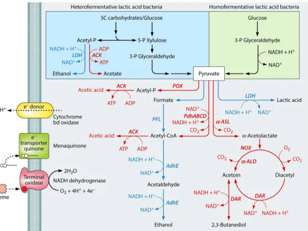
LAB are subjected to marked metabolic perturbations under en-vironmental stress conditions (Fig. 2and3). As a consequence of stress, cells lower their metabolic activities, which decreases en-ergy production and the generation of a proton motive force (PMF) and alters growth and viability (17,158). Overall, the re-sponses consist of the selection of alternative fates of pyruvate, the utilization of other carbon sources, the activation of the proteo-lytic system, and/or the catabolism of free amino acids (FAA) by cells. Metabolic adaptation is crucial for survival because it stim-ulates the production of additional energy and lowers the stress level, e.g., through alkalization of the cytosol under acidic condi-tions (159). The extent of metabolic perturbation and the main routes to reprogram pathways responsible for substrate catabo-lism in order to adapt to environmental stresses vary between LAB species (160).
Metabolism of Carbon Sources and Energy Production Under environmental stress conditions, LAB change metabolic and energy fluxes, modify the rate of growth, and adapt the me-tabolism of carbon sources to the new environment by modifying the synthesis of enzymes and metabolites (161). Environmental stresses inhibit the glycolytic pathway of Lc. lactis and decrease the synthesis of biochemical energy (17). Similar metabolic
perturba-tions are common in other LAB. Consequently, the ability of LAB to efficiently transport and metabolize carbohydrates and other carbon sources, such as malate and citrate, under environmental stress conditions is crucial for growth and persistence.
Transport and fermentation pathways of carbohydrates. LAB express numerous proteins responsible for carbohydrate trans-port and utilization. Proton-coupled active transtrans-port by proteins from the major facilitator superfamily (MFS), the glycoside-pen-toside-hexuronide (GPH) superfamily, and the ATP-binding cas-sette (ABC) superfamily and by group translocators, such as the phosphotransferase system– glucose-fructose-lactose (PTS-GFL) superfamily, is used most frequently (162). LAB modulate the synthesis of specific transporters depending on the type of carbo-hydrate available (163,164). Under acid stress conditions, Lb. ca-sei and S. mutans markedly decreased the synthesis of the phos-phoenolpyruvate phosphotransferase system (PEP-PTS) for glucose, which is the primary carbohydrate transport system be-longing to the PTS-GFL superfamily (165–167). Under the same conditions, an acid-resistant mutant of Lb. casei showed the high-est level of PEP-PTS and of the phosphocarrier protein (HPr). High levels of PEP-PTS may improve the acid resistance. The glu-cose PTS is upregulated at low pH in Streptococcus sobrinus (168) and Streptococcus macedonicus (34,36). Under optimal conditions and when cells are growing in glucose-rich media, HPr inhibits PEP-PTSs for carbohydrates other than glucose, preventing their
FIG 2 Schematic representation of changes of the carbohydrate metabolism, glycolysis, and fate of pyruvate in lactic acid bacteria. Colored arrows and enzymes indicate common reactions (black), those mainly induced during fermentation by unstressed cells (blue), and those induced in respiratory and/or environmen-tally stressed cells (red). LDH, lactate dehydrogenase; ACK, acetate kinase; POX, pyruvate oxidase; PFL, pyruvate formate lyase; PdhABCD, pyruvate dehydro-genase complex;␣-ASL, ␣-acetolactate synthase; AdhE, alcohol dehydrogenase; NOX, NADH oxidase; ␣-ALD, ␣-acetolactate decarboxylase; DAR, diacetyl reductase.
on May 22, 2020 by guest
http://mmbr.asm.org/
transport into the cell. In addition, glycolytic intermediates (e.g., fructose-1,6-bisphosphate) activate the phosphorylation of HPr at the serine residue at position 46 (169). The resulting P-Ser-HPr interacts with the global transcriptional regulator CcpA, and the complex prevents the catabolism of carbon sources other than glucose, the preferred sugar in most bacteria, by binding to the catabolic repression element (cre) upstream of the responsible genes and shutting them down. Under low-pH conditions, some LAB increase pyruvate kinase activity, which may accelerate the depletion of fructose-1,6-bisphosphate, relieving CcpA repression and allowing the use of alternative carbon sources (170).
Acid stress causes intracellular acidification, which decreases the activity of cytoplasmic enzymes (17). Transcriptomic and pro-teomic studies have highlighted that many LAB enhance the levels of glycolytic enzymes under acid, thermal, and osmotic stresses, but without increasing the synthesis of lactic acid (171,172). LAB such as Lactobacillus plantarum, Lactobacillus reuteri, Lactobacillus rhamnosus, and Lc. lactis modify pyruvate metabolism at the ex-pense of lactic acid, and they increase the synthesis of basic com-pounds (e.g., lysine and diacetyl/acetoin) (173,174), energy-rich
intermediates (such as ATP and NADH) (175), EPS, and/or gly-cogen (176). The level of lactate dehydrogenase (Ldh), which is responsible for the synthesis of lactic acid from pyruvate, mark-edly decreases. Pyruvate oxidase and phosphate acetyltransferase, used to synthesize acetyl-coenzyme A (acetyl-CoA), are induced in Lactobacillus delbrueckii subsp. bulgaricus and Lb. rhamnosus under acid stress conditions (161,170). Acetyl-CoA is rerouted toward the biosynthesis of fatty acids instead of butanoate (161, 170), which may enhance the rigidity and impermeability of the cytoplasmic membrane (177,178). Changes in pyruvate metabo-lism were also observed in S. mutans under acid stress (166,167). Metabolic adaptations in the presence of oxygen and ROS. Despite having a fermentative metabolism, several LAB species possess genes that code for a respiratory electron transport chain (179). They harbor the cydABCD operon, encoding a heme-de-pendent cytochrome with the capacity to generate PMF. Com-pared to when they are fermenting, LAB display an increase of extracellular pH and biomass under energetically favorable respi-ration conditions, that is, in the presence of oxygen, heme, and menaquinone (179–181) (Fig. 2). Lc. lactis growing anaerobically
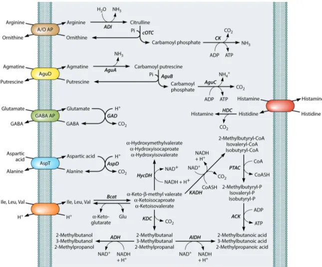
FIG 3 Schematic representation of the main free amino acid pathways of lactic acid bacteria induced under acid stress and/or starvation conditions. GABA, ␥-aminobutyric acid; ADI, arginine deiminase; cOTC, catabolic ornithine transcarbamoylase; CK, carbamate kinase; AguA, agmatine deiminase; AguB, pu-trescine carbamoyl transferase; AguC, carbamate kinase; AguD, agmatine/pupu-trescine antiporter; GAD, glutamate decarboxylase; HDC, histidine decarboxylase; AspD, aspartic acid decarboxylase; AspT, aspartate-alanine antiporter; Bcat,␣-ketoglutarate-dependent branched-chain aminotransferase; HycDH, hydroxy-acid dehydrogenase; KaDH, keto hydroxy-acid dehydrogenase; KDC, 2-keto hydroxy-acid decarboxylase; ADH, alcohol dehydrogenase; AIDH, aldehyde dehydrogenase; PTAC, phosphotransacylase; ACK, acetate kinase.