1
Alma Mater Studiorum –– Università di Bologna
DOTTORATO DI RICERCA IN
Scienze Chimiche
Ciclo XXIV
Settore Concorsuale di afferenza: CHIM/06
Settore Scientifico disciplinare: CHIMICA ORGANICA
TITOLO TESI
Synthesis of Modified Amino Acids and Insertion in Peptides and Mimetics.
Structural Aspects and Impact on Biological Activity.Presentata da: De Marco Rossella
Coordinatore Dottorato Relatore
Prof. Adriana Bigi Prof. Luca Gentilucci
2
Synthesis of Modified Amino Acids and Insertion
in Peptides and Mimetics.
Structural Aspects and Impact on Biological Activity.
by
Rossella De Marco
2012
3
4
Table of Contents
Cap 1. Chemical Modifications Designed to Improve Peptide Stability 1. Introduction
1.2. Enzymatic Degradation of Peptides
1.3. Structure Modifications to Improve Peptide Stability 1.3.1.Pseudopeptides
1.3.2. Reduced Peptide Bonds 1.3.3. Azapeptides
1.3.4. Retro-Inverso Peptides 1.3.5. Peptoids
1.4. Incorporation of Non-Natural Amino Acids 1.4.1. D-Amino Acids
1.4.2. N-Alkylated Amino Acids 1.4.3. α-Substituted α-Amino-Acids 1.4.4. β-Substituted α-Amino Acids 1.4.5. Proline analogues 1.4.6. β-Amino-Acids 1.5. Cyclization 1.6. β-Turn-Mimetics 1.7. Conclusion References
Chapter 2. Cyclopeptide Analogs for Generating New Molecular and 3D Diversity. 2. Introduction
2. 1. Matherial and Methods 2.2. General Methods.
2.3. General Procedure for Peptide Coupling 2.3.1. Boc group deprotection
2.3.2. Fmoc group deprotection
2.3.3. Cbz and benzyl group deprotection 2.3.4. General Procedure for Peptide Cyclization 2.4. Conformation Analysis
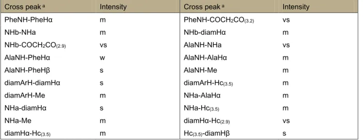
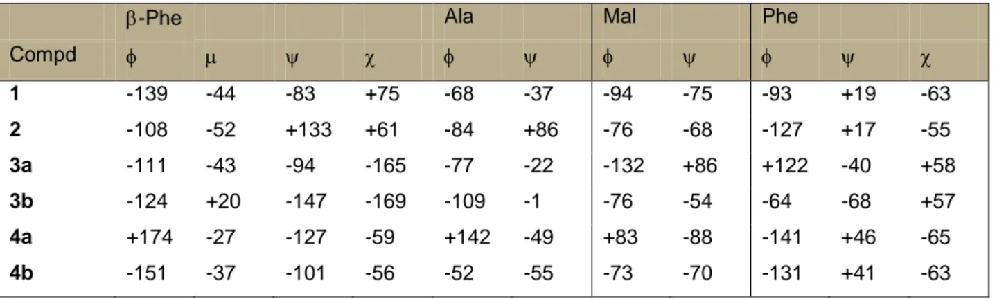
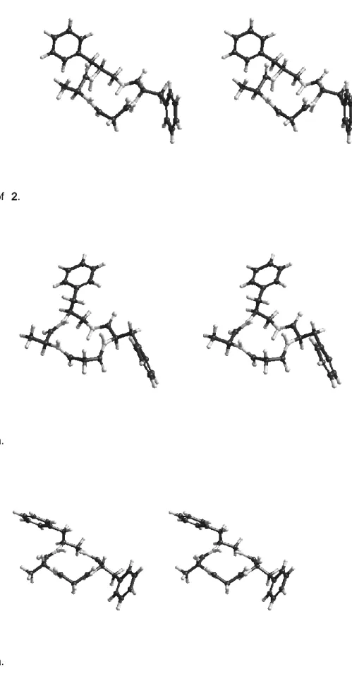
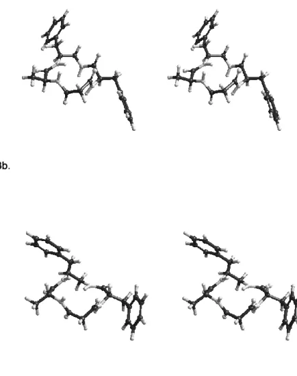
2.5.Results 2.6. VT-1H-NMR 2.7. 2D-ROESY
5 2.8. Discussion
2.9. Conclusions References
Chapter 3. Synthesis and Conformational Analysis of Cyclotetrapeptide Mimetic β-Turn Templates and Validation as 3D Scaffolds. 3. Introduction 3.1. Experimental section 3.1.1. General methods 3.1.2. Synthesis of 6 3.1.3. Synthesis of 9 3.1.4. Peptide cleavage 3.1.5. Cyclization 3.2. Conformational analysis 3.3. Cell adhesion assay 3.4. Supporting Information References
Chapter 4. Antiangiogenic Effect of Dual/Selective α5β1/αvβ3 Integrin Antagonists Designed on Partially Modified Retro-Inverso Cyclotetrapeptide Mimetics.
4. Introduction 4.1. Results
4.1.1. Inhibition of Cell Adhesion
4.1.2. Effect of Integrin Antagonists on in Vitro Elicited by Basic Fibroblast Grotw Factor (bFGF) 4.1.3. α5β1/αvβ3 Integrin Antagonists Do Not Affect Endothelial Cell Viability
4.1.4. Conformational Analysis of 2 and 3 in Solution 4.2. Molecular Docking
4.3. Discussion 4.4. Conclusions
4.5. Experimental Section 4.5.1. General Methods
4.5.2. Representative Synthetic Procedures and Analytical Characterization of PMRI RGD Mimetics 2 and 3.
4.5.3. 14 {c[βPheψ(NHCO)Asp(Ot-Bu)ψ(NHCO)Gly-Arg(Mtr)]} 4.5.4. 2 {c[βPheψ(NHCO)Aspψ(NHCO)Gly-Arg]}
4.5.5. 3 {c[(R)-βPheψ(NHCO)Aspψ(NHCO)Gly-Arg]} 4.6. Pharmacological Assays
6 4.6.2. Cell Culture
4.6.3. Cell Adhesion Assays 4.6.3. Flow Cytometry Assays
4.6.4. In Vitro Tubular Formation of HUVEC 4.7. Conformational Analysis 4.7.1. Molecular Docking 4.7.3. Protein Setup 4.7.4. Docking 4.8. Supporting Information References
Chapter 5. Molecular docking of opioid peptides and analogues, a powerful tool for the design of selective agonists and antagonists, and for the investigation of atypical ligand-receptor interactions. 5. Introduction
5.1. Structure and functions of the opioid receptors, and representative opioid ligands 5.2. Insights into the interactions between ligands and receptors
5.3. Principles of molecular docking.
5.4. Exploring the determinants of ligand affinity and selectivity: comparative docking studies 5.5. The docking of ligands lacking of the cationic amino group
5.5.1. Salvinorins
5.5.2. 6,6-Bicyclic enkephalin mimetics 5.5.3. Dhp-peptide
5.5.4. Fentanyl “carba”-analogues
5.6. The D-Trp-Phe β-turn MOR pharmacophoric motif References
Chapter 6. The Inverse Type II β-Turn on D-Trp-Phe, a Pharmacophoric Motif for MOR Agonist. 6. Introduction
6.1. Results
6.1.2. Cyclopeptide Design 6.1.3. Cyclopeptide Synthesis
6.1.3. Binding Affinity to the Cloned Human Opioid Receptors 6.1.4. Effects on Forskolin-Stimulated cAMP Production 6.2. Conformational Analysis
6.3. Molecular Backbone 6.4. Molecular Docking 6.5. Discussion
7 6.7. Experimental section 6.7.1. Chemistry 6.7.2. General Methods 6.7.3. Peptide Synthesis 6.7.4. Peptide Cleavage 6.7.5. Peptide Cyclization 6.8 Biology
6.8.1. Receptor Binding Assays to Cloned Human DOR and KOR 6.8.2. Determination of Inhibition of Cyclic AMP Accumulation 6.9. Conformational Analysis 6.9.1. Computational Methods 6.9.2. Molecular Docking 6.9.3. Hybrid QM/MM 6.9.4. Supporting Information References
Chapter 7. A simple route towards peptide analogues containing substituted (S)- or (R)-tryptophans. 7. Introduction
7.1. Results and Discussion 7.2. Conclusions
7.3. Supporting Information References
Chapter 8. Synthesis of Constrained Peptidomimetics Containing 2-Oxo-1,3-oxazolidine-4-carboxylic Acids. 8. Introduction
8.1. Results and Discussion 8.2. Conclusions
8.3. Experimental Section References
Chapter 9. Expedient Synthesis of Pseudo-Pro-Containing Peptides 9. Introduction
9.1. Results and discussion
9.1.1. Optimization of the Reaction Conditions 9.1.2. Synthesis of di-Oxd-peptides
9.2. Conformational Aspects of the Oxd peptides 9.3. Conclusions
8 9.4. Experimental part
9.4.1. Peptide Synthesis
9.4.2. Mono-Oxd-peptide Synthesis 9.4.3. Di-Oxd-peptide Synthesis
9.4.4. Di-Oxd-peptide Solid-Phase Synthesis 9.5. Theoretical Computations
9.5.1. Conformational Analysis 9.5.2.. Circular Dichroism 9.5.3. NMR Analysis
9.5.4. Roesy and Molecular Dynamics 9.6. Supporting information
References Conclusions Curriculum Vitae List of Publications
9
Abbreviations and acronyms
CNS = Central nervous systemBBB = Blood-brain barrier
ACE = Angiotensin-converting enzyme
ADMET = Absorption distribution metabolism excretion toxicity SPPS = Solid phase peptide synthesis
SAR = Structure-activity relationship RGD = Arg-Gly-Asp
CCK = Cholecystokinin
LHRH = Luteinizing hormone-releasing hormone TRH = Thyrotropin releasing hormone
PEG = Polyethylene glycol
PMRI = Partially modified retro inverso RCM = Ring-closing methatesis Boc = Tert-butyloxycarbonyl Fmoc = 9H-fluoren-9-ylmethoxycarbonyl EDCI = 1-ethyl-3-(3-dimethylaminopropyl)carbodiimide HOBt = Hydroxybenzotriazole PG = Protecting group Bz = Benzyl DIC = N,N'-diisopropylcarbodiimide DEAD = Diethylazodicarboxylate Cbz = Benzyloxycarbonyl Dpp = Diphenylphosphinamide LDA = Lithium diisopropylamide
Salen = Salicylic aldehyde ethylenediamine A* = Chiral auxiliary
TMS = Trimethylsilyl
PyBOP = Benzotriazol-1-yl-oxytripyrrolidinophosphonium hexafluorophosphate DIEA = Diisopropylethylamine
TFA = Trifluoroacetic acid Bom = Benzyloxymethyl
Pip = Pipecolic acid or piperidine TEA = triethylamine
DPPA = diphenylphosphorylazide DCM= dichlorometane
10 THF = tetrahydrofuran
DMF = dimethylformamide EM = endomorphin MOR, DOR, KOR,
-, δ- ,k- = opioid receptor, respectively EL = extracellular loop
TMH = transmembrane helix
2D, 3D = two-, three-dimensional, respectively
HBTU = 2-(1H-Benzotriazole-1-yl)-1,1,3,3-tetramethyluronium hexafluorophosphate DMSO = dimethyl sulfoxide
RP = reversed-phase
ES-MS = electrospray ionization mass spectrometry SE = standard error
DAMGO = H-T yr-D-Ala-Gly-N-MePhe-glyol DPDPE = [D-Pen2,D-Pen5]-enkephalin
JOM-6 = Tyr-c(S-Et-S)[D-Cys-Phe-D-Pen]NH2 MD = molecular dynamics
Dock = molecular docking DO = docking orientation VT = variable temperature
AMBER = assisted model building with energy refinement TIP3P = transferrable intermolecular potential three point DAD = diode array detector
HMBC = heteronuclear multiple-bond coherence HSQC = heteronuclear single-quantum coherence rt = room temperature
CPK = Corey, Pauling, and Koltun
CCP4 = Collaborative Computational Project 4 PBS = phosphate-buffered saline
bFGF = basic fibroblast growth factor CTP = cyclotetrapeptide
K562 = human erythroleukemic cells
SK-MEL-24 = human malignant melanoma cells HUVEC = human umbilical vein endothelial cells BSA = bovine serum albumin
ECM = extracellular matrix S.E.M. = standard error of mean ANOVA = analysis of variance test RMSD = root mean square deviation
11 MIDAS = metal-ion-dependent adhesion site
ODS = octadecyl silane
EDTA = ethylenediaminetetraacetic acid MEM = minimum essential medium RPMI = Roswell park Memorial Institute FBS = fetal bovine serum
PBS = phosphate buffered saline PMA = phorbol 12-myristate 13-acetate PI = propidium iodide.
12
Chapter 1
Chemical Modifications Designed to Improve Peptide Stability
1. IntroductionPeptides are amino acid derived compounds containing at least one amide (peptide) bound. Conventionally, peptides up to 20 amino acids are named oligopeptides, while the term polypeptatide refers to peptides up to 100 amino acids. From the structural point of view, peptides encompass diverse types such as linear, cyclic peptides, depsipeptides, and peptides modified with diverse nonpeptide moietes including phosphoryl groups or carbohydrate, polyketides or terpenoids, etc [1].
The evolution of enzymatic synthesis, recombinant DNA technology, and automated synthetic methodologies in particular SPPS, allow for the production of large libraries of diverse peptides characterized by a range of pharmacological effects. Peptide or peptidomimetic drugs are currently utilized for the treatment of prostate and breast cancer, as HIV protease inhibitors or as ACE inhibitors, against hypertension and heart failures, as antibiotics, hormone, neurotrasmetters, immunomodulators, and so on. Peptides have the potential to be potent pharmaceutical agents for the treatment of many diseases of the Central Nervous System (CNS)[2,3]. Unfortunately, the clinical use of these promising drugs is hampered by their rapid degradation and scarce permeation across biological barriers, such as the intestinal lumen, the intestinal mucosa, the blood-brain barriers (BBB), etc. these problems lead to short in vivo half-lives (generally <30 min) and low oral bioavailability (1-2%)[4,5].
The peptidomimetic strategy consists of altering the physico-chemical characteristics of a peptide without changing the biological activity [2]. Peptidomimetic compounds bear identifiable similarity to the parent peptides and imitate or inhibit their biochemical effects. Very often, they contain non peptidic structural elements [6], such as peptide bond-surrogates not cleaved by peptidases, a feature also found in natural peptidase inhibitors.
The design of a peptidomimetic based on the structure of a native peptide begins with a extensive SAR investigation of the parent peptide, aimed to identify the minimal sequence and the pharmacocophoric groups responsible for the bioactivity. Next, the most active 3D display of these key residues is determined by means of conformationally rigid analogues [7,8]. Moving from the resulting 3D model, the pharmacophores can then be recombined by the use of non-peptidic scaffolds. Finally, the resulting compounds are tested to demonstrate the biological activity, stability, bioavailability and conformational stability, and ADMET profile. Many non-peptide mimetics of bioactive peptides have been reported in the literature. The αvβ3 -integrin inhibitor (1), SB223245, with a 1-4 benzodiazepine nucleus as central ɣ-turn mimetic scaffold [9], the TRH (pGlu-His-Pro-NH2 ) analogue (2), containing a cis-1,3,5-trisubstituted cyclohexane scaffold [10], the glucose-derived non-peptidomimetic of somatostatin (3) [11], and the C2-symmetric cyclic urea as HIV protease inhibitor (4) [12], represent outstanding example of non-peptidomimetics with high biological activity and increased enzymatic stability, Fig (1). However, in several cases the non-peptidic compounds have failed to reproduce the biological activity of the natural peptide [13,14]. Therefore, peptidomimetics still preserve their attractiveness for replacing the original peptides.
13
The field of peptidomimetics has been very extensively reviewed. Some of the most relevant kinds of peptidomimetic therapeutic agents describe in the literature are the HIV protease inhibitors [15], anti thrombotic agents [16], ACE [17] and renin inhibitors [18], etc.
It is worth notice that many of the tricks used by chemists to enhance activity and stability, by protecting peptides against both endo and exo-peptidases, can also be found in active peptides of microbial or marine origin: D-amino acids or inusual amino acids instead of the natural L-residues, cyclization, glycosilation, deamination, complete removal of the first residue, N-acylation or N-formylation at the N-terminus, amidation of the C-terminus, etc [1,2].
Fig.(1) Selected examples of non-peptidomimetics
It is also possible to introduce temporary modifications using a prodrug approach [19]. A peptide prodrug can be obtained by combining a biolagically active peptide with additional elements which give the whole molecule increased resistance against enzymatic hydrolysis and/or bioavaibility [19,20] .
Proteolytic cleavage of the additional molecule, especially a short truncation at the N-terminus, in the proximity of the site of action, release the pharmacologically active drug. Prodrug or prodrug-enzyme inhibitor combinations may optimize the delivery of peptide or protein drugs to the CNS. For example, the N-terminal 4-imidazolidinone prodrugs of Leu-enkephalin, being metabolically stable and bioreversible, have been proposed as a suitable prodrug candidates for delivery of Leu-enkephalin to the brain [21].
Peptide stability can be achieved by conjugation to a polymer [22]. Polymer conjugation improves pharmacokinetics by increasing the molecular mass of protein and peptides, preventing the approach of antibodies or antigen processing cells and shielding them from proteolytic enzymes. The most promising polymer is PEG [23], which shows little toxicity and is eliminated from the body intact. Polymer conjugation allows an increase peptide stability, and reduce elimination. The conjugated PEG-DPDPE seems to act as a
14
pro-drug, enhancing peripheral stability, while undergoing hydrolysis in the brain and allowing nonconjugated DPDPE to act at the receptor [24].
Finally, proteolytic peptide degradation can be defeated using alternative routes of administration, including controlled-release parenteral route (subcutaneous, intramuscular or intravenous), mucosal route (nasal spray, pulmonary delivery, sublingual delivery), oral route (penetration enhancers, protease inhibitors,carriers) and transdermal route (patches) [25].
1.1. ENZYMATIC DEGRADATION OF PEPTIDES
The therapeutic efficacy of a peptide drug candidate is linked to its activity at the specific receptor,as well as to its pharmacokinetic properties (adsorption, transport, ability to cross biological barriers, excretion) and toxicity. Besides to aqueous solubility, lipophilicity, molecular size and weight, and ability to form H-bonds (Lipinski's Rule of 5), chemical and metabolic stability plays a major role in determining peptide bioavailability. Peptide degradation by protelytic enzyme is followed by rapid excretion of the metabolites from the circulation by the liver and kidneys [4,5].
The enzymatic stability of a peptide is dependent upon several factors, in particular kind and sequence of the amino acids, overall size, flexibility, and conformations. Side chain metabolism, such as the oxidation and reduction of disulfide bonds, can also play a important role. Amino acid composition and peptide structure also determine lipophilicity, the degreeof protein binding, cellular sequestration, uptake into non-target tissue, clearance rate, and affinity for carrier mechanisms.
Peptidase that are capable of cleaving the internal peptide bonds of a substrate are designated as endopeptidases ( e.g., serine proteinases, metalloproteinases). The peptidase which remove one or more residues from the termini of the peptide are classified as exopeptidases (e.g.,aminopeptidases, carboxipeptidases) [26,27.] Large peptides or peptides protected at the N-terminus, or at the C-terminus, require endopeptidases to initiate hydrolysis.
After administration, peptides meet proteolytic enzymes at many compartments [28]. In case of intravenous injection, the peptide is immediately subjected to numerous proteolytic enzymes such as esterases and peptidases in the human plasma [29]. In case of oral delivery, a part from the strong acidic gastric enviroment, the peptide encounters two main biophysical and biochemical barriers, the lumen of the small intestine, and the brush border membrane. For peptides drugs targeting the CNS, the BBB also costitutes a formidable barrier [30].
The metabolic activity in the intestinal lumen reduces the absorption of peptide-based drugs. The gastrointestinal tract degrades proteins and peptides by using variety of enzymes into smaller sequences, which can be easily absorbed across the intestinal mucosa [31,32].In the duodenum, the degradation can be mediated by pancreatic proteases. The contribution of luminal hydrolysis in the overall degradation process depends on the size and composition of the peptide. Most of the degradation of peptides requires at least contact with the brush-border membrane and/or uptake into the intestinal mucosal cells.
Among the most relevant peptidases which can be found in the intestine, it is possible to mention aminopeptidase P, aminopeptidase W, aminopeptidase N and dipeptidyl peptidase IV. The lumen of the small intestine contains a number of pancreatic peptidases, α-chymotrypsin, trypsin, pancreatic elastase,
15
carboxypeptidases A, B, D, N, U, etc., and cellular peptidases secreted by mucosal cells. The brush border membrane of the epithelial cells contains many different peptidases [33], dipeptidyl-peptidase IV, prolyl tripeptidylpeptidase, ACE, leucyl-aminopeptidase, aminopeptidase M, aminopeptidase A, neprilysin, etc. Many enzymes are also present in the liver [34], kidney and other organs, or in different tissues. For instance, lysosomal peptidases, leukocyte elastase, cathepsins B, D, etc., can be found in epithelial or endothelial cells; other enzymes are the interstitial collagenase (MMP-1), or carboxypeptidase C or Y.
The BBB is a unique physical and enzymatic barrier that segregates the brain from the systemic circulation. BBB capillary endothelia are sealed by tight junctions, which inhibit any significant paracellular transport [30.] Specific transporters exist at the BBB that permit nutrients to enter the brain and toxicants/waste products to exit. These transporters are potential routes for mimetic designed drugs. The main peptidases which can be found in the brain microcapillaries of the BBB are gamma-glutamyl transpeptidase alkaline phosphatase, monoamine oxidase catechol-O-methyl transferase, butyrylcholinesterase and aromatic-L-amino-acid carboxylase (or Dopa-decarboxylase or aromatic-L-amino-acid decarboxylase), epoxidehydrolase (or epoxide hydrolase), UDP-glucuronosyl-transferase, benzyloxyresorufin-O-deethylase (cytochrome P-450 CYP2B1), NADPH cytochrome P-450 reductase and glutathione-S-tranferase [22]. The protein-disulfide reductase, is also present in the brain and can alter peptide structures stable in plasma.
In many cases, the active peptides are enzymatically converted to products which retain some bioactivity. These bioactive metabolites may mimic but also counteract the action of the parent peptide.
The released fragment may serve as a modulator of the response of the original compound [35]. This phenomenon has been found to occur in several neuropeptide systems, including the opioid peptides, tachykinins, as well as peptides belonging to the renin-angiotensin system. Normally, the products interact with the same receptor as the native compound, but sometimes it appears that the released fragments interact with sites distinct from those of the original peptide.
1.2. STRUCTURE MODIFICATIONS TO IMPROVE PEPTIDE STABILITY
As mentioned in the introduction, peptidomimetics resemble native peptides or proteins but contain some synthetic element designed to reduce metabolism and to optimize the biological activity of the agent. Peptide bond hydrolysis in vivo can be limited by specifically protecting or replacing the targeted bond, by introducing atypical moieties, or by modifying the peptide conformation alltogether, in such a way that it is no longer recognized by the protease of concern. Even modest structural changes near the scissile peptide bond can result in significant conformational differences. Example are the introduction of a N-alkyl group, that increases the incidence of the cis configuration of the amide bond, the use of a D-amino acid, or of a residue containing an unnatural side chain. In many cases, the introduction of non-peptidic scaffolds to imitate the secondary structure that are thought to be especially involved in binding interactions, including the ɣ- and β-turn, β-sheet and the α-helix, proved to be a very effective strategy.
16 1.2.1. Pseudopeptides
The backbone of a peptide can be modified in various ways by changing at least one peptide bond with a isosteric or isoelectronic surrogate. Examples are shown in Fig. (2). The mostly utilized isosters are the reduced amides, azapeptides, retro-inverso peptides, and peptoids; these are discussed in detail in the next sections. Other isosters less frequently appeared in the literature, such as the urea peptide mimetics [36,37], sulphonamide peptides/peptoids [38], etc., are not reviewed here.
The replacement of a labile peptide bond with a isoster was of great help for designing therapeutic agents targeting proteases, like those associated with the HIV virus, as well as targets like ACE, renin, endothelin, interleukin-converting enzyme and others [15-18]. Very often, the isoster imitates the transition state of peptide bond cleavage, including the hydroxyl group resulting from enzyme nucleophilic addition, Fig. (2), hydroxyethylamino, hydroxyethylene isosters, etc.
Fig.(2) Isosteric surrogates of the peptide bond
1.2.2. Reduced Peptide Bonds
The incorporation of reduced peptide bonds (CH2-NH), Fig. (2), renders the native sequences of opioid peptides highly resistant towards enzymatic hydrolysis in the modified positions. Synthetic peptides containing reduced bonds have found applications as vaccines for their immunogenic properties, linear pseudooligolysines, containing multiple adjacent CH2-NH bonds have been designed as DNA carriers in gene delivery. Reduced amides have also seen use in the preparation of peptide nucleic acids and antibacterial peptides [2,6]. Representative examples in the field of opioid peptides are the TIPP-derived opioid antagonists with subnanomolar affinity and high δ-receptor selectivity, obtained by introduction of a
17
reduced peptide bond between Tic2 and Phe3 residues, to give H-Tyr-TicΨ[CH2NH]Phe-Phe-OH (TIPP[Ψ]) and H-Tyr-TicΨ[CH2NH] Cha-Phe-OH, Cha: cyclohexylalanine (TICP[Ψ]). The modification conferred the molecules a high stability against chemical and enzymatic degradation [39].
Introduction of the CH2-NH peptide bond isoster can be accomplished in solid phase. The free N-terminal amino group of the resin-bound peptide is reductively alkylated by the requisite protected α-aminoaldehyde in the presence of sodium cyanoborohydride (NaBH3CN) in DMF containing 1% AcOH. Microwave irradiation can be utilized to shorten the reaction times and improve the yields [40].
1.2.3. Azapeptides
In azapeptide isosters the α-CH group of the backbone is substituted by a isoelectronic nitrogen atom, the side chains remaining unaltered, Fig. (2). Azapeptides have been developed by several groups for the design of hormone analogues, protease inhibitors, etc [41]. Examples of therapeutically useful peptides incorporating the azapeptide modification can be found in the field of serine and cysteine proteases inhibitors [42]. Atazanavir (5), Fig. (3), BMS-232632, is a highly active azapeptide inhibitor of the HIV protease, that has recently received approval as a human immunodeficiency virus (HIV) treatment [43,44]. It inhibits the protease enzyme, thereby preventing the cleavage of the viral polyproteins and resulting in an immature, non-infectious virion. It is the first, and to date the only, protease inhibitor designed to be applied once daily, with comparable anti-HIV efficacy to nelfinavir, efavirenz and the combination of ritonavir and saquinavir [45]. The synthesis of azapeptides generally starts from substituted hydrazines or hydrazides [46].The preparation of Atazanavir (5) is shown in Fig. (3). The key building blocks are the hydrazino carbamate, obtained in turn by a Suzuki-Miyaura coupling, the amino acid-derived N-protected threo-3-amino-1,2 epoxybutane, and two equivalents of N-protected-tert-leucine [45].
18 1.2.4. Retro-Inverso Peptides
In these peptide-mimetics the normal sequence from N- to C- terminus is reversed, and the natural L-amino acids are changed by D-amino acids, Fig. (2) [47].This reversal warrants that the side chain topologies of the natural peptide and the peptidomimetic are the same. Peptide-bond reversal represents an important structural alteration for peptides, and proved to be useful to reduce the degradation rate of the peptides by peptidases. In a retro-inverso sequence the N- and C-termini are reversed, therefore the positive charge located at the N-terminus of the natural sequence is replaced by a negative one in the peptidomimetic, and vice-versa, unless modified termini are introduced. This may be the cause of the low biological activity observed in several cases. The introduction of end-group modifications, Fig. (2), increases the complementarity with the native peptide (see also PMRI).
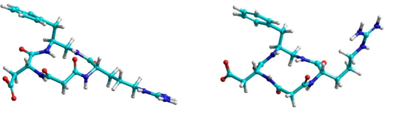
Retro-inverso peptides have found applications as immunogens, immunomodulators, immunostimulators; and as anti-inflammatory, antimicrobial, and diagnostic reagents, as well as modified isomers of membrane-penetrating peptides as delivery systems [48]. An evolution of the retro-inverso concept is the partially modified retro-inverso (PMRI) peptide, in which the retro-inverso structures is incorporated into a normal sequence; the retro inverso and the normal portions are connected by a diamine and/or a diacid.
The PMRI Tuftsin analogue 6 [49], Fig. (4), is degraded less than 2 % in 50 min, while maintaining the biological activity of Tuftsine, H-Thr-Lys-Pro-Arg-OH, a immune system stimulator, which is completely degraded in vivo in about 8 min. Other nice examples can be found as mimetics of enkephalin, CCK, RGD, gastrin antagonists, etc. [47,48]. In general, the analogues displayed higher activity than the parent peptides in an in vitro test. Another example is the angiotensin analogue incorporating aza-α'- homoamino acids of natural and inverted configuration, [Asn1, aza- α'-homoTyr4, Val5]angiotensin II (7), Fig. (4).
Fig.(4) PMRI analogues of Tuftsin (6) and Angiotensin II (7).
The synthesis of a PMRI peptide requires standard conditions. The principal concern of PMRI peptide synthesis is the construction of the gem-diaminoalkyl (8), Fig. (5), and C-2-substituted malonyl residues (9), Fig. (6).
The Curtius and Hofmann rearrangements remain the methods used most commonly for the syntheses of gem-diaminoalkyl derivatives (8); during these rearrangements the migrating groups retain the configuration, Fig. (5). Acyl azides undergo the Curtius rearrangement to yield the corresponding isocyanates, whose hydrolysis give the gem-diamines. Acyl azides can be prepared from the amino acids by treatment with diphenylphosphoryl azide (DPPA), or via the intermediate mixed anhydride [47].
19
The isocyanates can be reacted with a carboxylic acid derivative to yield a PMRI peptidic unit directly [50], the so-called “Goldschmidt and Wick type reaction”. The Hofmann rearrangement is extensively employed for the synthesis of PMRI peptides, using exclusively the mild oxidant iodobenzene bis(trifluoroacetate) (IBTFA). Other procedures to synthesize monoprotected gemdiaminoalkyl (8), are based on the Mannich reaction [51], on the formation of intermediate oxazolones, or nitriles, etc.
Fig.(5) Preparations of the gem-diaminoalkyl residues.
Concerning the preparation of C-2-substituted malonyl derivatives (9), the classical method is the alkylation and partial hydrolysis of malonic acid diesters, and of cyanoacetates. A very convenient method is based on the use of Meldrum’s acid, Fig. (6). The Knoevenagel reaction with aldehydes or some ketones and in situ reduction yields mono-C-5-substituted Meldrum’s acids. Subsequent alcoholysis gives C-2-substituted malonic acid monoesters. The acylation of the enolate of a tert-butyl carboxylate with a carbonate, a chloroformate, or a isocyanate, can be utilized to obtain C-2-substituted malonates. Special issues to take into consideration are the acidity of the C-2 hydrogen of malonates, and the configurational lability of the C-2 malonyl position during synthesis.
Fig.(6) Preparations of the C-2-substituted malonyls 1.2.5. Peptoids
In the early 1990’s Bartlett defined peptoids as pseudopeptides containing N-alkylated glycines linked by peptide bonds. Formally, the nitrogen atom of some residues is shifted to the α-CH position, and the NH-groups have been substituted by CH2-groups, Fig. (2).
Therefore, the side chains and the carbonyl groups remain at their positions, while the backbone CH- and NH-groups change their places. The sequence of peptoids are opposite to the ones of native peptides, and the stereogenic α-carbons of natural amino acids are lost; besides, they have higher conformational flexibility respect to natural peptides. Peptoid analogues of most natural amino acids are commercially available, or they can be easily prepared. Peptoids (10) can be routinely synthesized on Rink amide linker-derivatized solid supports using the submonomer synthesis method developed by Zuckermann et al., Fig. (7) [52,53].
20
Metabolically stable, successful compounds based on the peptoid concept are the α-amylase inhibitors Ac-Nhtrp-Nharg-Nhtyr- NH2, and Ac-Nhtyr-Nharg-Nhtrp-NH2 [14], active as or more active then their natural parent peptides (Nh indicates the peptoid homologue of the natural amino acid); other examples are the antimicrobial peptoids derived from pexiganan, protegrins, and melittin [54].
Fig.(7) Example of SPPS of peptoids.
1.3. Incorporation of Non-Natural Amino Acids
Peptide analogs containing non-natural residues have been obtained by diverse approaches [55], ranging from the simple replacement of the natural L-amino acids with their D-enantiomers, to the use of N-alkyl amino acids, α-substituted α-amino acids, β-substituted α-amino acids, proline analogues, ɣ- and β-amino acids, substituted α- or β-amino acids, and so on. For the huge number of non natural amino acids described in the literature [56,57], only these relevant families are discussed in the following paragraphs.
In some cases, it has been observed that peptide coupling with unusual amino acids can be troublesome. For instance, with N-methyl or Cα,α-disubstituted amino acids, racemisation, diketopiperazine formation, etc., are common side-reactions.
1.3.1. D-amino Acids
The introduction of D-amino acids [58] in a sequence can give the peptide an increased stability, since only a few enzymes that effectively hydrolyse peptide bonds involving D-amino acids have been discovered and characterized in multicellular organisms [59].
Moreover, D-residues often enforce a different conformation of the peptide [60], and strongly influence receptor affinity and selectivity [61]. Some of the first successes of this approach [62] in the field of opioid peptides have been the δ-receptor selective enkephalin analogues DADLE, H-Tyr-D-Ala-Gly-Phe-D-Leu-OH, and the μ-receptor selective DAMGO, H-Tyr-D-Ala-Gly-MePhe-Glyol, widely used as a radioligand for binding experiments in its [3H]-form [3,63].
1.3.2. N-Alkylated Amino Acids
N-alkylation (generally N-methylation) is present in a number of biologically active, natural peptides from different sources, in particular of marine or microbial origins, including antibiotics, monamycins, echinomycin, or insecticides, antitumor agents, such as bouvardin, or antiinflammatory peptides [64,65]. For instance, the cyclic undecapeptide cyclosporine A [66], isolated from Trichoderma polysporum, contains seven N-methylated amino acids.
21
Several N-alkyl amino acids are commercially available, allowing their direct use in solid phase peptide synthesis, while many others can be prepared [64,67-69]. N-methyl amino acids (11) can be synthesized from N-protected amino acids, by direct methylation of carbamate or diphenylphosphinamide (Dpp) protected amino acids, Fig (8)A, or by the Mitsunobu reaction, with arylsulfonyl protecting group, Fig (8)B. Oxazolidinones obtained from the N-carbamate amino acids and formaldehyde can be reduced to N-methylamino acid with triethylsilane, Fig (8)C. Alternatively, oxazolidinones obtained with hexafluoroacetone can be treated with paraformaldehyde and thionyl chloride; reduction and deprotection affords the N-methyl amino acid.
Fig. (8). Syntheses of N-methyl amino acids.
The alkylation of amino acids has been obtained by two successive reductive aminations of aldehydes, Fig. (9)A [64]. N-methylation has been performed via sultam-directed “enolate” hydroxyamination of non-chiral acyl chains, Fig. (9)B. The sultam chiral auxiliary served also for the alkylation of chiral enolate derived from sarcosine (N-methylGly).
Finally, the N-methylation of a peptide can be directly performed on solid support; for instance, the key step of Fig. (9)C is the selective deprotonation of the resin-bound free amine peptide protected as o-nitrobenzene sulfonamide with a non-ionic base, and alkylation with methyl p-nitrobenzenesulfonate. This strategy has been applied to the N-methyl scan of the thrombin receptor agonistpeptide H-SFLLRNNH2[70].
22
Fig. (9). Other syntheses of N-methyl amino acids.
Generally, peptides modified by the use of N-methyl amino acids resulted in analogues with improved pharmacological properties and stability. The role of the position to be N-methylated for peptide protection from proteolysis is essential. Substance P (Arg-Pro-Lys-Pro-Gln-Gln-Phe-Phe-Gly-Leu-Met-NH2) had been shown to be degraded in the human brain at peptide bonds 5-6, 7-8 and 8-9.
The heptapeptide analogue, Glu-Gln-Phe-NMePhe-NMeGly-Leu-Met-NH2, was almost completely resistant [71]. To mention another case, in comparison to endothelin (half-life circa 10-20 min), the N-methylated analogues revealed an increased stability by 500-800 fold [72]. In neurotensin(7-13) (Pro-Arg-Arg-Pro-Tyr-Ile-Leu), the scissile bonds are the positions Arg8-Arg9, Pro10-Tyr11 and Tyr11- Ile12. N-methylation in position 8 led to increased half-life in plasma [73]. Finally, the N-methyl modification has been applied also to enzyme inhibitors, enkephalin, LHRH, angiotensin, and CCK [64,65].
The presence of the N-alkyl group affects the conformational freedom of the backbone and of the side chain of the residues close to the N-alkyl group. In particular it eliminates the predominance of trans versus cis peptide bond configuration. Besides, the substitution of NH by N-alkyl groups eliminates some inter- and intramolecular hydrogen bonds. Finally, the adjacent carbonyl group increases basicity and decreases polarity [74].
Besides to their utility to protect biologically active peptides against enzymatic degradation without concomitant loss of biological activity, N-alkyl residues have been also widely utilized for SAR studies. By successively alkylating each backbone NH and evaluating the biological activity (N-alkyl-scan), the pharmacophoric residues can be identified. A prototypic example is represented by the N-methyl scan performed on the cyclo RGD analogues by Kessler et al., discussed in the paragraph dedicated to cyclization.
23 1.3.2. α-Substituted α-Amino Acids
α-Substituted α-amino acids, or α,α-disubstituted glycines, are present in many natural sequences, for instance in several peptide antibiotics [75], such as alamethicin, an antimicrobial membraneactive peptide [76]. Among the more represented α-alkyl α-amino acids it is possible to cite α-aminobutyric acid (Aib), diethylglycine (Deg), or isovaline (Iva), Cα-methyl-Cα-allylglycine (Mag), (αMe)- α,α-diphenylalanine (α MeDip), and several kinds of cyclic or heterocyclic derivatives, Fig.(10).
α-Substituted α-amino acids have been used for the synthesis of peptidomimetics as enzyme inhibitors, and to provide peptides with a higher resistance to biodegradation. For example, in contrast to angiotensin II, the [ α MeTyr4] analogue is resistant to chymotrypsin [77]. Aib-containing analogues of the insect kinin neuropeptide family also demonstrate resistance to an insect ACE [78]. Incorporation of Aib has been described also for enkephalin, bradykinin, angiotensin II [64].
Fig. (10). Examples of alfa-alkyl alfa-amino acids.
One of the more relevant features of α-substituted α-amino acids is the conformational constraint introduced into peptide backbones [79,80]. Aib, the most widely studied of the family, restricts ϕ and Ψ to angles present in α- or 310 helices. When Deg is utilized, the preferred conformation is extended with trans ϕ and Ψ angles. A noteworthy conformational restriction is obtained when residues having the two side chains in a ring are utilized, leading to a β-turn secondary structure or a helix 310. Interestingly, this introduction gives the peptides increased resistance against hydrolysis [81,82]. A nice example is represented by the family of the α-aminocycloalkane carboxylic acids, Acn c. For instance, the introduction of Ac6c into various positions of Leu-Enkephalin, resulted in peptide mimetics with greater in vivo activity.
The synthesis of α-substituted α-amino acids (12) [64] can be performed by the stereoselective alkylation of imidazolidinones, Fig. (11A). Variants based on the use of other intermediate heterocycles are the alkylation of bis-lactim ether (13), obtained by treatment of the L-alanine diketopiperazine, or the alkylation of metallated imidazolidinones (14), obtained by cyclization of chiral α-isocyanoamides. α-Methylamino acids can be prepared by alkylation of Schiff bases derived from chiral amino acids and Oppolzer’s sultam, Fig. (11B). The asymmetric alkylation of alanine enolates with chiral phase transfer catalysts, for instance with copper complexes of Salen, proceeded with ee up to 90%, Fig. (11C).
24 Fig. (11). Syntheses of alfa-substituted alfa-amino acids.
1.3.4. β-Substituted α-Amino Acids
Analogues of natural amino acids alkylated at the β -carbon have been often utilized to induce a conformational preference in side chains. Some β-Me analogues of Phe, Trp, and Tyr, are shown in Fig. (12). These β-substituted α-amino acids have also an additional β-stereogenic center, therefore four preferred configurations (-gauche, +gauche, and two enantiomeric trans geometries) are accessible from varying the two stereocenters, Fig. (13). 2-(Carboxycyclopropyl)- glycine (CCG) is a different kind of β-substituted amino acid.
25 Fig. (13). Preferred conformations of beta-substituted alfa-amino acids.
Replacement of the natural amino acids often resulted in a comparably higher activity and increased biological stability with respect to the modified peptides [83]. For instance, the activity of short peptides which are active at the δ-opioid receptor was successfully improved by exchanging phenylalanine by its β-Me analogue [84,85]. Also, the introduction of three methyl groups at the 2’-, 6’- and β-position of natural tyrosine hinders the free rotation around the χ an angle giving compounds with improved biological activity [86].
1.3.5. Proline Analogues
The cyclic structure of proline forces the ϕ angle to -65°+/-15°, thus preventing the formation of a α-helix, and promoting the formation of a β-turn. Besides, while the barrier to secondary amide cis/trans isomerization is about 10 kcal/mol, Fig. (14), the presence of Pro reduces the barrier to just 2 kcal/mol, hence influencing the biological behaviour of peptides [87,88].Many Pro derivatives were found in proteins of microbial or marine origins, Fig. (14), in antibiotics and cytotoxic peptides. Many other Pro derivatives were synthesized by the introduction of alkyl chains, aromatic groups, heteroatoms, or halogens in different positions of the fivemembered ring [89].
Some analogues are characterized by smaller or larger rings, such as azyline-2-carboxylic acid (Azy), azetine-2-carboxylic acid (Aze), or pipecolic-2-carboxylic acid (Pip). The difference among Azy, Aze and Pro is largely the steric bulk of the side chain rather then ϕ and Ψ angles, while Pip prefers a chair conformation in which the COOH group is axial. Finally, the 5,5-dimethylthiazolidine- 4-carboxylic acid (Dtc) allows angles in the β-turn region.
26 Fig.(14) Proline analogues.
It has been utilized in place of Pro in Angiotensin II, H-Asp-Arg- Val-Tyr-Ile-His-Pro-Phe-OH, a key octapeptide in blood pressure regulation, resulting in a peptidomimetic with about 40% greater activity respect to the native peptide [90].
1.3.6. β -Amino Acids
β- (and ɣ-) amino acids have been utilized to construct the mimetics of naturally occurring peptide hormones, MHC-binding beta-peptides, opioid peptides, somatostatin, or amphipathic betapeptide inhibitors of membrane-bound proteins [91]. There are different kinds of β-amino acids, the β2- or β3- versions, Fig. (15), which can be further distinguished in homologated β-amino acids, possessing an extra C atom, or isomeric β -amino acids, which maintain the same MW of the corresponding α-analogue.
Fig.(15) Beta-amino acids
The β3-amino acids are much more utilized than the β2 ones. All appropriately protected β3-derivatives with proteinogenic side chains, with a few exceptions, are commercially available. The enzymatic resolution of racemates with isolated immobilized enzymes or with cell cultures constitutes a cheap and easy method to obtain optically active β-amino acids [92]. Among the many enzymes which have been utilized, chymotrypsin, β-lactamases, nitrilases, hydantoinases, lipases, transferases and isomerases, one of the most general and substrate-tolerant is the penicillin acylase.
27
The β3-amino acids, with proteinogenic or non proteinogenic side chains, are readily obtained by direct Arndt–Eistert homologation of the Boc or Fmoc β-amino acids, Fig (16A). Other homologation procedures have been also proposed, but these are generally less efficient.
Concerning the preparation of substituted β3-amino acids [93], the best options are the functionalization of intermediate di- or perhydropyrimidin- 4-ones, Fig (16B), and the conjugate addition to α,β-unsaturated esters or imides Fig (16C). The latter procedure was developed in particular with lithium amides of chiral amines as nucleophiles, or with chiral auxiliaries.
Fig.(16) Synthesis of beta-amino acids
The β2-amino acids have been prepared by a number of different synthetic strategies [94].The use of chiral auxiliaries and catalysts for C(2)–C(3) bond formation is well documented. To mention a specific approach, aminomethylating agents or synthetic equivalents (for instance CbzNHCH2Oi-Pr [95] ) are utilized with enolates carrying chiral auxiliaries (e.g. Evans oxazolidinones), Fig. (17A).
β2-Amino acids can be obtained by formation of the C(2)–R bond,via classical β-alkylations of chiral enolates (with a chiral auxiliary) derived from N-protected β-aminopropanoic acid, Fig. (17B).
Alternatively, diastereoselective protonation, hydrogenation, or hydrogen- atom transfer of enols or enolates derived from 3- aminopropanoic acid afford β2-amino acids by stereoselective formation of the C(2)-H bond, Fig. (17C). In some cases, the enolate was generated in situ, by addition of an N-nucleophile to an acrylate carrying the side chain R in the a-carbonyl position.
28 Fig.(17) Other syntheses of beta-amino acids.
Most of the reactions required the presence of a chiral auxiliary. Conversely, the use of acrylates or nitroolefins allowed the convenient synthesis of β-amino acids by enantioselective hydrogenation, with rhodium or ruthenium or enzymatic catalyst. Also the enantioselective formation of the C(1)–C(2) bond by conjugate additions of carbon nucleophiles to the C=C bond of α,β-unsaturated carboxylic acid derivatives can be conducted catalytically Fig. (18).
Fig.(18) Synthesis of beta amino acids.
Peptides formed by homologated β-amino acids have been studied for years to discover stable secondary structures [96-98]. In general, the substitution of α-amino acids by their β-isomers in biologically active peptides gave increased activity and enzymatic stability [99].Tests with proteolytic enzymes of all types (from mammals, microorganisms, yeasts) and in vivo examinations (mice, rats, insects, plants) showed β- and ɣ-peptides to be completely stable towards proteolysis and, as demonstrated for two β-peptides, extraordinarily stable towards metabolism. Even the introduction of a single β-amino acid in a strategic position of a native peptide can confer stability towards hydrolysis. A few examples of opioid peptidomimetics
are discussed here. The introduction of β2-isomeric or β3-homologue amino acids in the native sequence of endomorphin-1, H-Tyr-Pro-Trp-PheNH2, gave μ-opioid receptor agonists whose affinity largely varied depending on the β-amino acid. In particular, the affinity of the modified endomorphins [β2-Pro3] [100], and [β3 -homo-Pro3]endomorphin-1 [101] were in the nanomolar range. It has been also determined that the modifications introduced allowed an enzymatic stability enhancement with respect to endomorphin-1 [101,102], and in vivo analgesic efficacy [103].
29 1.4. Cyclization
The first bioactive cyclopeptide, gramicidin S, was discovered in 1947. Subsequently, a growing number of cyclopeptides of marine or microbial origins attracted attention for their potential utility in medicinal chemistry. For instance, cyclodepsipeptides widely exist in marine sponges, tunicates, cyanobacteria, fungi, etc. and exhibit varieties of biological activities, such as anti-inflammatory, anti HIV, anti-tumor activities, etc [2]. Also worth of mention are the antimicrobial cyclopeptides defensins and their derivatives [104,105],and many other naturally occurring circular peptides, cyclotides, and proteins [106,107].
The interest in these compounds encouraged the development of cyclic mimetics of biologically active, naturally occurring linear peptides. In general, the cyclic analogues are much more stable with respect to the native peptides, conformationally more defined, and more selective towards the specific target. Some selected examples of pharmacologically relevant cyclic peptidomimetics are shown in Fig. (19).
Different kinds of connections have been utilized to restraint peptide structure into a cyclic framework, including macrolactons, ether bridges, biaryl bridges, or by disulfide bridges or mimics, etc.
Linkage of the N- with the C-terminus of the backbone is quite usual [108], but often the connection of two side chains that are not involved in the interaction with the targets (Lys, Ornitine), or eventually the connection of either the C- or the N-terminus with one of the side chains, is preferred [109]. A example is the selective and potent μ-opioid receptor agonist (15) (Tyr-c[-D-Orn-2-Nal-D-Pro-NMe-Ala]), analogue of the natural occurring β-casomorphin-5, a peptide derived from the milk protein β-casein, Fig. (19) [110,111].
The connection between Lys and Asp has been utilized in the 31 N-terminal residues of the human parathyroid hormone (hPTH) to give a therapeutic osteogenic agent. This analogue contains three lactam bridges, thus resulting in a peptide with a helical structure, much more active than the natural compound [112]. Disulfide bridges are key structural features of many peptides and proteins, playing a role in folding and stabilization of bioactive conformations. Several cyclic peptidomimetics active towards the opioid receptors have been prepared by linking Cys residues or penicillamine residues via the oxidation to give a disulfide bridge.
This method was utilized in the cyclic enkephalin analogue DPDPE, Fig. (19), which is active at the δ-opiate receptor [3,63].
The disulfide group is sensitive to reduction, so many efforts have been made to mimic this kind of conformational constraint, (e.g. thioether-bridges, dicarba analogues, RCM). The use of sulphur- based bridges is commonly found in the field of opioid peptidomimetics.
Selected examples are the μ-selective JOM-6, Fig. (19), and the δ-selective JOM-13 [3,113]. The 16-ammino acid peptide α-conotoxin MII, having two labile disulfide bonds between the Cys residues in positions 2-8, 3-16, was further stabilized by cyclization with a range of short peptide linkers. The cyclic MII analogue containing a seven-residue linker joining the N and C termini was as active and selective as the native peptide, and its resistance to proteolysis against a specific protease and in human plasma was significantly improved [114]. Another member of the conotoxin family is ziconotide, a cyclic synthetic analog of the ω-conotoxin containing three disulfide bonds, presently in the final stages of clinical development as non-opioid treatment for severe chronic pain [115].
30
Other kinds of cyclization strategies can be appreciated in the methylamine-bridged enkephalin derivatives MABE [116,117], or in the antiangiogenic compound (16), a dual inhibitor of α5β1/αvβ3 integrin-mediated cell adhesion, showing a RGD sequence embedded in a PMRI structure, Fig. (19) [118].
Selected examples of cyclopeptidemimetics obtained by connection of phenolic side chains are shown in Fig. (19). The analogue of K-13 (17), a natural non-competitive inhibitor of ACE [119], is a competitive inhibitor for aminopeptidase B. The compounds family 18 exhibits immunopotentiating activity and were confirmed to have antitumor activity, but they lack classical toxicity [120]. Another example is the inhibitor of HIV-1 protease (19); the tripeptide sequence Phe-Ile-Val from the natural peptide Ac-Leu- Val-Phe-CHOHCH2 -{Phe-Ile-Val}-NH2 was replaced by a cyclic motif consisting of a tyrosine, a leucine and an alkyl amine [121,122].
31
As for the synthetic methodologies and strategies [123], cyclization can be simply performed in solution starting from the linear precursor. Macrolactamization or cycloetherification are performed in the presence of carbodiimide activating agents, or phosphonium, uronium, or uronium/aminium-type coupling reagents, the latter being more efficient. The process is affected by many parameters, concentration, temperature, base, additives, ratio of substrates, time, and requires a careful retrosynthetic analysis to identify the peptide bond designed for cyclization, in order to reduce side reactions such as oligomerization and racemization.
Examples of cyclopeptidomimetics prepared by simple cyclization with diphenylphosphoryl azide (DPPA) are the RGD integrin inhibitors developed by Kessler for treatment of human tumor metastasis and tumor induced angiogenesis, bone remodelling and osteoporosis [124,125]. N-methylation scan on these cyclic peptides c[Arg-Gly-Asp-D-Phe-Val] provided c[Arg-Gly-Asp-D-Phe-NMeVal], Cilengitide, with enhanced biological activity and affinity.
These conformationally defined RGD mimetics have been utilized also for investigating the relationship between the 3D display of the pharmacophores and the different selectivity towards different kinds of RGD-binding integrins [126].
Another approach is the cyclization in solid phase. This process required to take into account for parameters such as the resin, resin load, the orthogonal protecting groups, and protection/deprotection steps. However, the problem of oligomerization is completely suppressed.
The most common way is through anchoring an amino acid on resin by its side chain or its main chain at the C-terminal, Fig. (20). The amino acids whose side chain can be attached onto the resin are Asp (protected as Fmoc-Asp-Oallyl, Fmoc-Asp-ODmab, etc.), Glu, Lys, Tyr, Ser. For instance, the antibacterial peptide (20) was prepared starting from Fmoc-Asp(resin)-ODmab [127].
Fig.(20) Peptide cyclization in solid phase.
In the cleavage-by-cyclization approach, the linear precursor anchored on the resin is subjected to concurrent cleavage and cyclization, by using special linkers such as Kaiser’s oxime, active esters, or safety-catch linkers. The linker should be stable during the SPPS, but should, at the same time, enable on-resin acid induced deprotection followed by nucleophilic displacement by the Nterminus.
32
The examples reported show the synthesis of the human calcitonin fragment analogue (21) by using the oxime linker [128], and the synthesis of (22) by the use of a safety-catch linker [129].
These linkers are masked variants of active esters, and can be activated by a specific chemical modification (Boc deprotection with HF, in the selected example), Fig. (21).
Fig.(21) Cleavage - by-cyclization
The arylsilane-based traceless linker strategy can be regarded as a variant of the side-chain anchoring method, but the preparation of the aryl silyl amino acid requires several steps, and is limited to Phe and other amino acids carrying a hydrophobic side chain. At the end of the peptide synthesis, the C-Si bond is cleaved with TFA. The backbone amide linker strategy does not require the side chain functionality, since the nitrogen of the C-terminal amino acid is connected to a handle by reductive amination, as shown in the synthesis of the cytotoxic stylostatin 1 (23) [130], Fig. (22).
33 Fig.(22) Synthesis of the cytotoxic stylostatin 1.
Bisaryl ether bonds, see for instance (17) and (18), Fig. (19), exist in different naturally occurring cyclopeptides including the well known glycopeptide antibiotic vancomycin [2], a effective clinical agent useful against bacterial infections caused by drugresistant pathogens. The ruthenium-catalyzed intramolecular nucleophilic aromatic substitution allowed to prepare (17) [119].
Other synthetic methodologies for the formation of such bond are based on intramolecular aromatic substitution, or the Ulmann reaction, the oxidative thallium trinitrate-mediated macrocyclization, or arylboronic acid-mediate ring closure [123].
A extremely powerful approach to peptide cyclization is the ring-closing methatesis (RCM)[131,132] of dienes. The reaction, which can be performed also in water, is catalyzed by the Grubbs catalysts, such as benzylidene-bis(tricyclohexylphosphine) dichlororuthenium, Fig. (23) [133,134]. Cross-links consisting of hydrocarbons are much more stable in vivo respect to disulphide or lactam bridges, as the latter also occur in natural sequences and are susceptible to degradation. To take advantage of the reaction, protected allylglycines, or in general amino acid residues bearing an alkene side chain, can be incorporated into one chain by solid phase peptide synthesis, and they can be cyclized by the use of Grubbs catalysts [38,135]. One example is the mimic of the domain BH3 of the pro-apoptotic sub-family of proteins, forced into a helical conformation through a metathesis reaction, resulting in a significantly enhanced stability and an altered in vitro and in vivo activity [136]. Other examples are shown in Fig. (23).
34 Fig.(23) Ring-closing methatesis.
1.5. β-Turn Mimetics
β-Turns are the most frequently mimicked protein secondary structures. They are defined as tetrapeptide sequences where the distance between the Cα of the residues i and i+3 is less or equal to 7 Å, Fig. (24). The turn can be stabilized by a ten membered ring intramolecular H-bond, or by chelation of a cation, such as Ca++. An ideal β-turn mimic has a rigid scaffold that orients the side chain residues in the same direction as the natural protein, while conferring good solubility and resistance to enzymatic degradation [137]. Unfortunately, many of the peptidomimetics synthesized by the use of these building blocks were inactive. Selected examples of β-turn mimetics are reported in Fig. (24) [138,139]. A nice example of turn mimetic is the compound (25), which has been utilized to prepare different biologically active peptidomimetics. In particular, a wide library of analogues was prepared on solid support and screened in binding assays against the fMLF receptor [137].
35 Fig.(24) Beta-turn mimetics.
The preparation of the scaffolds can be very tricky, in particular for the eventual presence of stereogenic centres. For instance, the bicyclic scaffold (24) was obtained by RCM, Fig (23) [140]. A very effective strategy to favour a geometry compatible with the β-turn requisites is to cyclize the peptide using a covalent linkage, by the amide nitrogen, the α-carbon or a side chain [141], Fig. (25). The Freidinger lactam (26) was designed as a mimic of Gly-Leu, and embedded in the backbone of LHRH [142,143].The new analog showed greater potency than its parent hormone, which was attributed to a higher binding affinity for its receptor and increased metabolic stability.
36
A β-turn peptidomimetic based on a spiro-lactam scaffold was introduced within the structure of Substance P, H-Arg-Pro-Lys-Pro-Gln-Gln-PhePhe-Gly-Leu-MetNH2, a tachykinin neuropeptide with therapeutic potential towards gastrointestinal inflammation, arthritis, Parkinson’s and Alzheimer’s diseases, in place of the Phe8- Gly9 portion. Indeed, SAR studies indicated the presence of a β- turn centered on the sequence Phe8-Gly9-Leu10, fundamental for receptor binding. The incorporation of the spiro-lactam peptidomimetic GR71251 (27) resulted in a potent antagonist of the NK1 Substance P receptor, Fig. (26) [144].
Fig.(26) Example of spiro-lactam scaffold.
1.6. CONCLUSIONS
In spite of their potential as therapeutic agents, natural peptides have found few practical applications, mainly due to their poor stability in physiological conditions. Therefore, many efforts have been profused to design peptide-derived compounds with improved stability and ability to mimic peptide functions. The peptidomimetic approach represents a well-established strategy for developing novel, effective non-toxic therapeutic agents. Apart from the many uses in pharmacology, recent evidence have stated that the peptidomimetic strategy is the front runner in biotechnology and nanotechnology, for creating new biomaterials and biodevices, biosensors, bioelectronics, to perform specific operations within a physiological environment. In this paper we have discussed the main classes of peptide modifications intended to increase peptide stability towards proteases. The pharmacokinetic profile of a bioactive natural peptide can be strongly improved by introducing peptide bond mimetics, unnatural amino acids, conformational constraints, or non-peptide scaffolds. Many of these modifications are currently considered routine, some others are still pioneering work. These classes have been illustrated by means of selected, representative examples, supported by a brief overview of the synthetic methodologies so far developed.
37 REFERENCES
[1] Jakubke, HD; Sewald, N. Peptides from A to Z. A concise encyclopedia, Weinheim: Wiley 2008; p. 387. [2] Gentilucci, L; Tolomelli, A; Squassabia, F. Curr Med Chem. 2006, 13, 2449-66.
[3] Gentilucci, L. Curr Topics Med Chem. 2004, 4, 19-38.
[4] Lee, VHL; Yamamoto, A. Adv Drug Deliv Rev .1990, 4, 171- 207. [5] Bocci, V. Adv Drug Deliv Rev. 1990, 4, 149-69.
[6] Hruby, VJ; Matsunaga, TO. In: Grant GA, Ed, Synthetic peptides 2nd ed. New York: Oxford University Press 2002, pp. 292-376.
[7] Kessler, H. Angew Chem Int Ed. 1982, 2, 512-23.
[8] Cowell, SM; Lee, YS; Cain, JP; Hruby, VJCurr Med Chem. 2004, 11, 2785-98. [9] Cacciari, B; Spalluto, G. Curr Med Chem 2005, 51, 12-70.
[10] Olson, GL; Cheung, HC; Chiang, E; et al. J Med Chem 1995, 38, 2866-79. [11]Hirschmann, R; Nicolaou, KC; Pietranico, S. J Am Chem Soc 1993, 115, 12550-8. [12] Lam, PY. Science 1994, 263, 380-4.
[13] Hruby, VJ; Balse, PM. Curr Med Chem 2000, 7,945-70. [14] Gante, J. Angew Chem Int Ed 1994, 33, 1699-720.
[15] Randolph, JT; DeGoey, DA. Curr Topics Med Chem 2004, 4, 1079-95. [16] Ojima, I; Chakravarty, S; Dong, Q. Bioorg Med Chem 1995, 3, 337-60.
[17] Kostis, JB; De Felice, EA; Liss, AR. In: Alan R, ed. New York: Liss Incorporated 1987, p. 285. [18] Stanton, A. Am J Cardiov Drugs 2003, 3, 389-94.
[19] Anderson, BD. Adv Drug Deliv Rev 1996, 19,171-202. [20] Bundgaar,d H. Adv Drug Deliv Rev 1992, 8,1-38.
[21] Bak, A; Fich, M; Larsen, B.D.; Frokjaer, S; Friis, G.J. Eur J Pharm Sci 1999, 7, 317-23. [22] Witt, K.A.; Gillespie, T.J.; Huber, J.D.; Egleton, R.D.; Davis, T.P. Peptides 2001, 22, 2329-43. [23] Veronese, F.M. Biomaterials 2001, 22, 405-17.
[24] Witt, K.A.; Huber, J.D.; Egleton, R.D.; et al. J Pharmacol Exp Ther 2001, 298, 848-56. [25] Pettit, D.K.; Gombotz, W.R. Trends Biotechnol 1998, 8, 343-9.
[26] Barrett, A.J.; McDonald, J.K. New York NY: Academic Press 1980, vol. 1. [27] McDonald, J.K.; Barrett, A.J. New York, NY: Academic Press 1986, Vol. 2.
[28] Paulettia, G.M.; Gangwara, S; Siahaana, T.J.; Aube´, J; Borchardt, R.T. Adv Drug Deliv Rev 1997, 27, 235-56.
[29] Powell, M.F. In: Bristol JA, Ed. Annual Re ports in Medicinal Chemistry. London: Academic Press Ltd. 1993, 28, pp. 285-94.
[30] Witt, K.A.; Davis, T.P. AAPS J 2008, 8, E76-E88.
[31] Erickson, R.H. In: Taylor MD, Amidon Gl, Eds. Peptide-based drug design: controlling transport and metabolism. DC: American Chemical Society Washington, 1995, pp. 23-45.
[32] Krishnamoorthy, R; Mitra, A.K. In: Taylor MD, Amidon GL Eds, Peptide-based drug design: controlling transport and metabolism. American Chemical Society. Washington, DC 1995, 47-68.
38
[34] Marks, D.L.; Gores, G.J.; La Russo, N.F. Hepatic processing of peptides. In Taylor MD, Amidon GL, Eds. Washington, DC: American Chemical Society 1995, pp. 221-48.
[35] Nyberg, F.; Hallberg, M. Curr Drug Targets 2007, 8, 147- 54. [36] Bakshi, P.; Wolfe, M.S. J Med Chem 2004, 47, 6485-9.
[37] Boeijen, A; Liskamp, R.M.J. Eur J Org Chem 1999, 9, 2127-35. [38] Brouwer, A.J.; Liskamp, R.M.J. J Org Chem 2004, 69, 3662-8.
[39] Schiller, P.W.; Weltrowska, G.; Berezowska, I.; et al. Biopolymers 1999, 51, 411-25. [40] Matej, Z.; Ziga, J.; Stanislav, G. Curr Med Chem 2009, 16, 2289-304.
[41] Gante, J.; Krug, M.; Lauterbach, G.; Weitzel, R.; Hiller, W. J Pept Sci 1995, 1, 201-6. [42] Magrath, J.; Abeles, R.H. J Med Chem 1992, 35, 4279-83.
[43] Orrick, J.J.; Steinhart, C.R. Ann Pharmacother 2004, 38,1664-74. [44] von Hentig, N.; Johann, W. Drugs Today 2008, 44, 103-32.
[45] dos Santos Pinheiro, E.; Ceva Antunes, O.A.; Fortunak, J.M.D. Antiviral Res 2008, 79, 143-65. [46] Zega, A. Curr Med Chem 2005, 12,589-97.
[47] Fletcher, M.D.; Campbell, M.M. Chem Rev 1998, 98, 763-96. [48] Chorev, M. Biopolymers 2005, 80, 67-84.
[49] Verdini, J. Med Chem 1991, 34, 3372-9.
[50] Chorev, M.; Goodman, M. Int J Pept Protein Res 1983, 21,258-68. [51] Katritzky, A.R.; Urogdi, L.; Mayence, A. J Org Chem 1990, 55, 2206-14.
[52] Zuckermann, R.N.; Kerr, J.M.; Kent, S.B.H.; Moos, W.H.. J Am Chem Soc 1992, 114, 10646-7. [53] Fowler, S.A.; Blackwell, H.E. Org Biomol Chem 2009, 7, 1508-24.
[54] Chongsiriwatana, N.P.; Patch, J.A.; Czyzewski, A.M.; et al. Proc Natl Acad Sci USA 2008,105, 2794- 9. [55] Cardillo, G.; Gentilucci, L.; Tolomelli, A. Aldrich Acta 2003, 36,39-50.
[56] Cardillo, G.; Gentilucci, L.; Tolomelli, AMini Rev Med Chem 2006, 6, 293-304. [57] Perdih, A.; Dolenc, M.; Sollner. Curr Org Chem 2007, 11, 801-32.
[58] Luthman, K.; Hacksell, U. In Krogsgaard-Larsen P, Liljefors T, Madsen U, Eds. Textbook of drug design and discovery. 3rd Ed. London: Taylor & Francis 2002, pp. 459-85.
[59] Yamada, R.; Kera, Y. EXS 1998, 85, 143-55. [60] Durani, S. Acc Chem Res 2008, 41,1301-1308.
[61] Gentilucci, L.; Cardillo, G.; Squassabia, F.; et al. Bioorg Med Chem Lett 2007,17, 2329-33. [62] Schiller, P.W. Handb Exp Pharm 1993, 104/1(Opioids I), 681-710.
[63] Eguchi, M. Med Res Rev 2004, 24, 182-212.
[64] Sagan, S.; Karoyan, P.; Lequin, O.; Chassaing, G.; Lavielle, S. Curr Med Chem 2004, 11, 2799-822. [65] Wipf, P. Chem Rev 1995, 95, 2115-34.
[66] Wenger RM. Synthesis Helv Chim Acta 1984, 67, 502 25.
[67] Gilon, C.; Dechantsreiter, M.A.; Burkhart, F.; Friedler, A.; Kessler, H. New York: Georg Thieme Verlag Stuttgart 2003, vol. 22, pp. 215-71.
[68] Aurelio, L.; Brownlee, R.T.C.; Hughes, A.B. Chem Rev 2004, 104, 5823-46. [69] Fairlie, D.P.; Abbenante, G.; March, D.R. Curr Med Chem 1995, 2, 654-86.