UNIVERSITA’ DEGLI STUDI DI VERONA
DIPARTIMENTO DI BIOTECNOLOGIE
SCUOLA DI DOTTORATO IN SCIENZE DELLA VITA E DELLA SALUTE
DOTTORATO DI RICERCA IN
BIOTECNOLOGIE MOLECOLARI INDUSTRIALI ED AMBIENTALI
CICLO XXVII / 2012-2014
Photoprotection in oxygenic photosynthesis:
A reverse genetic study
S.S.D. BIO/04
Coordinatore
:
:
Ch.mo Prof. Roberto Bassi
Supervisore:
Ch.mo Prof. Roberto Bassi
Contents
Summary ... 3
Introduction ... 13
Section A. Role of carotenoids in photoprotection and composition of photosynthetic
complexes. ... 61
A1. A quadruple mutant of Arabidopsis thaliana reveals a β-carotene hydroxylation activity
for LUT1/CYP97C1 and a regulatory role of xanthophylls on determination of the PSI/PSII
ratio. ... 61
A2. The Arabidopsis thaliana nox mutant lacking carotene hydroxylase activity reveals a
critical role for xanthophylls in photosystem I biogenesis. ... 83
A3. The Arabidopsis thaliana szl1 Mutant Reveals a Critical Role of β-Carotene in
Photosystem I Photoprotection. ... 111
A4. Zeaxanthin protects plant photosynthesis by modulating chlorophyll triplet yield in
specific light-harvesting antenna subunits. ... 133
Section B. Disturbed excitation energy transfer in Arabidopsis thaliana mutants lacking
minor antenna complexes of photosystem II. ... 161
Section C. Role of Chloroplast relocation in photoprotection and its contribution in
defining NPQ kinetic. ... 181
C1. Interaction between avoidance of photon absorption, excess energy dissipation and
zeaxanthin synthesis against photooxidative stress in Arabidopsis thaliana. ... 181
C2. On the origin of a slowly reversible fluorescence decay component in the Arabidopsis
thaliana npq4 mutant. ... 203
Appendix A. Domestication of the green alga Chlorella sorokiniana: reduction of
antenna size improves light-use efficiency in a photobioreactor. ... 219
Appendix B. Biogenesis of photosynthetic complexes in the chloroplast of
Chlamydomonas reinhardtii requires ARSA1, a homolog of prokaryotic arsenite
transporter and eukaryotic TRC40 for guided entry of tail-anchored proteins... 243
Conclusions ... 265
Summary
Light is essential for photosynthesis and life on earth and yet it is harmful for plants. When photons
are absorbed in excess with respect to the capacity of photosynthetic electron transport, reactive oxygen
species are produced that causes photoinhibition, limiting plant growth and productivity. Oxygenic
photosynthetic organisms have evolved photoprotective mechanisms to prevent/avoid photodamage.
Among these, the Non-Photochemical Quenching (of chlorophyll fluorescence) or NPQ is of particular
interest. NPQ has been reported to quench the chlorophyll excited states thus catalyzing the thermal
dissipation of energy absorbed in excess. Over the past decades many efforts have been made to elucidate
the mechanisms underlying these processes. Besides academic curiosity, manipulation of thermal
dissipation rate and its regulation in response to environmental cues appears to be the key for both
enhancing stress resistance and productivity for food and fuels.
In my PhD I used a reverse genetic approach on the model organism Arabidopsis thaliana to
disentangle and characterize the role of different components of photoprotective mechanisms as well as
their contribution to acclimation to abiotic stresses. Of particular interest have been the generation and
analysis of mutants defective in carotenoids biosynthesis, specific xanthophyll binding proteins and in the
chloroplast light avoidance mechanism.
Section A. Role of carotenoids in photoprotection and composition of
photosynthetic complexes.
Carotenoids fulfill several important functions in photosynthesis. They have a major role in
photoprotection, contribute to the assembly and stability of photosynthetic complexes and act as
photoreceptors. Photoprotection is catalyzed through (i) the quenching of chlorophyll triplets, (ii) the
scavenging of singlet oxygen and other ROS, and (iii) the heat dissipation of excess singlet excited states
(NPQ). In higher plants carotenoids can be grouped in two major classes: carotenes which are polyenes
with cyclic groups in both ends and xanthophylls, oxygenated derivatives of carotenes. I studied
biosynthesis mutants with altered levels of individual carotenoid species.
A1. A quadruple mutant of Arabidopsis thaliana reveals a β-carotene hydroxylation activity for
LUT1/CYP97C1 and a regulatory role of xanthophylls on determination of the PSI/PSII ratio.
Xanthophylls play a crucial role in the photosynthetic apparatus of higher plants. Their composition
is remarkably conserved and consists of five major xanthophylls: lutein, violaxanthin, neoxanthin,
antheraxanthin and zeaxanthin.
Xanthophylls biosynthesis in plants is organized in two distinct branches: the α branch leads to the
formation of the ε-β-hydroxylated xanthophyll lutein from α-carotene, while the β branch leads to the
production of β-β-hydroxylated xanthophylls (zeaxanthin, antheraxanthin, violaxanthin and neoxanthin)
from β-carotene. The first step consists into the hydroxylation of α- and β-carotene. Two different classes
of enzymes are involved: the ferredoxin-dependent di-iron oxygenases (CHY1 and CHY2) which are active in
β-ring hydroxylation, and the cytochromes P450 (LUT1/CYP97C1, LUT5/CYP97A3), which are active in
hydroxylation of both the ε-ring and β-ring of α-carotene.
We have introduced the lut2 mutation in the chy1chy2lut5 background of A. thaliana. LUT2 is the
lycopene epsilon-cyclase enzyme that converts lycopene to α-carotene; the mutant lut2 is thus blocked in
lutein biosynthesis.
Surprisingly, the chy1chy2lut2lut5 mutant showed increased abundance of β-β-xanthophylls with
respect to chy1chy2lut5. This evidenced that the LUT1 protein, previously reported to act in α-carotene
hydroxylation only, in fact had a major β-carotene hydroxylation activity in the absence of α-carotene.
The chy1chy2lut2lut5 showed a higher photosensitivity with respect to chy1chy2lut5. NPQ
amplitude was strongly reduced in the chy1chy2lut2lut5 mutant, its amplitude being close to zero,
supporting the correlation between xanthophyll content and the efficiency of quenching reactions.
The analysis of the pigment-protein complexes in the chy1chy2lut2lut5 mutant showed that while
LHCB proteins are strongly decreased with respect to PSII, the LHCI proteins are maintained with the same
stoichiometry with respect to PSI reaction center. Unexpectedly, in spite of its correct folding, the
abundance of PSI reaction center is drastically reduced in chy1chy2lut2lut5 with respect to wild type. Upon
analysis of genotypes having different xanthophyll/carotenoid ratios, we showed that xanthophyll
availability correlates with PSI/PSII ratio within a wide range, controlling either PSI synthesis or
degradation.
A2. The Arabidopsis thaliana nox mutant lacking carotene hydroxylase activity reveals a critical
role for xanthophylls in photosystem I biogenesis.
Each xanthophyll species has a specific role in photoprotection but their collected importance as a
class of compounds distinct from carotenes had not been assessed. During my PhD, we isolated and
characterized the A. thaliana chy1chy2lut1lut5 quadruple mutant (referred to as nox), which lacks all
xanthophylls but retains carotenes.
Knockout of the four hydroxylase genes completely abolished xanthophyll biosynthesis, thus
confirming that CHY1, CHY2, LUT1, and LUT5 constitute the full complement of carotenoids hydroxylases in
A. thaliana. The phenotype included depletion of light-harvesting complex subunits and impairment of
NPQ, two effects consistent with the location of xanthophylls in photosystem II antenna. The biogenesis of
the photosynthetic apparatus was strongly affected in nox plants, and this resulted in reduced
photosynthetic electron transport and increased photosensitivity. The nox was unable to sustain
photoautotrophic growth in low light and rapidly underwent photoinhibition in moderate light. Thus,
xanthophylls appeared to be essential not only for photoprotection but also for biogenesis of the
photosynthetic machinery.
In section A1 the decrease in the xanthophylls/carotenoids ratio was shown to cause a proportional
decrease in the abundance of PSI core units with respect to PSII. We showed that nox leaves fail to
accumulate PSI complexes, thus confirming the need for xanthophylls in PSI biogenesis. This result was
surprising, since there is no evident reason for the preferential effect of xanthophyll depletion on PSI versus
PSII core complexes; PSI core complexes bind chlorophyll a and β-carotene as the only pigments, which are
not limited in nox. Biochemical analysis revealed that the nox mutant was specifically depleted in
photosystem I function due to a severe deficiency in PSAA/B subunits. While the stationary level of PSAA/B
transcripts showed no major differences between genotypes, the stability of newly synthesized PSAA/B
proteins was decreased and translation of PSAA/B mRNA was impaired in nox with respect to wild type
plants. Xanthophylls, besides their role in photoprotection and LHC assembly, are also needed for
photosystem I core translation and stability. We suggest that a linear relation between the abundance of
LHCB proteins connected to PSII, controlling its antenna size, and the total amount of PSI-LHCI complex is
functional to the maintenance of physiological redox poise of plastoquinone pool during acclimative
response to light intensity.
A3. The Arabidopsis thaliana szl1 Mutant Reveals a Critical Role of β-Carotene in Photosystem I
Photoprotection.
While xanthophyll biosynthesis mutants of A. thaliana and Chlamydomonas reinhardtii have
revealed distinct photoprotective roles in vivo for xanthophyll species, until recently no photoautotrophic
mutant had been described showing a selective β-carotene loss, thus hampering the elucidation of function
for this species. Recently, the A. thaliana mutant szl1, that carries a point mutation of lcyB gene, decreasing
lycopene β-cyclase activity with respect to the wild type, was identified. Due to the cooperative action of
the four carotene hydroxylase enzymes that catalyze the downstream reactions leading to xanthophylls
synthesis, a depletion in β-carotene with respect to wild type plants is produced in the mutant, offering the
opportunity of specifically probing carotene function in vivo in the presence of a level of xanthophylls
similar to wild type.
The szl1 plants, besides lower carotene content, also showed a lower β,β/ε,β-xanthophylls and a
slight accumulation of α-carotene with respect to wild type. For these reasons we included the chy1chy2
and lut5 genotypes in this characterization as controls. The chy1chy2 double mutant has a reduced
conversion of β-carotene into β,β-xanthophylls, yielding the same β,β/ε,β-xanthophylls ratio as the szl1
plants. The lut5 genotype had a level of carotenes similar to wild type but accumulates α-carotene.
When exposed to High Light (HL) at low temperature, szl1 plants showed the highest levels of
photodamage measured as pigment bleaching and lipid peroxidation. This effect was not due to
β,β-xanthophylls content or α-carotene accumulation, since szl1 was more photoinhibited than chy1chy2 and
lut5. The increased photoinhibition was specifically due to the decreased β-carotene content.
The szl1 plants were specifically affected in PSI complex, leading to a dramatic photosensitivity of
PSI activity at all light intensities. It is particularly remarkable that PSI, which in the literature has been
considered to be far more resistant than PSII, was preferentially affected by the deficiency of carotenes. In
fact, while the PSII photoinhibition and PSII repair efficiency were very similar for the different genotypes,
the szl1 was far more sensitive to PSI photoinhibition, as shown by a 6-fold-faster PSI photoinhibition rate.
The
1O
2yield was 2-fold higher in the PSI-LHCI from szl1 with respect to that from the wild type, while PSII
core complexes from all genotypes showed a similar yield in
1O
2.
The regulation of PSI Chl excited states under HL and cold stress is crucial for protection of the
photosynthetic apparatus. While PSII has an efficient repair machinery, the recovery of PSI from
photoinhibition takes several days and the damage to PSI is considered to be essentially irreversible thus
involving degradation and re-synthesis of the whole complex. Taken together, the above results showed
that carotene ligands to PSI are crucial in ensuring its photoprotection.
A4. Zeaxanthin protects plant photosynthesis by modulating chlorophyll triplet yield in specific
light-harvesting antenna subunits.
Xanthophylls are involved in a number of photoprotection mechanisms, being active in preventing
over-excitation of reaction centers by quenching
1Chl* states and quenching
3Chl* by carotenoid triplet
(
3Car*) formation, thus avoiding
1O
2generation. Moreover, they scavenge ROS whenever formed. Among
xanthophylls, zeaxanthin (Zea) is of particular interest because it is accumulated in the excess light only and
it increase high light stress resistance.
Earlier reports have emphasized some effect of zeaxanthin accumulation, including the enhancing
effect on NPQ, the PSBS-dependent thermal dissipation of
1Chl* excited states. However, genetic dissection
showed that thermal dissipation of excess energy accounts for a relatively small fraction of Zea
photoprotection activity as assessed by comparing npq1npq4 to npq4 (npq1 constitutively lacks Zea, npq4
lacks PSBS). Thus, understanding the Zea-dependent photoprotection mechanism(s), their location and
dependence on
1Chl*/
3Chl* quenching, ROS scavenging or other factors was to be obtained.
First, we evaluated the photoprotective effect of lipid-free versus LHC-bound Zea using a mutant,
Zea had a small effect on photoprotection which was strongly enhanced by Zea binding to LHC proteins. In
this condition a strong negative effect on
1O
2production was observed.
By using time-resolved differential spectroscopy in vivo, we studied
3Car* optical spectra and
identified a Zea-dependent spectral form red shifted in triplet-minus-singlet spectra of leaves and, upon
fractionation, on specific pigment-binding LHC proteins. This signal was found in the sub-family of
monomeric CP24, CP26 and CP29 subunits of PSII and the LHCA1–4 subunits of PSI but it was not present in
the major LHCII antenna proteins. The red spectral shift was correlated to resistance to excess light
conditions, i.e. with the dominant component of Zea-dependent photoprotection: monomeric LHCB,
without Zea, were preferentially destroyed in excess light. The hypothesis that Zea could quench
chlorophyll triplets specifically in the monomeric antenna complexes of Photosystem II was confirmed by
fluorescence-detected magnetic resonance spectroscopic analysis. These results showed that the high
light-induced binding of Zea to key proteins located in between the major antenna proteins and PSII reaction
centers plays a major role in enhancing photoprotection by modulating the yield of potentially dangerous
chlorophyll-excited states and preventing the production of singlet oxygen in vivo.
Section B. Disturbed excitation energy transfer in Arabidopsis thaliana
mutants lacking minor antenna complexes of photosystem II.
The “minor” antenna protein of PSII, CP24, CP26 and CP29, have been proposed to be involved in
the mechanism of thermal dissipation of excitation energy in excess. Elucidating the molecular details of
NPQ induction in higher plants has proven to be a major challenge. The role of individual subunits has been
investigated using reverse genetics but depletion of single monomeric LHCB proteins could not completely
abolish NPQ, implying redundancy within the subfamily members. The making of a mutant lacking all three
monomeric proteins was important in order to verify whether NPQ can be sustained in the absence of this
class of gene products sharing the common properties of binding Zea in site L2 and having intermediate
location between LHCII and the Core Complex.
Upon extensive breeding, we were able to isolate a mutant completely deleted of minor antenna
that we called NoM for No Minor antenna. In order to isolate knock-out lines of A. thaliana lacking two or
three minor antennae, kolhcb4.1, kolhcb4.2 (CP29), kolhcb5 (CP26) and kolhcb6 (CP24) homozygous KO
lines were identified in seed pools using specific antibodies raised against single antenna proteins. KO
double mutants kolhcb5kolhcb6 retained CP29 as the only minor antenna, while deletion of both CP29
isoforms in the kolhcb4.1kolhcb4.2 double mutant results in a plant retaining CP26 as the only minor
antenna, since accumulation of CP24 is hampered in this genotype. Triple mutant
kolhcb4.1kolhcb4.2kolhcb5 actually lacked all minor antennae: indeed, deletion of both lhcb4.1 and lhcb4.2
KO only retains subunits of the major antenna complex LHCII. In this section I present preliminary results on
this genotype.
When grown in control conditions NoM plants were much smaller than wild type. The pigment
content of mutant thylakoids showed a significant decrease in the Chl a/Chl b ratio with respect to the
membranes from wild type, reflecting the relative decrease in outer antenna. The PSI/PSII ratio was
essentially the same as in the wild type, while the LHCII/PSII showed an increase by ~45%. The NoM mutant
lacked the antenna complex CP29–CP24–LHCII and was completely devoid of PSII supercomplexes. The
missing minor complexes are not replaced by other LHCs, implying that they are unique among the antenna
subunits and crucial for the functioning and macro-organization of PSII.
Thylakoid membranes of wild type, the double knock-out mutants koCP26/24 and koCP29/24 (as
controls) and NoM were been studied by time-resolved fluorescence spectroscopy. The lifetime of PSI
component was similar in all the mutants while that of PSII-LHCII was far slower in the three mutants and,
especially, in the NoM. Using a two excitation wavelength analysis of the fluorescence decay upon
picoseconds excitation, it was possible to determine that a large part of the LHCII trimer was detached from
PSII core and was found in a quenched state in the NoM, possibly aggregated in LHCII-only clusters. The
same measure, when performed on koCP26/24 and koCP29/24 mutants, showed that only one LHCII trimer
was directly (specifically) connected to the PSII core (or two LHCII trimers per PSII core dimer) in these
genotypes whereas all other trimers are interspersed between the supercomplexes and still lead to
relatively good excitation energy transfer, not hampering plant growth.
A key consideration for the efficiency of primary productivity in plants and algae is the size of the
light-harvesting system: theoretical simulation of net CO
2uptake suggested that a smaller antenna size
would significantly improve photosynthetic efficiency on crop canopies. However, strategies to improve
light penetration must ensure that truncated antenna mutants are not photosynthetically impaired. The
present results showed that depletion of even a sub-group of LHCs strongly affects the PSII light-harvesting
efficiency and thus limits photoautotrophic growth.
Section C. Role of Chloroplast relocation in photoprotection and its
contribution in defining NPQ kinetic.
Besides relying in dissipative mechanisms located within the chloroplasts, plants can also avoid
over-excitation by decreasing light absorption. This is obtained by relocating chloroplasts within the cell:
the ‘avoidance response’ relocates chloroplasts alongside cell walls where they shade each other and
decrease overall leaf photon absorption. In low light, instead, the ‘accumulation response’ directs
chloroplasts toward the cytosolic layer along the periclinal cell walls maximizing light harvesting. In A.
phot2 mutant lacks PHOT2, a membrane-bound serine/threonine kinase receptor activated by blue light: its
chloroplasts remains always aligned on periclinal cell walls regardless of light intensity, making phot2 plants
more susceptible to photodamage than wild type when exposed to HL. Although the photoprotective
actions of chloroplast avoidance, NPQ or Zea synthesis have been previously investigated, their relative
contribution to photosynthetic efficiency in HL is unknown. We evaluated their relative photoprotective
effect under excess light. During these analyses we also found out new insight for the interpretation of the
fluorescence decay kinetics of leaves.
NPQ curves identified three kinetic components: qE, the fast phase (τ
½~1 min); a middle phase (τ
½~10–20 min); qI, the slow phase of relaxation (τ
½> 1 h). As the rapidly-reversible qE component provides a
major contribution to NPQ amplitude, it has been investigated more thoroughly than the other
components. It depends on ΔpH gradient across the membrane, PSBS protein and Zea. The intermediate
kinetic component was alternatively attributed to state transitions and PSBS-independent Zea quenching. I
found out that the intermediate phase of the apparent NPQ kinetics strongly depended on the chloroplast
avoidance movement rather than on genuine fluorescence quenching effects.
C1. Interaction between avoidance of photon absorption, excess energy dissipation and
zeaxanthin synthesis against photooxidative stress in Arabidopsis thaliana.
In order to evaluate the relative photoprotective effect of NPQ and chloroplast relocation, we have
produced double mutants impaired in the chloroplast avoidance movement (phot2) and in either the qE
activity (npq4), or the Zea synthesis (npq1), and analyzed their photoprotection performance in vivo.
Suppression of avoidance response resulted in oxidative stress under excess light at low temperature, while
removing either Zea or PSBS had a milder effect. The double mutant phot2npq1 and phot2npq4 showed the
highest sensitivity to photooxidative stress, indicating that xanthophyll cycle and qE have additive effects
over the avoidance response. Our results highlight a crucial role of chloroplast photorelocation response as
compared to other protective mechanisms.
The interactions between non-photochemical quenching and avoidance responses were studied by
analyzing fluorescence decay and recovery at different light intensities in wild type and phot2. The rapidly
induced qE activity was the same in both genotypes but the NPQ kinetics evidenced that phot2
fluorescence decay lacked the intermediate (τ
½~10–20 min) component. The same component was
induced in wild type by white light but not by red actinic light. We showed that the intermediate phase of
NPQ kinetics strongly depended on the chloroplast avoidance movement, while it was not affected by Zea
synthesis, photoinhibition or state1-state2 transitions. On these bases, we suggested that chloroplast
photorelocation, rather than xanthophyll cycle, is the main process contributing to the quenching
component previously described as qT or qZ. This decay kinetic component represented a light-induced
decrease in photon absorption which leads to a decrease in fluorescence yield rather than the building up
of a genuine quenching process. During illumination, chloroplast movement towards the anticlinal cell walls
changed the distribution of pigments with the formation of areas with extremely high absorption due to
the formation of localized chloroplast stacks. This produces a ‘sieve effect’ which reduces the photon dose
absorbed by the ensemble of chloroplasts, thus yielding into a reduced Chl fluorescence emission. When
using fluorometry this event can easily be interpreted as a fluorescence quenching. Thus, we decided to
rename the intermediate component of NPQ decay curves as “qM”, for chloroplast Movement.
C2. On the origin of a slowly reversible fluorescence decay component in the Arabidopsis
thaliana npq4 mutant.
We proceeded to a further characterization of qM using the mutant npq4. The npq4 mutant lacks
the qE component but maintain the same qM of wild type thus allowing dissecting qM without the qE
“background”. The middle phase component is not related to PSII photoinhibition or ΔpH slow relaxation
after illumination. Instead, we found that it was uncoupler-sensitive and that the fluorescence decline was
prevented in leaves infiltrated with the ionophore nigericin.
In order to search for the molecular basis of this process, the npq4 genotype was crossed with
others, which blocked different mechanisms known to alter NPQ activity, and the fluorescence quenching
kinetic was analyzed on the double mutants obtained. Zea and Lute did not affect the amplitude of qM.
Mutants depleted in LHC proteins showed the same fluorescence decay as npq4. The npq4stn7 mutant
(blocked in state transition) had the same qM as npq4.
The npq4phot2 mutant confirmed that qM is affected by chloroplast relocation and absent when
using red light actinic excitation. Light microscopy analysis confirmed that movement of chloroplasts was
inhibited in the presence of nigericin, consistent with the depletion in qM. Nigericin wrecks all the
transmembrane electrochemical gradients, thus blocking several signal transduction events. The double
effect of nigericin in collapsing the thylakoid pH gradient and in blocking chloroplast relocation can easily
lead to misinterpretation of qM as a slow qE response in the absence of PSBS. Although chloroplast
relocation is the major factor affecting the amplitude of qM in npq4, the fluorescence recovery kinetics of
npq4phot2 were not completely devoid of qM. The residual component accounts for about 18% of total
reversible quenching in wild type and reflects a mechanism sensitive to uncouplers and yet distinct from
the avoidance response.
Previous reports had hypothesized that npq4 plants lacking PSBS were nevertheless competent in
quenching, although the process was slower than in wild type plants. Our results showed that no qE occurs
in npq4 leaves within a wide range of actinic light intensities. Moreover, light-induced fluorescence decline
was always far lower in npq4 than in wild type plants, even upon one hour of exposure to high light.
Overall, these results point to a crucial role of PSBS in the modulation of NPQ and show that sensing of
trans-thylakoid ΔpH by protonatable residues in the LHC is not enough to induce wild type levels of NPQ in
the absence of PSBS.
While most of my thesis work focused on fundamental aspects of photosynthesis, I devoted a fraction of
my time to attempt exploiting the concepts established in basic research to applied problems: the
utilization of biomass from microalgae for feed and fuel production. This is one of the key elements for the
development of a sustainable and secure energy supply and I would greatly be satisfied by contributing to
solving such a crucial problem for society.
Appendix A. Domestication of the green alga Chlorella sorokiniana: reduction
of antenna size improves light-use efficiency in a photobioreactor.
Among the different microalgae, Chlorella species are of interest because of their high productivity,
high lipid content, and resistance to the high light conditions typical of photobioreactors. However, the
economic feasibility of growing algae at an industrial scale is yet to be realized, in large part because of
biological constraints that limit biomass yield. A key issue is the inefficient use of light due to uneven light
distribution in photobioreactors, and the dissipation of excess absorbed light as heat. The successful
implementation of biofuel production facilities requires the development of algal strains with enhanced
light use efficiency in photobioreactors. Such domestication strategies include decreasing the absorption
cross section in order to enhance light penetration, increasing the size of metabolic sinks per chlorophyll
and minimizing feedback energy dissipation.
During my PhD we applied random mutagenesis and phenotypic selection to Chlorella species C.
sorokiniana. Truncated antenna mutants (TAMs) were selected that exhibited a lower fluorescence yield
than the wild type strain. Six putatively interesting mutants were selected by high throughput fluorescence
video imaging, two of which, TAM-2 and TAM-4, were found to have approximately half the chlorophyll
content per cell and LHCII complement per PSII with respect to the wild type. In batch culture, TAM-2
showed an increased photon use efficiency, yielding a higher P
maxat saturating irradiances with respect to
the wild type. Cultivation of TAM-2 in both laboratory-scale and outdoor photobioreactors showed higher
productivity than wild type, with a 30% higher biomass yield in dense cell suspensions typical of industrial
photobioreactors.
Appendix B. Biogenesis of photosynthetic complexes in the chloroplast of
Chlamydomonas reinhardtii requires ARSA1, a homolog of prokaryotic
arsenite transporter and eukaryotic TRC40 for guided entry of tail-anchored
proteins.
The generation and screening of pale green mutant was also performed in C. reinhardtii, a model
organism for microalgae that is suitable for random genetic transformation and which genome sequence is
available, thus allowing the identification of the mutations. Random insertion mutagenesis of C. reinhardtii
and phenotype screening identified a mutant severely affected in chlorophyll content, down to about 8% of
the wild type level, named as1, for antenna size mutant 1. The mutant was found to carry an insertion into
a gene homologous to prokaryotic arsenite transporter (ARSA), whose yeast and mammal counterparts
were found to be involved in the targeting of tail-anchored (TA) proteins to cytosol-exposed membranes,
essential for several cellular functions. During my PhD we characterized the first insertion mutant in an
ARSA-homolog gene and showed it has a strong effect on photosynthesis.
This mutant showed a light-harvesting antenna size of both photosystems significantly reduced
with respect to wild type and a smaller chloroplast size. It showed a general reduced level of
photosynthetic polypeptides.
ARSA1 protein was localized in the cytosol and we demonstrated that it is necessary for the
insertion of the TA-protein TOC34 into the outer chloroplast membrane. TOC34 is a key component of the
outer chloroplast membrane translocon complex that performs the physical task of translocating the
nuclear proteins across the double membrane envelope of the chloroplast. In the as1 mutant, no trace of
TOC34 can be detected thus explaining the extreme pale phenotype since many genes of photosynthetic
complexes are nuclear encoded.
Introduction
1.1 Oxygenic Photosynthesis
Photosynthesis is the process that converts the light energy of the sun into chemical energy in
plants, green algae and cyanobacteria. In oxygenic photosynthesis, the initial substrates are water, used as
electron donor, and carbon dioxide that is converted in carbohydrates like sucrose, glucose or starch,
following this reaction:
nH2O + nCO2 + light → (CH2O)n + nO2
Oxygen is generated as secondary product. This is one of the most important chemical processes on
Earth: the production of oxygen and assimilation of carbon dioxide into organic matter determines the
composition of our atmosphere and provides all heterotrophic organisms with essential food and fuel. All
organisms depend directly or indirectly from the solar energy conversion.
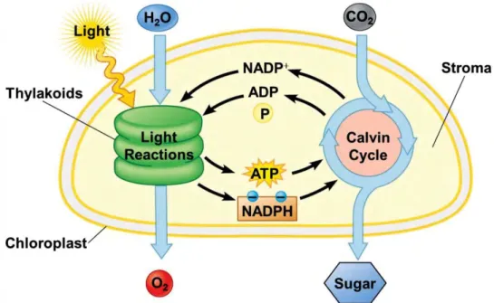
Photosynthesis consists of both light-dependent and light-independent reactions (fig. 1). In the
light-depended phase, sunlight is absorbed by pigment molecules and the energy was transferred to the
reaction centre of the two photosystems where charge separation occurs. This event induces a set of
electron transfer reactions leading to the formation of a proton gradient across the thylakoid membrane
and finally to the generation of free energy and reducing power, in the form of ATP and NADPH.
Figure 1. Schematic representation of light and dark reactions in photosynthesis (Pearson Education 2012).
Meanwhile, each chlorophyll molecule replaces its lost electron with an electron from water, generating
oxygen as consequence:
2 NADP
++2H2O + light → 2 NADPH + O2 + 2H
+ADP + Pi + energy → ATP
In the light-independent phase energy from the ATP and NADPH molecules generated by the light
reactions are used to reduce carbon dioxide from the atmosphere to a three-carbon sugar called
glyceraldehyde-3-phosphate (GAP). Cells then use GAP to build a wide variety of other sugars (such as
glucose) and organic molecules:
3 CO2 + 9 ATP + 6 NADPH → GAP + 9 ADP + 8 Pi + 6 NADP+
In photosynthetic eukaryotes, photosynthesis occurs in organelles called chloroplasts in which both
light-dependent and independent phases take place. Chloroplasts have a diameter of 5-10 μm and a depth
of 3-4 μm. They are limited by two membranes (together called envelope): the first one is highly
permeable, while the second one contains specific transporters which mediate the flux with the cytoplasm.
The soluble phase delimited by the envelope membranes is called stroma and contains all enzymes
Figure 2. Transmission Electron Microscopy merged to a schematic reconstruction of a chloroplast structure (Pearson Education 2006).
catalyzing the light-independent reactions and the plastidial DNA, RNA and ribosomes. A third membrane
system, the thylakoids, is found in the stroma and it confines a second compartment, the lumen. This
membrane presents an extensive folding and an inhomogeneous structure. They consist of two main
domains: the grana, which are stacks of thylakoids, and the stroma lamellae, which are unstacked
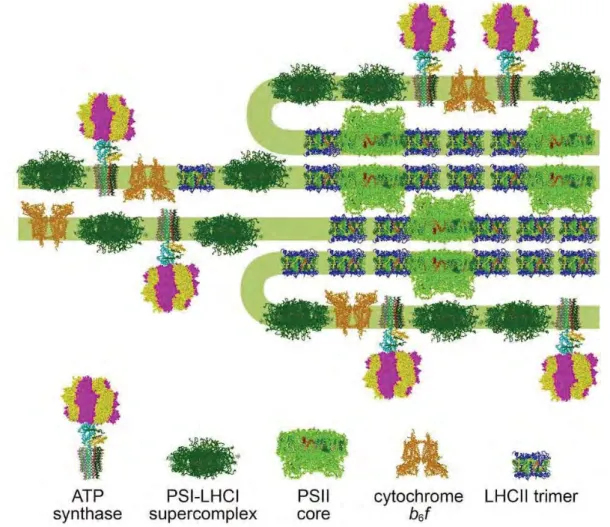
thylakoids and connect the grana stacks (fig. 2) (Barber 1980). Complexes that catalyze the light reaction,
embedded into thylakoid membrane, are not evenly distributed throughout it: PSII and LHCII reside mainly
in the grana membranes, while PSI and ATPase reside predominantly in the stroma and the cytochrome
b6/f complex is distributed in grana and grana margins (fig. 3). Protein–protein interactions determine for a
major part the shape and folding pattern of the thylakoid membrane.
Figure 3. Organization of protein complexes in thylakoid membranes (Nagy et al. 2014).
1.1.1 The light-dependent phase
In thylakoid membranes there are four large membrane-protein complexes called photosystem I
(PSI), photosystem II (PSII), cytochrome b6/f (Cyt-b6/f) and ATP synthase (ATPase) that drive
light-dependent phase of oxygenic photosynthesis (fig. 4). These complexes catalyze the processes of light
harvesting, electron transport and photo-phosphorylation, leading to the conversion of light energy to
chemical free energy (ATP and NADPH). According to the partial reactions that they catalyze, PSII is defined
as a water–plastoquinone oxidoreductase, the cytochrome b6/f complex as a plastoquinone-plastocyanin
oxidoreductase, PSI as a plastocyanin-ferredoxin oxidoreductase and the ATPase as a pmf (proton motive
force) driven ATP synthase.
Figure 4. The light phase of photosynthesis. A schematic organization of the major protein complexes in thylakoid membranes and electron transport chain is shown.
PSI and PSII binds a large number of pigments that harvest light within the visible region. The
excitation energy is transferred among individual pigment molecules via a mechanism called “Forster’s
transfer”. The energy transfer requires that pigment molecules are in close contact with each other. This is
an energetically down-hill reaction, and energy is thus preferentially transferred from chlorophyll b
(max≈647 nm) to chlorophyll a (max≈663 nm). Due to difference in the redox potential, larger than the
energy content of a red photon, between the electron donor (oxygen in a water molecule) and final
electron acceptor during the light phase of photosynthesis (NADP+), two photosystems work in series in
order to accumulate the energy of two photons, as described in the so called Z-scheme (fig. 5)(Hill and
Bendall 1960).
After absorption of light by light-harvesting antenna of PSII, the excitation energy is transferred to a
special pair of chlorophylls in the reaction centre (RC), named P680 (Primary electron donor absorbing at
680 nm) (fig. 5). Upon receiving the first energy quantum, an electron is released from P680 through an
accessory chlorophyll and a pheophytin (Pheo) molecule to the tightly bound quinone Q
A, and this is
followed by the reduction of a mobile quinone PQ at the Q
Bsite. P680
+, which has a high redox potentials,
oxidizes a nearby tyrosine (Tyr
z); Tyr
zextracts an electron from a cluster of four manganese ions (OEC,
oxygen-evolving complex), which binds two substrate water molecules (fig. 6A) (Zouni et al. 2001). After
another photochemical cycle, the doubly reduced plastoquinone (PQ
2-) takes up two protons from the
stromal space to form plastoquinol (PQH
2), which diffuses into the membrane toward the Cyt-b6/f complex
and it’s replaced by an oxidized quinone from the pool (fig. 7A). After two more photochemical cycles, the
manganese cluster accumulates a total of four oxidizing equivalents, which are used to oxidize two water
molecules leading to the formation of O
2, the release of protons in the inner thylakoid space and the return
of manganese cluster to the reduced state (Ferreira et al. 2004).
Figure 5. The Z-scheme of Bendall and Hill. Cofactors involved in electron translocation between H2O and NADP+ are indicated
Figure 6. Electron transfer reactions of PSII (A) and PSI (B) reaction centre (Caffarri et al. 2014)
In Cyt-b6/f the electrons from the PQH
2are transferred to plastocyanin (PC), a small,
copper-containing protein (fig. 7). The resulting PQ is recycled to PSII while two protons are released into the inner
thylakoid space increasing the pmf formed across the membrane. These reactions are called Q-cycle. The
Q-cycle oxidizes two plastoquinols, reduces one PQ and one PC, and translocates 4 H
+for every 2 electrons
transported to PSI (Trumpower 1990).
In PSI, light is absorbed by the antenna pigments and the excitation energy is transferred to the RC.
As in PSII, a special pair of Chls is present in the PSI-RC defined as P700 (Primary electron donor absorbing
at 700 nm) (fig. 6B). P700 upon excitation releases an electron that reduces ferredoxin (Fd) on the stroma
side. Reduced Fd is subsequently used in numerous regulatory cycles and reactions, like nitrate and CO
2assimilation, fatty-acid desaturation and NADPH production through a NADP
+oxidoreductase (Buchanan
1991). Both photosystems operate with a very high quantum yield but while PSII operates with a lower
efficiency (about 0.85), PSI works with an almost perfect quantum yield of 1.0. Cyclic electron flow (CEF) or
cyclic photophosphorylation is an alternative electron-transfer pathway that, unlike the prevailing linear
flow (LEF), does not involve PSII (Harbinson and Foyer 1991). In this process, electrons are circled around
PSI, Fd and the Cyt-b6/f complex; no NADPH is formed in this pathway but a pmf is generated by
plastocyanin reduction.
Figure 7. Cytochrome b6f complex. (A) Electron and proton transfer pathway through the b6f complex
and distances between redox cofactors. (B) Side view showing bound cofactors and protein subunits (Kurisu et al. 2003)
The charge separation in PSI and PSII, together with the electron transfer through the Cyt-b6/f and
cyclic electron transport, leads to the formation of an electrochemical potential gradient, between the
stromal and the lumenal side of the membrane, which powers ATP synthesis by the ATPase (Mitchell 1961).
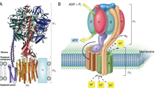
The ATPase enzyme is a multimeric complex with a stromal (CF1) and transmembrane regions (CF0). Proton
transport through CF0 is coupled to ATP synthesis/hydrolysis in the β-subunits of CF1. The whole CF0-CF1
complex is thought to function as a rotary proton-driven motor, in which the stationary subunits are I, II, IV,
δ, α and β, and the rotary subunits are III (c), γ and ε (fig. 8)(McCarty et al. 2000).
Figure 8. Structure of ATPase. (A) 3D model created using available structural data for mitochondrial F-ATPase subcomplexes (Nelson and Ben Shem 2004). (B) Schematic model of ATPase.
1.1.2 The dark phase
The dark phase of photosynthesis includes different reactions, on the whole indicated as Calvin
cycle (Benson and Calvin 1950): through these reactions, atmospheric CO
2is reduced to carbohydrates,
using the chemical free energy (ATP and NADPH) produced during the light reactions (fig. 9).
1.2 Photosynthetic pigments
Photosynthetic pigments are categorized into two chemical groups or “chromophores”: the
chlorophylls (Chls) and carotenoids (Cars). The Chls are the pigments of the RCs and also occur in the core
and light harvesting antennae. Specialized Chls at the reaction centre serve to trap the excitation energy
and convert the electronic energy to chemical energy through charge separation. The Cars are accessory
pigments that help collecting light and serve to protect Chls against photodamage.
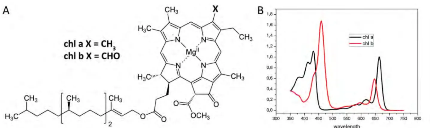
1.2.1 Chlorophylls
Figure 10. Chlorophyll a and b. (A) Structure and (B) absorption spectra in acetone 80%.
The most abundant light-harvesting pigments are Chls. Their structure consists of a cyclic tetrapyrrole
(porphyrin) in which the four nitrogen atoms of the pyrroles coordinate an Mg atom and a long phytol
chain esterified to the ring (fig. 10). The characteristic ability of Chls to absorb light in the visible region is
due to the high number of conjugated double bonds present in these molecules. Chls are synthesized
starting from the amino acid glutamic acid. In the first phase glutamic acid is converted to 5-aminolevulinic
acid (ALA) than two molecules of ALA are then condensed to form porphobilinogen (PBG), which ultimately
form the pyrrole rings in Chls. The next phase is the assembly of a porphyrin structure from four molecules
of PBG. This phase consists of six distinct enzymatic steps, ending with the product protoporphyrin IX. Then
the insertion of an Mg atom and the attachment of a fifth ring and of a phytol tail completes the
biosynthesis (fig. 11).
Figure 11. Main steps of biosynthetic pathway of chlorophyll a.
In photosynthetic organism 5 different types of Chls are present differing in their substitutions but in
vascular plants only Chl a and Chl b are present. These two pigments are almost identical but Chl a has a
methyl group on second pyrrole ring while Chl b has a formil group (fig. 10A). The Chl a and Chl b
absorption spectra in solution do not completely overlap, this increase the spectral range over which light is
absorbed, thus increasing the efficiency of light-harvesting (fig. 10B). The absorption spectra of the Chls
present two main bands: the Qy transition is the red-most band, which peaks around 640-670 nm,
respectively in Chl b and Chl a in organic solvent. It corresponds to the transition of an electron from S
0to
S
1(the first excited state). The Soret band, on the contrary, corresponds to transitions to higher states. Its
maximum is around 430 and 460 nm for Chl a and Chl b, respectively. The last absorption band of the
spectrum is the weak Qx transition that appears around 580-640 nm and is partly masked by the Qy
vibronic transitions. It corresponds to the transition from a ground state (S
0) electron to the second excited
state (S
2). The strong absorption of both red and blue/violet light by Chls causes the green colors of most
plants. The absorbance spectra of Chls is influenced by the protein complexes in which they are bound,
such that there can be variations in the same type of pigment and the peak absorbance in vivo tend to be
broadened and shifted compared to those of the pure pigments extracted in solution.
1.2.2 Carotenoids
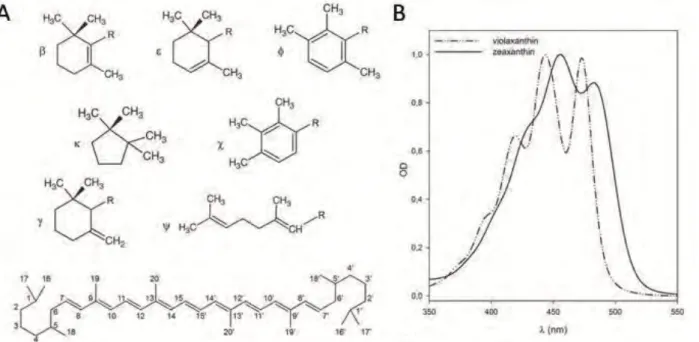
Figure 12. Carotenoid structure. (A) Characteristic end groups of carotenoids and structure of a generic carotenoid with common numbering system. (B) Visible absorption spectra of violaxanthin and zeaxanthin in acetone.
Cars are a class of more than 600 naturally occurring pigments synthesized by all photosynthetic
organisms and some non-photosynthetic bacteria and fungi (Kull and Pfander 1995). They absorb in the
blue-green region with pronounced absorption bands between 450 and 550 nm, where Chls do not absorb
efficiently (fig. 12). Cars fulfill several important functions in photosynthesis. They contribute to the
assembly and stability of photosynthetic complexes (Plumley and Schmidt 1987, Paulsen et al. 1993), act as
photoreceptors (Mimuro and Katoh 1991, Gradinaru et al. 2000), and have a main role in photoprotection
(Havaux and Niyogi 1999). Carotenoids protect the photosynthetic apparatus in different ways, including
the quenching of chlorophyll triplets, scavenging of singlet oxygen, and the dissipation of excess light
energy absorbed by the antenna pigments by non-photochemical quenching of chlorophyll fluorescence.
Plant carotenoids are tetraterpenes derived from the 40-carbon isoprenoid phytoene. These molecules
consist of a polyene chain of alternating single and double bonds, with two rings at the end of the molecule
(fig 12). Different levels of hydrogenation and introduction of oxygen-containing functional groups create a
large family of carotenoids. In higher plants, they can be grouped in two major classes: carotenes which are
hydrocarbons with linear structure and with cyclic groups in one or both extremities and xanthophylls
which are oxygenated derivatives of carotenes. The conjugated double bond system of carotenoid
molecules determines their photochemical properties. The π-electrons delocalization in the conjugated
double bonds system leads to the light absorption in the visible range 400-500 nm. When Cars absorb light,
electrons are transferred from ground state S
0to the second excited singlet state S
2; this strongly
dipole-dipole allowed transition is responsible for the characteristic absorption spectrum. The first excited singlet
state S
1cannot be populated from the ground state by photon absorption due to symmetry reasons. The
absorption spectra of Cars are strongly red-shifted in vivo, compared to their spectra in organic solvents.
This shift represents a lowering of the S
2energy level, which has been ascribed to the mutual polarizability
of the carotenoid and protein environment (Andersson et al. 1991). In higher plants the most abundant
carotenoids associated with thylakoid membranes are the α- and β-Carotene (α-Car, β-Car) and the
xanthophylls Lutein (Lute), Violaxanthin (Viola), Neoxanthin (Neo) and Zeaxanthin (Zea). Chloroplasts have
a remarkably similar carotenoid composition in all plants, with Lute (45% of the total), β-Car (25–30%),
Viola (10–15%) and Neo (10–15%) as the most abundant carotenoids (Britton 1995). Carotenes (mainly
β-Car) are enriched in the photosystem reaction centre, whereas xanthophylls are most abundant in the
light-harvesting complexes (Niyogi et al. 1997, Dall'Osto et al. 2007).
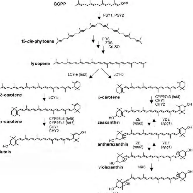
Like all isoprenoids, Cars are synthesized from the five-carbon units isopentenyl diphosphate (IPP)
and its double-bond isomer dimethylallyl diphosphate (DMAPP) (fig. 13). Addition of three IPP molecules to
DMAPP generates geranylgeranyl diphosphate (GGPP). The condensation of two GGPP molecules produces
the 40-carbon phytoene. Phytoene is then desaturated to create the chromophore-bearing chain of
conjugated double bonds that forms the backbone of plant carotenoids and determines their physical and
biological properties (Britton 1995). Desaturation and isomerization of uncolored phytoene eventually
results in the production of lycopene, a red carotenoid. The cyclization of the ends of the lycopene polyene
chain is the first branch point in the pathway and results in the production of carotenes either with one ß
ring and one ε ring (α-Car) or with two β rings (β-Car). Carotenoids with two ε rings do not exist in A.
thaliana and are uncommon in plants. Hydroxylation of the carotene rings generates xanthophylls such as
Lute (from α-Car) and Zea (from β-Car). Zea is epoxidated twice to make Viola, which can be subsequently
modified to make Neo. In Arabidopsis four enzymes provide for Chls hydrogenation: CHY1 and CHY2, two
non-heme di-iron monoxygenase, which catalyze the hydroxylation of β ring only and CYP97A3 and
CYP97C1, two heme-containing cytochrome P450 hydroxylases, which catalyze the hydroxylation of β and ε
ring respectively.
Figure 14. Schematic representation of xanthophyll cycle.
While hydroxylation of α-Car produces Lute, a carotenoid end-product that accumulates at high
levels, hydroxylation of β-Car produce zeaxanthin that, under light conditions that do not saturate
photosynthesis or in the dark, is readily converted to violaxanthin via antheraxanthin in a two-step reaction
catalyzed by the enzyme zeaxanthin epoxidase (ZE). When light is strong and exceeds the photosynthetic
capacity, Viola is de-epoxidated back into Zea by the activity of the enzyme violaxanthin de-epoxidase
(VDE) (Yamamoto and Kamite 1972, Demmig-Adams et al. 1996) (fig. 14). The interconversion of Zea and
Viola is known as the xanthophyll cycle and has a key role in the adaptation of plants to different light
intensities (Dall'Osto et al. 2005). VDE is activated when light-driven proton translocation across the
thylakoid membrane exceeds the dissipation rate of the proton gradient by ATPase, leading to a decrease
in pH in the thylakoid lumen while ZE is always active. The xanthophyll cycle is uniquely separated on
opposite sides of the thylakoid membrane; VDE activity takes place on the thylakoid lumen side of the
membrane, whereas ZE occurs on the chloroplast stromal side (Hieber et al. 2000). Xanthophyll cycle is a
key component of several photo-protective mechanisms as scavenging of ROS, thermal dissipation of
excitation energy in excess or Chls triplets excited state quenching (Niyogi 1999, Holt et al. 2004).
1.3 Photosystems
PSI and PSII are multi-protein complexes binding the pigments responsible for light harvesting and
charge separation. Both are composed of a core complex, where Chls special pairs and cofactors involved in
electron transport are located and a peripheral antenna system, composed by Chls binding proteins
responsible for light harvesting and energy transfer to the reaction centre (RC). Chls and Cars are bound
both by core complex and antenna system; core complexes bind only Chl a and carotenes, while antenna
proteins bind Chl a, Chl b and xanthophylls. The core complexes have been well conserved during the
evolution, as most of the subunits are similar in prokaryotic and eukaryotic photosystems and only a few
are specific to each group. On the contrary, the peripheral antenna system displays great variability, being
composed of peripheral associated membrane proteins in cyanobacteria, the phycobilisomes, and integral
light harvesting complexes (LHC) membrane proteins in eukaryotic cells.
1.3.1 PSII Core complex
Figure 15. (A) Schematic model for PSII and (B) 3D crystal structure of PSII core complex (Ferreira et al. 2004).
Core complex of PSII is composed by the polypeptides denominated PSB encoded from both
nuclear and plastidial genes. The core of PSII is a multi-subunit complex composed of about 25-30 subunits;
it contains four large membrane-intrinsic subunits (PSBA-D), three membrane-extrinsic subunits (PSBO–Q)
and a large number of small subunits (fig. 15), most of which span the membrane once and are involved in
the dimerization or in Chls and Cars binding stabilization, but they do not all have a well-clarified function
(Shi et al. 2012). PSBA (D1) and PSBD (D2) bind six Chl a and two Pheo a molecules and constitute the
photochemical reaction centre in which the charge separation and primary electron transfer reactions take
place. PSBB (CP47) and PSBC (CP43) bind 16 and 14 Chl a molecules respectively and have a light-harvesting
function: they absorb light and transfer the excitation energy to the reaction centre and also accept
excitation energy from the peripheral antenna and transfer this to the reaction centre (Barber et al. 2000).
There are no indications yet that any of the extrinsic or small proteins binds chlorophyll. On the lumenal
side of the complex, three extrinsic proteins of 33, 23 and 17 KDa (OEC1-3) compose the OEC, and have a
calcium, chloride and bicarbonate ion as necessary cofactors (Zouni et al. 2001).
1.3.2 PSII peripheral antenna
Figure 16. Molecular model of LHCII monomer (A) and CP29 (B) showing chlorophyll and xanthophyll chromophores bound to different binding sites (Ballottari et al. 2012).
In higher plants and eukaryotic algae, the peripheral antenna of PSII consists of a number of
pigment-protein complexes belonging to the LHC super-gene family. In green plants two types of peripheral
antenna proteins associated to PSII can be distinguished: the “major” LHCII antenna complex (Thornber et
al. 1967) and the minor antenna complexes (Bassi et al. 1996). LHCII occurs in a trimeric association state
and is not unique in composition; it consists of various combinations of three very similar proteins named
LHCB1-3. In addition, there are three “minor” antenna complexes, which are called LHCB4 (CP29), LHCB5
(CP26) and LHCB6 (CP24) and usually occur in monomeric aggregation states. All these complexes bind
various molecules of Chl a and Chl b and of the xanthophylls Lute, Viola and Neo (fig. 16). All the peripheral
antennae are encoded by nuclear genes. In Arabidopsis thaliana LHCB3 is encoded by a single gene while
LHCB1 and LHCB2 are encoded by high homologous multiple genes. Single genes encode CP26 and CP24,
while three lhcb4 genes are present (lhcb4.1-3) (Jansson 1999). Recently the expression profile of the third
gene coding for CP29, lhcb4.3, was shown to be different from lhcb4.1 and lhcb4.2 and was then renamed
lhcb8. An additional isoform for minor complexes was identified and named LHCB7 (Klimmek et al. 2006).
However LHCB8 and LHCB7 proteins are not present in detectable amount into thylakoids membranes and
their role is still unclear.
LHCII trimers are heterotrimers constituted by the subunits LHCB1, LHCB2 and LHCB3; the three
polypeptides, however, are not equimolar, with LHCB1 found in larger amounts (Caffarri et al. 2004, Dekker
and Boekema 2005). High resolution structural models of antenna complexes have been obtained for LHCII
(Liu et al. 2004) (fig. 16A). In the trimeric complex of LHCII, each monomer is constituted of 3
transmembrane domains with α-helix conformation (helices A, B and C). The N-terminal is fully hydrophilic,
thus protruding into the stroma space and the C-terminal peptides is exposed on the lumenal space. Two
amphipathic helixes, named D and E, were found respectively on the C-terminal peptide and in the BC loop
region; both helices lie on the lumenal surface. The trimerization domain covers: the amino-terminal
domain, the carboxyl terminus, the stromal end of helix B, several hydrophobic residues from helix C and
also pigments and lipid as phosphatidylglycerol (PG), bound to these parts of the polypeptide chain. Six Chl
a (two from each monomer) constitute the core of the trimer (Liu et al. 2004). Each monomer binds 14 Chls
and 4 xanthophylls. Part of the chlorophylls binding sites are selective for Chl a or Chl b, while in other
cases Chls binding sites with mixed occupancy are present. Two central lutein molecules are bound in the
grooves on both sides of the helices A and B cross-brace, forming the internal L1 and L2 carotenoid binding
sites (Caffarri et al. 2001); the polyene chains are firmly fixed in two hydrophobic cavities, providing strong
linkage between helices A and B. The third xanthophyll, Neo is located in the Chl b-rich region around helix
C in the carotenoid binding site N1 (Remelli et al. 1999); side chains from helices B and C as well as phytyl
chains form a hydrophobic space that accommodates the hook-shaped polyene chain of Neo, while ring on
the other ends stretches into the exterior solvent region. The fourth carotenoid is located in a peripheral
site named V1 (Ruban et al. 1999). V1 site is constituted by a hydrophobic pocket at the interface
monomer-monomer, formed by several Chls, hydrophobic residues and the PG; part of the xanthophyll is
located inside this pocket, while the opposite end group is located outside, toward the stromal surface.
Three Chl a, b-and xanthophyll-binding proteins constitute monomeric minor antennae: CP29, CP26
and CP24, named from their apparent mass in non-denaturing SDS-PAGE gels (Bassi et al. 1987). The
transcription levels of minor antennae are similar and the proteins are found in equimolar amounts in the
thylakoids (Nield et al. 2000).
CP29 is composed by 256-258 amino acids in its mature form in A. thaliana, and it is the largest
among LHC encoded proteins. The overall sequence identity between CP29 and LHCII is 34%, but most of
the substitutions are conservative, especially in the helix regions. It is necessary for PSII organization and a
key component for the stability of the PSII-LHCII supercomplex (van Oort et al. 2010). And since is located
between the outmost antenna LHCII and the inner antenna CP47, CP29 plays a bridging-type role in
transferring the excitation energy to the reaction centre (Caffarri et al. 2009). A recent crystal structure of
spinach CP29 (Pan et al. 2011) shows that it contains 13 Chl-binding sites: eight Chls a, four Chls b and one
possible mixture of Chl a and Chl b (fig. 16B). Moreover, the purified CP29 sample used for crystallization
contains three species of carotenoids: Lute, Viola and Neo. Each carotenoid molecule occupies a separate
site in CP29: Lute at L1 site, Viola at L2 site and Neo at the N1 site. Compared to LHCII, CP29 does not bind
any pigments at Chls b601 and b605 sites that are located at the periphery of the LHCII monomer. Instead,
a new Chl-binding site, a615, which is absent in LHCII, has been discovered on the surface of CP29 and close
to the previous Chl b601 site in LHCII. Regarding the Cars binding sites, CP29 and LHCII show great
differences at site L2, which binds Lute in LHCII but Viola in CP29. In addition, CP29 does not contain the
fourth Car-binding site at the monomer-monomer interface of the LHCII trimer (V1). The L1 and N1 sites are
conserved between CP29 and LHCII. Nevertheless, the Neo at the N1 site in CP29 rotates slightly and forms
an increased angle between its transition dipole moment and the membrane normal (60.2° versus 58° in
LHCII). The structural differences between CP29 and LHCII, especially their pigment composition and spatial
arrangement, may account for their different functions in photosynthetic light harvesting and regulatory
processes. CP29 can be phosphorylated, especially when plants are exposed to low temperature and high
light stress (Croce et al. 1996). CP29 phosphorylation is supposed to induce conformational rearrangement
or modification in the PSII supercomplex that could facilitate thermal energy dissipation.
CP26 is 243 amino acids long in A. thaliana, shows 48% identity with respect to LHCII and
coordinates 7 Chl a, 3 Chl b and 2-3 xanthophylls (Lute, Viola and Neo) (Bassi et al. 1996, Croce et al. 2002).
CP26, as LHCB1 and CP29, presents a Tyr residue which is suggested to stabilize the third carotenoid
binding site N1 (Caffarri et al. 2007).
CP24 is the smallest of LHC proteins (211 amino acids in A. thaliana), due to the lack of the major
part of the C-terminal region of the protein. Sequence homology and absorption spectra suggest that 5 Chl
a, 5 Chl b and 2 xanthophylls are bound (Bassi et al. 1996, Pagano et al. 1998). CP24, differently from other
1.3.4 PSII supercomplex
Figure 17. (A) PSII-LHCII supercomplex (C2S2M2) and of the (B) PSI-LHCII supercomplex in State II (Caffarri et al. 2014)
A variable number of peripheral antenna proteins can associate with dimeric PSII core complexes to
form PSII-LHCII supercomplexes (fig. 17A). A dimeric core (C
2) associate with up to four copies of peripheral
LHCII trimers. Connection of the first two LHCII-S trimers (strongly bound) extends a C
2complex to a C
2S
2supercomplex, and two further M trimers (moderately strong bound) are bound in a C
2S
2M
2supercomplex.
Spinach supercomplexes may bind a third type of L trimer (loosely bound), but the resulting C
2S
2M
2L
1–2supercomplexes are rare. Recent studies suggest that LHCII-L trimer may migrate between PSII and PSI to
balance the excitation level of two photosystems in response to light fluctuations (Galka et al. 2012). The
C
2S
2M
2supercomplex of A. thaliana was analyzed by single particle electron microscopy at 12 Å resolution
(Caffarri et al. 2009), permitting a more accurate fitting of the peripheral antenna proteins, based on the
known LHCII structure. C
2S
2M
2contains a dimeric core (C
2), four LHCII trimers, two strongly bound (S) and
two moderately strongly bound (M) to the core, and two monomeric copies each of CP29, CP26, and CP24.
In particular, LHCII-S trimers are attached to a dimeric PSII via CP29 to one PSII core monomer and CP26 to
the other and LHCII-M trimers are attached via CP24 and CP29. Biochemical experiments show that LHCB3
is present in trimer M only; instead, LHCB2 is a specific component of trimer S. LHCB1 is present in both
trimers (Dekker and Boekema 2005). CP24 and LHCB3 are necessary for binding the M trimer; A. thaliana
plants depleted of one of these subunits do not form C
2S
2M
2supercomplexes or the small pentameric
complex LHCII-M/CP24/CP29 (Kovacs et al. 2006). In the absence of CP26 no bands containing high
molecular weight supercomplexes are visible and the amount of the fractions containing the smaller
supercomplexes (C
2S, C
2M, C
2S
2, C
2SM) is extremely reduced (Caffarri et al. 2009). C
2S
2M
2were still
detectable in a mutant without CP29, albeit their amounts were reduced compared with the wild type. An
empty space was observed within this supercomplex at the CP29 position, implying that the missing CP29
was not replaced by other LHC subunits. This suggests that CP29 is unique among PSII antenna proteins and
determinant for its macro-organization and photoprotection (de Bianchi et al. 2011).
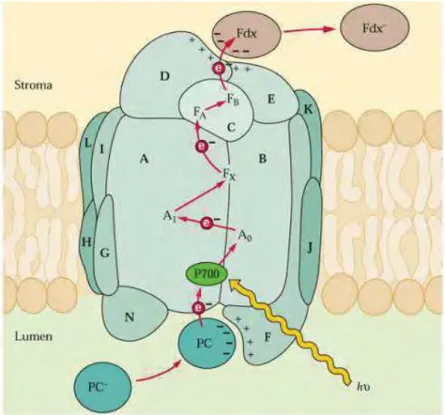
1.3.5 PSI core
Figure 18. Schematic model of photosystem I. Subunits organization and cofactors involved in electron transfer are indicated.