Bovine pericardium membrane,
gingival stem cells, and
ascorbic acid: A novel team
in regenerative medicine
Jacopo Pizzicannella,1Guya D. Marconi,2 Sante D. Pierdomenico,2 Marcos F.X.B. Cavalcanti,3
Francesca Diomede,2Oriana Trubiani2 1ASL02 Lanciano-Vasto-Chieti, “Ss. Annunziata” Hospital, Chieti, Italy 2Department of Medical, Oral and Biotechnological Sciences, University “G. d’Annunzio” Chieti-Pescara, Italy 3Laser in Dentistry Program, Cruzeiro do Sul University (UNICSUL), Sao Paulo, Brazil
Abstract
Recently, the development and the application of 3D scaffold able to promote stem cell differentiation represented an essential field of interest in regenerative medicine. In particular, functionalized scaf-folds improve bone tissue formation and promote bone defects repair. This research aims to evaluate the role of ascorbic acid (AS) supplementation in an in vitro model, in which a novel 3D-scaffold, bovine peri-cardium collagen membrane called BioRipar (BioR) was functionalized with human Gingival Mesenchymal Stem Cells (hGMSCs). As extensively reported in the literature, AS is an essential antioxidant molecule involved in the extracellular matrix secretion and in the osteogenic induction. Specifically, hGMSCs were seeded on BioR and treated with 60 and 90 μg/mL of AS in order to assess their growth behavior, the expression of bone specific markers involved in osteogenesis (runt-related transcription factor 2, RUNX2; col-lagen1A1, COL1A1; osteopontin, OPN; bone morphogenetic protein2/4, BMP2/4), and de novo deposition of calcium. The expression of COL1A1, RUNX2, BMP2/4 and OPN was evaluated by RT-PCR, Western blotting and immunocytochem-istry, and proved to be upregulated. Our results demonstrate that after three weeks of treatment AS at 60 and 90 μg/mL operates as an osteogenic inductor in hGMSCs. These data indicate that the AS supplemen-tation produces an enhancement of osteogenic phenotype commitment in an in
vitro environment. For this reason, AS
could represent a valid support for basic and translational research in tissue engineering and regenerative medicine.
Introduction
Tissue engineering is an interesting strategy to promote new bone tissue forma-tion. In bone tissue engineering, the combi-nation of several parameters such as cell type, growth factors or bioactive factors, mechanical stimuli, and 3D-scaffold materi-al, is fundamental in order to obtain positive results.1
The cell source for tissue engineering can be obtained from different tissue niches, one of this is obtained from the oral cavity, and in particular from the gingival tissue. Gingival mesenchymal stem cells (GMSCs) are obtained from stromal tissue with a rel-atively easy isolation procedure and with ability to produce high level of cells num-ber.2,3 In tissue engineering field, scaffold materials mimic the role of extracellular matrix (ECM) and provide a mechanical support for cells. An appropriate scaffold should be osteoinductive, osteoconductive and able to enroll MSCs derived from human tissues, and support the growth of the bone tissue.4Latest study demonstrated that cell proliferation and differentiation can be controlled by bioactive factors inside tissues.5 Recently, to provide a favorable microenvironment for MSCs differentia-tion, several molecules or ions have been incorporated into 3D scaffolds.
Ascorbic acid (AS) is an essential antioxidant molecule which works as a cofactor for many enzymes. Since humans have lost the ability to synthesize ascorbate due to the increase of mutations in the cod-ing sequence of the L-gulono-1,4-lactone oxidase,6-8this antioxidant molecule should be included as a supplement in the diet to assure tissue homeostasis. AS is present in the bloodstream at approximately 50-100 µM concentration in plasma of healthy sub-jects.9It can regulate adult stem cell differ-entiation to some mesenchymal tissues derivatives, such as adipocytes, osteocytes, myocytes, and chondrocytes.10AS plays a key role as co-factor in post-translational modification of collagen molecules,11which are components of the ECM of mes-enchyme-derived tissues.12 Moreover, this vitamin reduces the detrimental effect induced by methacrylates in clinical den-tistry leading to cell growth repair, proin-flammatory cytokine reduction, ROS level and NFkB/pERK/ERK signaling pathway down-regulation (data submitted).
In the present study, the role of AS as a bioactive factor for osteogenic differentia-tion was investigated in an in vitro model in which human GMSCs were grown on a novel 3D-scaffold, i.e. the bovine pericardi-um collagen membrane called BioRipar (BioR). Cell growth, de novo deposition of calcium as well as the expression of some
bone specific markers involved in osteoge-nesis (namely, the runt-related transcription factor-2, RUNX2; collagen1A1, COL1A1; and osteopontin, OPN) were evaluated.
As extensively reported in the literature, RUNX2 is a transcriptional factor essential for the activation of osteoblast-associated genes and also it has been reported to be an important early indicator of osteoblast dif-ferentiation and bone formation.13
OPN codes for one of the most predom-inant non-collagenous proteins in bone ECM produced by osteoblasts, and it also promotes cell adhesion to the bone sur-face.14 This protein regulates cell–matrix interactions and signaling through binding to integrins and CD44 receptors.15 Furthermore, receptors of OPN integrins and CD44 have been described on host stro-mal cells and in hGMSCs.16 OPN is pro-duced by mature osteoblasts in the process of bone formation and is recognized as a major marker of osteogenic differentiation, it also plays a main role in the regulation of vessel regeneration.17During bone resorp-tion, OPN plays an important role in the attachment of osteoclasts and in osteogene-sis regulating crystal size.18Upregulation of non-collagenous proteins such as OPN and osteonectin (SPARC), exhibited a vital role in osteogenesis, confirmed by the osteogen-esis-promoting effect of the AS on hGMSCs during bone mineralization.19
Correspondence: Oriana Trubiani, Department of Medical, Oral and Biotechnological Sciences, University “G. d’Annunzio” Chieti-Pescara, Via dei Vestini 31, 66100 Chieti, Italy. E-mail: [email protected]
Keywords: Pericardium membrane; osteogenic differentiation; human gingival derived stem cells; COL1A1; RUNX2; BMP2/4; OPN.
Contributions: JP, GDM, SDP, FD, OT, con-ception and design; JP, GDM, MFXBC, FD, analysis and interpretation of data; JP, SDP, FD, OT drafting the article; JP, MFXBC, SDP, FD, OT, revising critically for important intel-lectual content. All Authors read and approved the final version to be published.
Conflict of interest: The authors declare no conflict of interest.
Received for publication: 30 July 2019. Accepted for publication: 13 September 209. This work is licensed under a Creative Commons Attribution-NonCommercial 4.0 International License (CC BY-NC 4.0).
©Copyright: the Author(s), 2019 Licensee PAGEPress, Italy
European Journal of Histochemistry 2019; 63:3064 doi:10.4081/ejh.2019.3064
Collagen 1, known as an early marker of osteoprogenitor cells, fundamental for extracellular matrix synthesis and to pro-mote Bone morphogenetic protein2 (BMP2) release.20BMP2 is a member of the TGF-β superfamily, detected in cartilage and bone,21 that through activation of the Wnt pathway, promotes bone formation.22-24
Materials and Methods
Cell culture
This research study was approved by the “G. d’Annunzio” University Ethics Committee (n°266 / University of Chieti). Gingival tissue was collected from six healthy patients scheduled to dentistry sur-gical procedure as previously described,25in order to obtain hGMSCs. Tissue explants were washed several times with sodium phosphate buffer (PBS, Lonza, Basel, Switzerland), then samples were plated and maintained in Petri dish with Mesenchymal Stem Cells Growth Medium-Chemically Defined (MSCGM-CD) (Lonza). 26 The medium was replaced twice a week with a fresh one. After two weeks of culture, cells spontaneously migrated from tissue biop-sies. All experiments were performed using cells at 2ndpassage.
Cell characterization
To characterize hGMSCs, used in the present study, Dominici’s criteria have been followed.27First, to evaluate the mesenchy-mal features of hGMSCs cytofluorimetric detection and mesengenic differentiation have been performed. Expression of CD13, CD14, CD29, CD34, CD44, CD45, CD73, CD90 and CD105 was evaluated by cyto-fluorimetric analysis as previously described.28The analysis was performed by using FACStarPLUS flow cytometry sys-tem and the FlowJo™ software (TreeStar, Ashland, OR, USA).
An inverted light microscope Leica DMIL (Leica Microsystem, Milan, Italy) was used to evaluate cell morphology and the capacity to adhere to a plastic substrate. To assess the ability to differentiate into osteogenic and adipogenic commitment hGMSCs were maintained under osteogenic and adipogenic conditions for 21 and 28 days, respectively, as previously reported.29 To evaluate the formation of mineralized precipitates and lipid droplets, after the dif-ferentiation period, Alizarin Red S and Adipo Oil Red staining were performed on undifferentiated and differentiated cells. For Alizarin Red S, staining cells were washed with PBS, fixed in 10% (v/v) formaldehyde (Sigma-Aldrich, Milan, Italy) for 30 min, and washed twice with abundant distilled
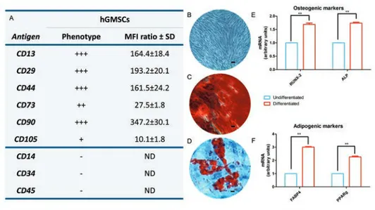
Figure 1. Cell characterization. A) Cytofluorimetric analysis; −, negative expression (0%); +, moderate expression; ++, positive expression; +++, high expression (100%); MFI ratio is the average of three different biological samples ± standard deviation. B) Plastic adher-ent hGMSCs observed at inverted light microscopy. C) Alizarin Red S positive staining in hGMSCs culture. D) Oil Red O staining in hGMSCs culture, cells showed a lipid droplet at cytoplasmic level. E) RT-PCR of osteogenic related markers, RUNX2 and ALP. F) RT-PCR of adipogenic related markers, FABP4 and PPAR (undifferentiated vs differ-entiated). **P<0.01 was considered statistically significant. Scale bars: 20 µm.
Figure 2. Cell proliferation and viability. A) MTT assay. (B) Trypan blue exclusion test. Treatment with AS 60 µg/mL and AS 90 µg/mL showed no statistical differences in terms of cell proliferation and viability after 24, 48 and 72 h of culture when compared to the CTRL group. DIFF OSTEO group showed a decrease in cell proliferation and viability when compared to all experimental groups (**P<0.01) at 48 and 72 h of culture. The results are expressed as mean ± SD.
water (dH2O) before addition of 0.5% Alizarin Red S in H2O, pH 4.0, for 1h at room temperature. After cells incubation under gentle shaking, cells were washed with dH2O four times for 5 min. For stain-ing quantification, 800 μL of 10% (v/v) acetic acid were added to each well. Cells were incubated for 30 min with shaking, then scraped from the plate, transferred into a 1.5 mL vial, and vortexed for 30 s. The obtained suspension, overlaid with 500 mL mineral oil (Sigma-Aldrich), was heated to 85°C for 10 min, then transferred to ice for 5 min, carefully avoiding opening of the tubes until fully cooled, and centrifuged at 20,000 g for 15 min. Five hundred μL of the supernatant were placed into a new 1.5 mL vial, and 200 μL of 10% (v/v) ammonium hydroxide were added (pH 4.1-pH 4.5). One hundred fifty μL of the supernatant obtained from differentiated and undifferentiated hPDLSCs were read in triplicate at 405 nm by a spectrophotometer (Synergy HT, BioTek Instruments, Bad Friedrichshall, Germany). For adipogenic specific staining, cells were fixed in 10% formalin for 15 min and washed with dH2O. Subsequently, the cells were stained with Oil Red O working solution (300 mg of Oil Red O/100 mL of isopropanol) for 5 min and counterstained with hematoxylin. The differentiation into adipogenic lineage was evaluated by AdipoRed assay reagent hydrophilic Nile Red fluorescence (Lonza). After differentia-tion, the plates were rinsed with PBS and 140 mL/well of AdipoRed was added; after 10 min, the fluorescence with an excitation at 485 nm and an emission at 572 nm was measured with a fluorimeter (Synergy HT). To validate the ability to differentiate into osteogenic and adipogenic lineages, the expression of RUNX-2, ALP, FABP4, and PPARγ were evaluated by reverse transcrip-tion polymerase chain reactranscrip-tion (RT-PCR) as described by Diomede et al.29 Commercially available TaqMan Gene Expression Assays (RUNX-2 Hs00231692_m1; ALP Hs01029144_m1; FABP4 Hs01086177_m1; PPARγ Hs01115513_m1) and the Taq-Man Universal PCR Master Mix (Applied Biosystems, Foster City, CA, USA) were used according to standard protocols. Beta-2 microglobulin (BBeta-2M Hs99999907_m1) (Applied Biosystems) was used for tem-plate normalization. Real-time PCR was performed in three independent experi-ments, and duplicate determinations were carried out for each sample.
Biomaterial
The BioRipar® (BioR; Assut Europe SpA, Magliano dei Marsi, AQ, Italy) is a collagen membrane derived from bovine pericardium. Purified pericardium,
com-Figure 3. Osteogenic differentiation. Alizarin Red S staining were used to evaluate the calcium depositions (red) in (A) CTRL, (B) AS 60 µg/mL, (C) AS 90 µg/mL and (D) DIFF OSTEO. E) Quantitative analysis of Alizarin Red S staining was performed by measuring the absorbance of Alizarin Red S. *BioR membrane. The results are expressed as mean ± SD (AS 60 µg/mL vs AS 90 µg/mL; 60 µg/mL vs DIFF OSTEO). **P<0.01 was recognized to be significant. Scale bars: 20 µm.
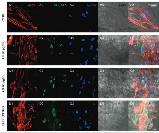
Figure 4. Expression of COL1A1. Immunofluorescence detection of COL1A1 in CTRL (A1-A5), AS 60 µg/mL (B1-B5), AS 90 µg/mL (C1-C5) and DIFF OSTEO (D1-D5). COL1A1 expression were upregulated in AS 90 µg/mL and DIFF OSTEO when com-pared to the AS 60 µg/mL. No significant differences have been evidenced between AS 90 µg/mL and DIFF OSTEO groups. Red fluorescence, cytoskeleton actin; green fluores-cence, specific marker; blue fluoresfluores-cence, cell nuclei; grey scale, membrane observed at transmission light channel; merge pictures showed the overlapping of abovementioned channels. Scale bar: 10 µm.
posed by type I Collagen and Elastin, repre-sented a new tool for fibroblasts growth and new blood vessel formation. Sterile scissors were used to cut the membrane in small piece size. PBS (Lonza) was used to rehy-drate the BioR membrane before use.
Experimental design
Cells at 2nd passage, seeded on BioR, have been divided in four experimental groups:
1. CTRL: BioR/hGMSCs cultured with basal medium (MSCGM-CD); 2. AS 60 µg/mL: BioR/hGMSCs treated
with ascorbic acid at a concentration of 60 µg/mL;
3. AS 90 µg/mL: BioR/hGMSCs treated with ascorbic acid at a concentration of 90 µg/mL;
4. DIFF OSTEO: BioR/hGMSCs cultured with osteogenic differentiation medium as following reported.
Cell proliferation and viability assay
Cell proliferation was evaluated by means of MTT assay as previously described;162×103 cells per well were seed-ed into 96-well plates with BioR in a mseed-edi- medi-um volmedi-ume of 200 μL to test all experimen-tal groups at different endpoint, 24, 48 and 72 h. Twenty μL of MTT (Promega, Milan, Italy) solution were added to each well. Absorbance at 490 nm was measured with a reference wavelength of 630 nm.30Cell viability was assessed by trypan blue exclusion test. At this purpose all sam-ples were incubated with trypan blue solu-tion at same endpoint used for MTT test (24, 48 and 72 h) and subsequently analysed with Burker’s chamber at inverted light microscopy as previously described.31
Osteogenic differentiation
evalua-tion
To perform osteogenic differentiation, hGMSCs were seeded in a 6-well plate at approximately 2.5×10³ cells/cm2 on BioR membrane. Upon reaching confluence the culture medium was replaced with osteogenic differentiation medium kit (Lonza).32 Osteogenic induction was per-formed for 21 days.
Evaluation of calcium deposition was obtained by a biochemical analysis, Alizarin Red S (ARS) staining assay, performed after 21 days. Cells were washed with PBS, fixed in 10% (v/v) formaldehyde (Sigma-Aldrich, Milan, Italy) for 30 min and washed twice with abundant dH2O prior to addition 0.5% Alizarin red S in H2O, pH 4.0, for 1 h at room temperature. After cell incubation under gentle shaking, cells were washed with dH2O four times for 5 min. For staining quantification, 800 μL of 10% (v/v) acetic acid was added to each well. Cells
incubated for 30 min were scraped from the plate, transferred into a 1.5 mL vial and vor-texed for 30 s. The obtained suspension, overlaid with 500 μL of mineral oil (Sigma-Aldrich), was heated to 85 °C for 10 min, then transferred to ice for 5 min, carefully avoiding opening of the tubes until fully cooled, and centrifuged at 20,000× g for 15 min.33In addition, 500 μL of the supernatant were placed into a new 1.5 mL vial and 200 μL of 10% (v/v) ammonium hydroxide was added (pH 4.1-pH 4.5). Furthermore, 150 μL of the supernatant obtained from cul-tures were read in triplicate at 405 nm by a spectrophotometer (Synergy HT).
Confocal laser scanning microscope
analysis
For immunofluorescence detections BioR/hGMSCs were fixed using 4% paraformaldehyde diluted in 0.1 M PBS (Lonza). After the fixation step, cells were permeabilized with 0.5% Triton X-100 in PBS for 10 min, followed by blocking with
5% skimmed milk in PBS for 30 min.28 Primary antibodies used for immunofluo-rescence were purchased from Santa Cruz Biotechnology (Santa Cruz Biotechnology, Santa Cruz, CA, USA). COL1A1 (1:200, Santa Cruz Biotechnology), RUNX2 (1:100, Santa Cruz Biotechnology), BMP2/4 (1:200, Santa Cruz Biotechnology) and OPN (1:200, Santa Cruz Biotechnology) were used as primary anti-bodies. Then cells were incubated by Alexa Fluor 568 red fluorescence conjugated goat anti-rabbit as secondary antibodies (1:200, Molecular Probes, Invitrogen, Eugene, OR, USA). Alexa Fluor 488 phalloidin green fluorescence conjugate (1:400, Molecular Probes) has been used to mark the cytoskeleton actin. After immunofluores-cence labelling cells were washed and incu-bated with TOPRO (1:200, Molecular Probes) for 1 h at 37°C.26 Samples were observed under Zeiss LSM800 confocal system (Zeiss, Jena, Germany). All the experiments were performed in triplicate.
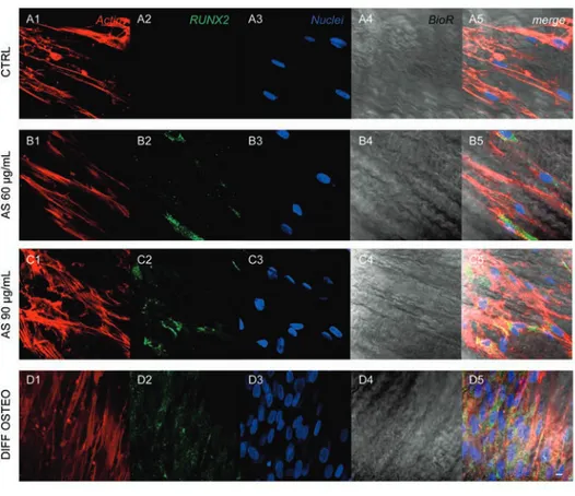
Figure 5. Expression of RUNX2. Immunofluorescence detection of RUNX2 in CTRL (A1-A5), AS 60 µg/mL (B1-B5), AS 90 µg/mL (C1-C5) and DIFF OSTEO (D1-D5). RUNX2 expression were overexpressed in AS 90 µg/mL and DIFF OSTEO samples when compared to the AS 60 µg/mL. No significant differences have been demonstrated when compared AS 90 µg/mL and DIFF OSTEO groups. Red fluorescence, cytoskeleton actin; green fluorescence, specific marker; blue fluorescence, cell nuclei; grey scale, membrane observed at transmission light channel; merge pictures showed the overlapping of above-mentioned channels. Scale bar: 10 µm.
Gene expression
Total RNA was isolated from all exper-imental groups used in the present study through the RNeasy Plus Universal Mini Kit (Qiagen, Valencia, CA, USA). ABI PRISM 7900 HT Sequence Detection System (Applied Biosystems, Foster City, CA, USA) was used for qPCR of studied markers (COL1A1 Hs00164004_m1; RUNX2 Hs00231692_m1; BMP2/4 Hs00154192_m1; OPN Hs00959010_m1; ThermoFischer, Milan, Italy). Beta-2 microglobulin (B2M Hs00187842_m1, Hs99999907_m1; ThermoFischer) was used for template normalization.34 Comparative 2-ΔΔCt relative quantification method has been used to analyze the mRNA expression.
Western blot analysis
Proteins (30 μg) derived from all exper-imental groups were processed as previous-ly described.35All antibodies used for west-ern blot procedure were purchased to Santa Cruz Biotechnology. After protein separa-tion, saturated sheets were incubated overnight at 4°C with COL1A1 (1:200, Santa Cruz Biotechnology), RUNX2 (1:1000, Santa Cruz Biotechnology), BMP2/4 (1:750, Santa Cruz Biotechnology), OPN (1:750, Santa Cruz Biotechnology) and β-Actin (1:1000, Santa Cruz Biotechnology).36Then samples were washed and incubated in secondary anti-body diluted 1:1000 in 1x TBS, 5% milk, 0.05% Tween-20. Protein specific bands were visualized by means the electrochemi-luminescence method.37
Statistical analysis
Graph Pad Prism 5.0 (GraphPad Software, La Jolla, CA, USA) was used to perform the statistical evaluation. To evalu-ate the differences in the osteogenic differ-entiation, gene and protein expression Student’s t-test has been used to analyze the differences between the experimental groups. Obtained results were reported as means ± SEM. A P-value <0.05 was consid-ered statistically significant.
Results
Human gingival mesenchymal stem
cells characterization
The expression of different markers as CD13, CD29, CD44, CD73, CD90 and CD105 were analyzed in hGMSCs, whilst, cells were negative for the subsequent mol-ecules CD14, CD34 and CD45 (Figure 1A). Cells were able to adhere on the plastic sub-strate showing a fibroblast-like
morpholo-gy. To evaluate the mesenchymal feature cells were induced to adipogenic and osteogenic commitment (Figure 1B). To evaluate the ability to differentiate into osteogenic lineage cells were stained with Alizarin Red S to observe the calcium dep-ositions (Figure 1C). The analysis of tran-scripts RUNX-2 and ALP confirmed the ability of both cell types to differentiate (Figure 1E). To evaluate the adipogenic dif-ferentiation of hGMSCs, cells were stained with Oil Red O and observed at light invert-ed microscopy. Cells showinvert-ed several intra-cellular lipid droplets at cytoplasmic level (Figure 1E). These data were validated by the upregulation of the FABP4 and PPARγ (Figure 1F).
Ascorbic acid effects on cell
prolifer-ation and viability
MTT and Trypan blue assay were per-formed to evaluate cell proliferation and viability on all samples. Cells treated with AS 60 µg/mL and AS 90 µg/mL showed no significance differences when compared to
the CTRL. DIFF OSTEO showed a decreased in cell proliferation and viability when compared to all others experimental groups (P<0.01) at 48 and 72 h of culture (Figure 2).
Ascorbic acid effects on the
osteogenic differentiation
Alizarin Red S staining was performed to evaluate the osteogenic differentiation process on all considered samples after 21 days of culture. Calcium depositions were evident in AS 90 µg/mL and in DIFF OSTEO when compared to the CTRL and to the AS 60 µg/mL. Calcium deposits were stained in red with high levels in AS 90 µg/mL and in DIFF OSTEO samples (Figure 3A-D). The in vitro staining of all samples showed a quantitative result report-ed in the bar graphs (Figure 3E).
Ascorbic acid treatment modulated
the osteogenic markers in hGMSCs
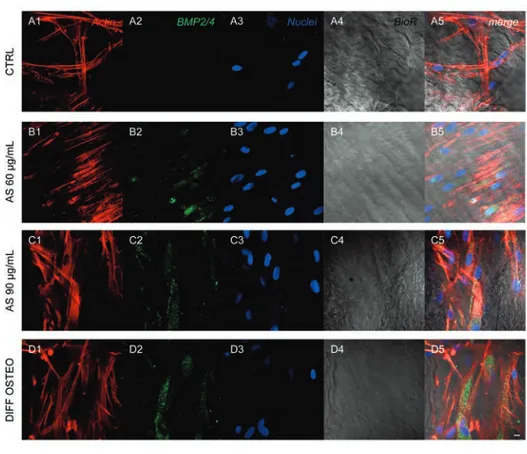
Confocal laser scanning microscopy acquisitions showed the expression ofFigure 6. Expression of BMP2/4. Immunofluorescence detection of BMP2/4 in CTRL(A1-A5), AS 60 µg/mL (B1-B5), AS 90 µg/mL (C1-C5) and DIFF OSTEO (D1-D5). BMP2/4 expression showed a higher expression in AS 90 µg/mL and DIFF OSTEO if compared to the AS 60 µg/mL. No significant differences have been demonstrated when compared AS 90 µg/mL and DIFF OSTEO groups. Red fluorescence, cytoskeleton actin; green fluorescence, specific marker; blue fluorescence, cell nuclei; grey scale, mem-brane observed at transmission light channel; merge pictures showed the overlapping of abovementioned channels. Scale bar: 10 µm.
markers related to the osteogenic differenti-ation, as COL1A1, RUNX2, BMP2/4 and OPN that have been showed an increased expression in DIFF OSTEO and in AS 90 µg/mL, when compared to the CTRL group and with AS 60 µg/mL (Figures 4-7).
Ascorbic acid treatment modulates
markers related to the osteogenic
commitment
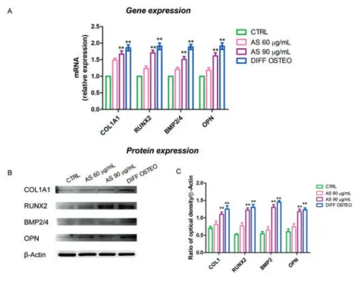
Osteogenic differentiation process was regulated in the early stages from COL1A1, RUNX2, BMP2/4 and OPN. RT-PCR analyses have been performed in order to evaluate their expression. The mRNA levels of COL1A1, RUNX2, BMP2/4 and OPN were at similar range in AS 90 µg/mL and DIFF OSTEO (Figure 8A), while the osteogenic markers showed no significance differences in AS 60 µg/mL compared to the CTRL. At the same time, the compari-son between AS 90 µg/mL and DIFF OSTEO groups showed an upregulation in mRNA levels of the above-mentioned markers when compared to the AS 60 µg/mL and CTRL groups. The protein expression of COL1A1, RUNX2, BMP2/4 and OPN confirmed the results obtained by RT-PCR (Figure 8 B,C).
Discussion
Nowadays, a variety of strategies have been developed to reduce bone loss and increase patients’ life quality. Tissue engi-neering is an innovative area for tissue regeneration and in particular oral derived MSCs represent a new source of adult stem cells that can be taken from tissue with min-imal invasive procedures.38 In the current study, BioR, a biocompatible, bio-absorbable, and osteoconductive collagen membrane from pericardium bovine enriched with hGMSCs, have been used as an in vitro model. Human GMSCs express features such as positivity for CD13, CD29, CD44, CD73, CD90 and CD105, negativity for CD14, CD34 and CD45 markers, as demonstrated by cytofluorimet-ric detection; moreover, they are able to dif-ferentiate into mesenchyme cell lineages with high ability to adhere to a plastic sub-strate, as evidenced by RT-PCR and mor-phological observations.39
Collagen is the major structural compo-nent of bone matrix and alterations of colla-gen properties can therefore affect the mechanical properties of bone and increase fracture susceptibility.40Collagen-base scaf-folds have been extensively investigated and used in bone tissue regeneration as a promising approach to achieve the same hierarchical structure of bone.41 Colla -genous membranes were reported to induce
Figure 7. Expression of OPN. Immunofluorescence detection of OPN in CTRL(A1-A5), AS 60 µg/mL (B1-B5), AS 90 µg/mL (C1-C5) and DIFF OSTEO (D1-D5). OPN expres-sion showed a higher expresexpres-sion in AS 90 µg/mL and DIFF OSTEO if compared to the AS 60 µg/mL. No significant differences have been demonstrated when compared AS 90 µg/mL and DIFF OSTEO groups. Red fluorescence, cytoskeleton actin; green fluores-cence, specific marker; blue fluoresfluores-cence, cell nuclei; grey scale, membrane observed at transmission light channel; merge pictures showed the overlapping of abovementioned channels. Scale bar: 10 µm.
Figure 8. Gene and protein expression. A) Bar graph of RT-PCR showed the expression of mRNA levels in CTRL, AS 60 µg/mL, AS 90 µg/mL and DIFF OSTEO of specific osteogenic related markers. B) COL1A1, RUNX2, BMP2/4 and OPN specific bands of CTRL, AS 60 µg/mL, AS 90 µg/mL and DIFF OSTEO samples. C) Graph bar showed the densitometric analysis of specific band of the markers related to the osteogenic process. The results are expressed as mean ± SD; AS 60 µg/mL vs AS 90 µg/mL; 60 µg/mL vs DIFF OSTEO; **P<0.01.
osteogenesis in situ (42). Several studies showed that collagen may be efficiently used as a scaffold for 3D cultures of MSCs and for subjecting MSCs to mechanical strains, without inducing cell death.43In our recent work, it was demonstrated that a 3D coculture platform using a bovine pericardi-um collagen membrane (BioR) loaded with human periodontal ligament stem cells (hPDLSCs) and endothelial differentiated cells from hPDLSCs (E-hPDLSCs) were able to undergo the osteoangiogenesis dif-ferentiation process.39Moreover, the use of BioR as substrate enriched with hPDLSCs and E-hPDLSCs in coculture, has been con-sidered a resourceful model able to activate the osteoangiogenesis phenotype through ERK1/2 signaling pathway, representing a new potential engineered platform for bone defects repair.39 Actually, functionalized scaffold with conditioned medium, extra-cellular vesicles, or natural molecules are taking a new role in the regenerative medi-cine processes.44
AS, a natural molecule and an essential dietary nutrient, required for various biologi-cal functions, can be considered a master reg-ulator of the collagen biosynthesis.20 Deficiencies in AS can lead to conditions such as scurvy, which, among other ailments, bone pain and impaired wound healing.45The role of AS is primarily related to the develop-ment and maintenance of bone tissues. Investigations of different epidemiological studies and genetic mouse models regarding the effect of vitamin C exhibit a positive out-come on bone health. Overall, vitamin C exerts a positive outcome on trabecular bone formation by influencing expression of bone matrix genes in osteoblasts.45
In the present study, the biological response of BioR/hGMSCs cultured for three weeks in the presence of 60 and 90 µg/mL of AS, was evaluated in terms of osteogenic differentiation. Alizarin Red S staining indicated a massive mineralization in the living construct BioR/hGMSCs sup-plemented with 90 µg/mL AS after three weeks of treatment; furthermore, a similar result has been evidenced in BioR/hGMSCs under osteogenic medium (DIFF OSTEO). The mRNA levels of several classic osteogenic genes such as COL1A1, RUNX2, BMP2/4 and OPN, were analyzed.
Mature bone, in vivo, is composed of proteins and minerals, collagen is the most abundant bone protein. AS is able to stimu-late cell proliferation, enhance the collagen synthesis, activate alkaline phosphatase, and induce the bone cell differentiation.46,47 AS is also an inhibitor of collagenase and the addition of AS into culture medium will enhance the collagen synthesis to promote the bone formation.48,49AS has been used in the supplementation of other scaffold types
as polyurethane scaffold; the supplementa-tion enhanced bone formasupplementa-tion, in vitro and
in vivo, and it can be used for bone tissue
engineering.50
In literature, it has been reported that the use of AS increased the mRNA level of collagen type I, osteocalcin, bone sialopro-tein, and alkaline phosphatase in association with the development of bone nodules in an
in vitro system. The expression analysis of
COL1A1, BMP2/4 and OPN shows an identical trend of RUNX2 after three weeks in osteogenic living construct BioR/hGMSCs supplemented with 60 and 90 µg/mL of AS. These results supported the critical role played by AS in the in vitro model BioR/hGMSCs to induce osteogenic process. This antioxidant molecule is important for collagen hydroxylation, fold-ing, and secretion; it also increases gen gene expression and enhances the colla-gen synthesis stabilizing its tertiary struc-tures. Results obtained through multipara-metric analysis evidenced an upregulation of the osteogenic markers involved in the osteogenic process underlying the pivotal and strategic role of AS in living construct BioR/hGMSCs in osteogenesis induction.
In conclusion, given its potential bene-fits, low cost, and safety profile, AS supple-mentation could play a key role in the regenerative medicine. In bone tissue regen-eration field, biomaterials supplemented with AS could represent an interesting approach in orthopedic and dental surgery procedures.51,52The proposed in vitro model BioR/hGMSCS supplemented with AS showed an interesting role in the osteogenic process inducing the expression of markers related to the bone formation leading to speed up and ameliorate the regeneration process.
References
1. Kock L, van Donkelaar CC, Ito K. Tissue engineering of functional articu-lar cartilage: the current status. Cell Tissue Res 2012;347:613-27. doi: 10.1007/s00441-011-1243-1.
2. Diomede F, Zini N, Pizzicannella J, Merciaro I, Pizzicannella G, D'Orazio M, et al. 5-Aza exposure improves reprogramming process through embry-oid body formation in human gingival stem cells. Front Genet 2018;9:419. doi: 10.3389/fgene.2018.00419
3. Libro R, Scionti D, Diomede F, Marchisio M, Grassi G, Pollastro F, et al. Cannabidiol modulates the immunophenotype and inhibits the acti-vation of the inflammasome in human gingival mesenchymal stem cells. Front Physiol 2016;7:559. doi: 10.3389/
fphys.2016.00559.
4. Diomede F, Gugliandolo A, Cardelli P, Merciaro I, Ettorre V, Traini T, et al. Three-dimensional printed PLA scaf-fold and human gingival stem cell-derived extracellular vesicles: a new tool for bone defect repair. Stem Cell Res Ther 2018;9:104. doi: 10.1186/s13287-018-0850-0.
5. Tabata Y. Biomaterial technology for tissue engineering applications. J R Soc Interface 2009;6:S311-24. doi: 10.1098/rsif.2008.0448.focus.
6. Yuan Y, Shi M, Li L, Liu J, Chen B, Chen Y, et al. Mesenchymal stem cell-conditioned media ameliorate diabetic endothelial dysfunction by improving mitochondrial bioenergetics via the Sirt1/AMPK/PGC-1alpha pathway. Clin Sci (Lond) 2016;130:2181-98. 7. Pawitan JA. Prospect of stem cell
con-ditioned medium in regenerative medi-cine. Biomed Res Int 2014;2014: 965849. doi: 10.1155/2014/965849. 8. Ando Y, Matsubara K, Ishikawa J, Fujio
M, Shohara R, Hibi H, et al. Stem cell-conditioned medium accelerates dis-traction osteogenesis through multiple regenerative mechanisms. Bone 2014;61:82-90. doi: 10.1016/j. bone. 2013.12.029.
9. Osugi M, Katagiri W, Yoshimi R, Inukai T, Hibi H, Ueda M. Conditioned media from mesenchymal stem cells enhanced bone regeneration in rat calvarial bone defects. Tissue Eng Part A 2012;18: 1479-89. doi: 10.1089/ten.TEA. 2011.0325.
10. Barlian A, Judawisastra H, Alfarafisa NM, Wibowo UA, Rosadi I. Chondrogenic differentiation of adi-pose-derived mesenchymal stem cells induced by L-ascorbic acid and platelet rich plasma on silk fibroin scaffold. Peer J 2018;6:e5809. doi: 10.7717/ peerj.5809.
11. Geesin JC, Darr D, Kaufman R, Murad S, Pinnell SR. Ascorbic acid specifical-ly increases type I and type III procolla-gen messenger RNA levels in human skin fibroblast. J Invest Dermatol 1988;90:420-4. doi: 10.1111/1523-1747.ep12460849.
12. De Witte TM, Fratila-Apachitei LE, Zadpoor AA, Peppas NA. Bone tissue engineering via growth factor delivery: from scaffolds to complex matrices. Regen Biomater 2018;5:197-211. doi: 10.1093/rb/rby013.
13. Ducy P, Zhang R, Geoffroy V, Ridall AL, Karsenty G. Osf2/Cbfa1: A tran-scriptional activator of osteoblast differ-entiation. Cell 1997;89:747-54. doi: 10.1016/s0092-8674(00)80257-3. 14. Sodek J, Ganss B, McKee MD.
Osteopontin. Crit Rev Oral Biol Med 2000;11:279-303.
15. Zhang X, Xu WR. Neutrophils diminish T-cell immunity to foster gastric cancer progression: the role of GM-CSF/PD-L1/PD-1 signalling pathway. Gut 2017;66:1878-80. doi: 10.1136/gutjnl-2017-313923.
16. Gugliandolo A, Diomede F, Cardelli P, Bramanti A, Scionti D, Bramanti P, et al. Transcriptomic analysis of gingival mesenchymal stem cells cultured on 3D bioprinted scaffold: A promising strate-gy for neuroregeneration. J Biomed Mater Res A 2018;106:126-37. doi: 10.1002/jbm.a.36213.
17. Filipowska J, Tomaszewski KA, Niedzwiedzki L, Walocha JA, Niedzwiedzki T. The role of vasculature in bone development, regeneration and proper systemic functioning. Angiogenesis 2017;20:291-302. doi: 10.1007/s10456-017-9541-1
18. Itzstein C, Coxon FP, Rogers MJ. The regulation of osteoclast function and bone resorption by small GTPases. Small GTPases 2011;2:117-30. doi: 10.4161/sgtp.2.3.16453
19. Shi AY, Heinayati A, Bao DY, Liu HF, Ding XC, Tong X, et al. Small molecule inhibitor of TGF- signaling enables robust osteogenesis of autologous GMSCs to successfully repair minipig severe maxillofacial bone defects. Stem Cell Res Ther 2019;10:172. doi: 10.1186/s13287-019-1281-2.
20. Ferrazzo PC, Niccoli S, Khaper N, Rathbone CR, Lees SJ. Ascorbic acid diminishes bone morphogenetic protein 2-induced osteogenic differentiation of muscle precursor cells. Muscle Nerve 2019;59:501-8. doi: 10.1002/mus. 26415.
21. Nomura S, Takano-Yamamoto T. Molecular events caused by mechanical stress in bone. Matrix Biol 2000;19:91-6.
22. Westendorf JJ, Kahler RA, Schroeder TM. Wnt signaling in osteoblasts and bone diseases. Gene 2004;341:19-39. doi: 10.1016/j.gene.2004.06.044. 23. Johnson ML, Harnish K, Nusse R, Van
Hul W. LRP5 and Wnt signaling: A union made for bone. J Bone Miner Res 2004;19:1749-57. doi: 10.1359/JBMR.040816.
24. van der Horst G, van der Wert SM, Farih-Sips H, van Bezooijen RL, Lowik CWGM, Karperien M. Downregulation of Wnt signaling by increased expres-sion of Dickkopf-1 and-2 is a prerequi-site for late-stage osteoblast differentia-tion of KS483 cells. J Bone Miner Res 2005;20:1867-77. doi: 10.1359/JBMR.050614
25. Diomede F, Gugliandolo A, Scionti D, Merciaro I, Cavalcanti MF, Mazzon E, et al. Biotherapeutic effect of gingival stem cells conditioned medium in bone tissue restoration. Int J Mol Sci 2018;19. pii:E329. doi: 10.3390/ijms19020329.
26. Pizzicannella J, Diomede F, Merciaro I, Caputi S, Tartaro A, Guarnieri S, et al. Endothelial committed oral stem cells as modelling in the relationship between periodontal and cardiovascular disease. J Cell Physiol 2018;233:6734-47. doi: 10.1002/jcp.26515.
27. Dominici M, Le Blanc K, Mueller I, Slaper-Cortenbach I, Marini F, Krause D, et al. Minimal criteria for defining multipotent mesenchymal stromal cells. The International Society for Cellular Therapy position statement. Cytotherapy 2006;8:315-7. doi: 10.1080/14653240600855905
28. Pizzicannella J, Gugliandolo A, Orsini T, Fontana A, Ventrella A, Mazzon E, et al. Engineered extracellular vesicles from human periodontal-ligament stem cells increase VEGF/VEGFR2 expres-sion during bone regeneration. Front Physiol 2019;10:512. doi: 10.3389/ fphys.2019.00512.
29. Diomede F, Rajan TS, Gatta V, D'Aurora M, Merciaro I, Marchisio M, et al. Stemness maintenance properties in human oral stem cells after long-term passage. Stem Cells Int 2017;2017: 5651287. doi: 10.1155/2017/5651287. 30. Diomede F, Merciaro I, Martinotti S,
Cavalcanti MFXB, Caputi S, Mazzon E, et al. miR-2861 is involved in osteogenic commitment of human peri-odontal ligament stem cells grown onto 3d scaffold. J Biol Regul Homeost Agents 2016;30:1009-18.
31. Rajan TS, Giacoppo S, Trubiani O, Diomede F, Piattelli A, Bramanti P, et al. Conditioned medium of periodontal ligament mesenchymal stem cells exert anti-inflammatory effects in lipopolysaccharide-activated mouse motoneurons. Exp Cell Res 2016;349: 152-61. doi: 10.1016/j.yexcr. 2016.10.008.
32. Bianchi M, Pisciotta A, Bertoni L, Berni M, Gambardella A, Visani A, et al. Osteogenic differentiation of hDPSCs on biogenic bone apatite thin films. Stem Cells Int 2017;2017: 3579283. 10.1155/ 2017/3579283. Corrigendum in: Stem Cells Int 2017; 2017:6587384. doi: 10.1155/2017/ 6587384.
33. Diomede F, D'Aurora M, Gugliandolo A, Merciaro I, Orsini T, Gatta V, et al. Biofunctionalized scaffold in bone tis-sue repair. Int J Mol Sci 2018;19. pii:
E1022. doi: 10.3390/ijms19041022. 34. Pizzicannella J, Cavalcanti M, Trubiani
O, Diomede F. MicroRNA 210 medi-ates VEGF upregulation in human peri-odontal ligament stem cells cultured on 3Dhydroxyapatite ceramic scaffold. Int J Mol Sci 2018;19. pii: E3916. doi: 10.3390/ijms19123916.
35. Diomede F, D'Aurora M, Gugliandolo A, Merciaro I, Ettorre V, Bramanti A, et al. A novel role in skeletal segment regeneration of extracellular vesicles released from periodontal-ligament stem cells. Int J Nanomedicine 2018;13:3805-25. doi: 10.2147/IJN .S162836.
36. Trubiani O, Ballerini P, Murmura G, Pizzicannella J, Giuliani P, Buccella S, et al. Toll-like receptor 4 expression, interleukin-6,-8 and Ccl-20 release, and Nf-Kb translocation in human peri-odontal ligament mesenchymal stem cells stimulated with Lps-P-gingivalis. Eur J Inflamm 2012;10:81-9. doi: 10.1177/1721727X1201000109 37. Mammana S, Gugliandolo A, Cavalli E,
Diomede F, Iori R, Zappacosta R, et al. Human gingival mesenchymal stem cells (GMSCs) pre-treated with vesicu-lar moringin nanostructures as a new therapeutic approach in a mouse model of spinal cord injury. J Tissue Eng Regen Med. 2019;13:1109-1121. doi: 10.1002/term.2857.
38. Trubiani O, Toniato E, Di Iorio D, Diomede F, Merciaro I, D'Arcangelo C, et al. Morphological analysis and inter-leukin release in human gingival fibrob-lasts seeded on different denture base acrylic resins. Int J Immunopath Ph 2012;25:637-43. doi: 10.1177/039463201202500310. 39. Pizzicannella J, Pierdomenico SD,
Piattelli A, Varvara G, Fonticoli L, Trubiani O, et al. 3D human periodontal stem cells and endothelial cells promote bone development in bovine pericardi-um-based tissue biomaterial. Materials (Basel) 2019;12. pii: E2157 doi: 10.3390/ma12132157.
40. Viguet-Carrin S, Garnero P, Delmas PD. The role of collagen in bone strength. Osteoporos Int 2006;17:319-36. doi: 10.1007/s00198-005-2035-9. 41. Ferreira AM, Gentile P, Chiono V,
Ciardelli G. Collagen for bone tissue regeneration. Acta Biomater 2012;8: 3191-200. doi: 10.1016/j. actbio.2012. 06.014.
42. Taguchi Y, Amizuka N, Nakadate M, Ohnishi H, Fujii N, Oda K, et al. A his-tological evaluation for guided bone regeneration induced by a collagenous membrane. Biomaterials 2005;26:6158-66. doi: 10.1016/j.biomaterials.2005.
03.023.
43. Sumanasinghe RD, Osborne JA, Loboa EG. Mesenchymal stem cell-seeded collagen matrices for bone repair: Effects of cyclic tensile strain, cell den-sity, and media conditions on matrix contraction in vitro. J Biomed Mater Res A 2009;88:778-86. doi: 10.1002/ jbm.a.31913.
44. Pizzicannella J, Diomede F, Gugliandolo A, Chiricosta L, Bramanti P, Merciaro I, et al. 3D printing PLA/gingival stem cells/ EVs upregu-late miR-2861 and -210 during osteoan-giogenesis commitment. Int J Mol Sci. 2019;20). pii: E3256. doi: 10.3390/ ijms20133256.
45. Aghajanian P, Hall S, Wongworawat MD, Mohan S. The roles and mecha-nisms of actions of vitamin C in bone: New developments. J Bone Miner Res 2015;30:1945-55. doi: 10.1002/ jbmr.2709.
46. Peterkofsky B. The effect of ascorbic acid on collagen polypeptide synthesis and proline hydroxylation during the growth of cultured fibroblasts. Arch Biochem Biophys 1972;152:318-28. doi: 10.1016/0003-9861(72)90221-4. 47. Berg RA, Steinmann B, Rennard SI,
Crystal RG. Ascorbate deficiency results in decreased collagen produc-tion: under-hydroxylation of proline leads to increased intracellular degrada-tion. Arch Biochem Biophys 1983;226:681-6. doi: 10.1016/0003-9861(83)90338-7.
48. Langenbach F, Handschel J. Effects of dexamethasone, ascorbic acid and beta-glycerophosphate on the osteogenic dif-ferentiation of stem cells in vitro. Stem Cell Res Ther 2013;4:117. doi: 10.1186/scrt328.
49. Park JB. The effects of dexamethasone, ascorbic acid, and beta-glycerophos-phate on osteoblastic differentiation by
regulating estrogen receptor and osteo-pontin expression. J Surg Res. 2012;173:99-104. doi: 10.1016/j.jss. 2010.09.010.
50. Wang C, Cao X, Zhang Y. A novel bioactive osteogenesis scaffold delivers ascorbic acid, beta-glycerophosphate, and dexamethasone in vivo to promote bone regeneration. Oncotarget 2017;8:31612-25. doi: 10.18632/onco-target.15779.
51. Iaquinta MR, Mazzoni E, Manfrini M, D'Agostino A, Trevisiol L, Nocini R, et al. Innovative Biomaterials for Bone Regrowth. Int J Mol Sci. 2019;20. pii: E618. doi: 10.3390/ijms20030618. 52. Qi YJ, Lohman J, Bratlie KM,
Peroutka-Bigus N, Bellaire B, Wannemuehler M, et al. Vitamin C and B-3 as new biomaterials to alter intes-tinal stem cells. J Biomed Mater Res A 2019;107:1886-97. doi: 10.1002/, jbm.a.36715.