©2008 LANDES BIOSCIENCE
. DO NO
T DISTRIBUTE
.
Research Paper
Carnitine Palmitoyltransferase I in Human Carcinomas
A Novel Role in Histone Deacetylation?
Paola Mazzarelli
1,†Sabina Pucci
1,†Elena Bonanno
1Fabiola Sesti
1Menotti Calvani
2Luigi Giusto Spagnoli
1,*
1Institute of Anatomic Pathology; Dept. of Biopathology, University of Rome Tor
Vergata; Rome, Italy
2Scientific Department, Sigma Tau S.p.A.; Pomezia, Italy. †These authors contributed equally to this work.
*Correspondence to: Prof. Luigi Giusto Spagnoli; Department of Biopathology; University of Rome Tor Vergata; Via Montpellier 1; Rome 00133 Italy; Tel.: +39.6.2090.3957; Fax: +39.6.2090.2209; Email: [email protected] Original manuscript submitted: 05/14/07
Revised manuscript submitted: 07/13/07 Manuscript accepted: 07/13/07
Previously published as a Cancer Biology & Therapy E-publication: http://www.landesbioscience.com/journals/cbt/article/4742
KEy wordS
carnitine palmitoyltransferase I (CPT1), fatty acid synthase (FAS), histone deacety-lase activity, cancer, immunohistochemistry, metabolism, histone deacetylase inhibitors.
ABBrEviAtionS
ALGD adenomas with low grade dysplasia CPT1 carnitine palmitoyl transferase I CRC colorectal adeno-carcinomas FAS fatty acid synthase
HDAC histone deacetylase
HDACI histone deacetylase inhibitor H&E haematoxylin-eosin
IDC breast invasive ductal carcinomas TSA trichostatin A
UDH usual ductal hyperplasia
ACKnowLEdGEMEntS
We thank Antonio Volpe for the technical assistance in the acquisition of images. This work was supported in part by grant from Sigma Tau Pharmaceutical S.p.A.
ABStrACt
Carnitine palmitoyl transferase I (CPT1) catalyzes the transport of long‑chain fatty acids into mitochondria for b‑oxidation. A link between CPT1 and apoptosis has been
suggested on the basis of several experimental data. Nevertheless, results are contradic‑ tory about the effective role of CPT1 in cell survival control and cancer development. Conversely, Fatty acid synthase (FAS) enzyme, required for the synthesis of fatty acids, is found over‑expressed in tumors and inhibition of FAS triggers apoptosis in human cancer cells.
We have studied the tumor‑specific modulation of CPT1 and FAS in human colorectal cancer (n = 11) and breast carcinomas (n = 24). CPT1 was significantly decreased in the cytoplasm of tumoral samples (p < 0.04), whereas FAS was increased (p < 0.04). A striking CPT1 nuclear localization was evident in the tumors (p < 0.04). In the nuclear environment the protein would modulate the levels of acetyl/acyl‑CoA implicated in the regulation of gene transcription. At this purpose, we performed in vitro experiments using epithelial neoplastic (MCF‑7, Caco‑2, HepG2 cells) and non neoplastic cell lines (MCF‑12F) confirming a nuclear localization of CPT1 protein exclusively in neoplastic cells. Moreover histone deacetylase (HDAC) activity showed significantly higher levels in nuclear extracts from neoplastic than from control cells. HDAC1 and CPT1 proteins coimmunoprecipitated in nuclear extracts from MCF‑7 cells. The treatment with HDAC inhibitors such as trichostatin A and butyrate significantly decreased nuclear expression of CPT1 and its bond to HDAC1. We also identified the existence of CPT1A mRNA tran‑ script variant 2 in MCF‑7, beside to the classic isoform 1.
The peculiar localization of CPT1 in the nuclei of human carcinomas and the disclosed functional link between nuclear CPT1 and HDAC1 propose a new role of CPT1 in the histonic acetylation level of tumors.
introduCtion
Mitochondrial dysfunction contributes to a number of human disorders and may aid cancer progression. In particular, functional alteration of enzymes involved in the mitochondrial oxidative metabolism has been found during apoptosis and neoplastic transformation.1-3 The altered metabolism of neoplasia would favor an increased cell
survival, promoting the increase of tumor size and cancer aggressiveness. Biochemical studies have demonstrated that one of the multiple differences between tumoral cells and healthy counterparts consists in the metabolic pathway, the uptake of glucose and the glycolytic process.4,5 In fact, the overall metabolic demand on the neoplastic cell is
significantly higher than most other tissues and the cancer cell depends more on glycolysis, even in the presence of available oxygen.5 The glycolytic process would be mediated by
the AMP-activated protein kinase (AMPK) that functions as a low energy checkpoint: in the neoplastic cells it would restore depleted ATP levels through fatty acid oxidation.5 The
reliance of tumor cells on glycolysis may be a potential means by which these cells evade programmed cell death.6
Carnitine Palmitoyl transferase 1 (CPT1) resides at the outer mitochondrial membrane and is a site for intracellular regulation of fatty acid metabolism, transporting long-chain fatty acids into mitochondria for b‑oxidation (together with CPT2 and Carnitine/ acyl-Carnitine Translocase).7,8 Two isoforms of CPT1 have been characterized, known
as L-CPT1 (CPT1A) and M-CPT1 (CPT1B) in liver (L-) and muscle (M-) respectively, showing overlapping tissue specific expression. While CPT1B is expressed in skeletal muscle, heart, testis and adipose tissue, CPT1A has a more widespread distribution.7
©2008 LANDES BIOSCIENCE
. DO NO
T DISTRIBUTE
.
A link between CPT1 and apoptosis has been suggested on the basis of several experimental data. It was demonstrated that CPT1 interacts with Bcl-2 protein, that regulates programmed cell death in several systems and it is also expressed at the outer mitochondrial membrane.9-11
On the other hand, increased levels of Fatty acid synthase (FAS), the major enzyme required for the anabolic conversion of dietary carbohydrate to fatty acids, are found in a wide array of solid tumors12-15 and FAS inhibition triggers apoptosis in human cancer
cells,8 supporting the “metabolic oncogene” theory for FAS
over-expression in cancer cells.15,16
Taking into account the remodulation of the metabolic pathway in the cancerous cells and the unclear role of CPT1 in cell survival control and cancer development, we analyzed the protein expression of CPT1 and FAS in human tumor tissues (n = 35) and healthy counterparts (n = 35). Here we show that CPT1 expression decreases in the cytoplasm of tumoral tissues, whereas FAS increases in the same human tumors. In contrast, a nuclear localization of CPT1 is evident in the tumoral tissues. CPT1 was previously described on the nuclear membrane,17 but its specific function in this cell
compartment has not been clarified yet. In the nuclear environ-ment, the protein would modulate the levels of acetyl/acyl-CoA and carnitine, both of which have been implicated in the regulation of gene transcription.18 Then, we performed in vitro experiments using
epithelial neoplastic (MCF-7, Caco-2, HepG2) and non transformed (MCF-12F) cell lines.
Two transcript variants have been recently described for CPT1-L (CPT1A) isoform, named variant 1 and 2, corresponding to the mRNA sequences NM_001876 and NM_001031847, respec-tively.19,20 The variant 2 differs in the 3’UTR and codes for an
isoform with a shorter C-terminus.
Immunocytochemical experiments confirm a peculiar nuclear localization of CPT1A protein in the neoplastic cells. Moreover, we identify the presence of the CPT1A transcript variant 2 in the same cells, beside to the classic isoform 1.19
To better clarify the role of nuclear CPT1A we studied its correla-tion with the histone deacetylase (HDAC) protein complex in the same cells. The histone deacetylase (HDAC) activity is significantly higher in nuclear extracts from neoplastic cells, as compared to the HDAC activity detected in MCF-12F cells. Moreover, HDAC1 and CPT1 proteins coimmunoprecipitate in nuclear extracts from neoplastic cells.
Inhibitors of histone deacetylase activity (HDACI) such as trichostatin A and sodium-butyrate are able to cause multiple effects in cultured mammalian cells, including inhibition of proliferation and induction of apoptosis, via de-repression of genes involved in cell growth and apoptosis.21,22 The treatment with such HDACI
significantly decreases both the nuclear expression of CPT1 and the HDAC1-CPT1 bond, in MCF-7 cells.
In vitro results match the histonic and not histonic acetylation status in the tumoral samples: the acetylation of histone H4 protein is much lower in the tumors analyzed, as compared with controls.
Here we identify a new role of CPT1 in cancer development, further than the physiological role in the transport of long-chain fatty acids into mitochondria. In fact, the presence of CPT1 in the nuclei of cancerous tissues and neoplastic cells and the disclosed functional link between CPT1 and HDAC1, show that CPT1 plays a role in the histone acetylation status of the tumors. As a consequence, it could
be involved in the transcriptional regulation of specific genes related to cancer development. Hence, the nuclear form of CPT1 might represent a new highly specific target for more effective anti-cancer therapies.
MAtEriALS And MEthodS
Patient characteristics and tissue samples. Eleven colorectal
adeno-carcinomas (CRC) and twenty four breast invasive ductal carcinomas (IDC) were collected in the authors’ department, irre-spectively of the clinical staging. Informed consent was obtained from all the patients included in the study. Ethical approval was obtained by the authors’ institutional review board. The mean age of the patients at the time of surgery was 69 ± 5 and the male/female ratio was 2:6. Distant normal mucosa (NM, n = 11) and adenomas with low grade dysplasia (ALGD, n = 6) were collected and analyzed for the colorectal cases. Histological classification was carried out on H&E-stained slides.23-25
Tissue arrays. Tissue arrays (TA) were performed for breast cancer
cases. Three 0.6 mm2 cores of breast cancer tissue were removed from
representative tumor areas on each block identified by the patholo-gist (EB). These cores were used to construct recipient array blocks in triplicate. Each TA was composed of 12 infiltrating ductal carci-nomas (IDC) (3G1; 6G2; 3G3), and non neoplastic breast tissues (NB, n = 12: 5 fibro-adenomas, 4 usual ductal hyperplasia or UDH, 3 adenosis) as controls. Adjacent normal breast tissue was occasion-ally present and evaluated in the IDC cores. If one or more core were uninformative because of loss or lack of tumor in core, then the scoring results were taken from the remaining core(s).
The tissue array was sectioned, stained with haematoxylin-eosin and received a final pathology review. For immunohistochemistry, the TA was cut in 5 mm sections and stained with appropriate anti-bodies and controls.
Immunohistochemistry (IHC) and immunocytochem‑ istry (ICC). Serial four micron sections from formalin-fixed and
paraffin embedded specimens were immunostained for CPT1A, FAS, acetyl-lysine and acetyl-histone-H4 following the streptoa-vidin-biotin method, as previously described.26 In brief, sections
were deparaffinezed and rehydrated in decreasing alcohol. Tissue antigen retrieval was performed in citrate buffer by microwave. Endogenous peroxidase activity was quenched with 0.03% hydrogen peroxide in absolute methanol, for 30 min at room temperature. The primary antibodies used were rabbit polyclonal anti-CPT1A anti-FAS antibodies (H95 raised against a recombinant protein, mapping near the aminoterminus of CPT1A and H300, Santa Cruz Biotechnology Inc. CA 95060, USA) validated for immu-nohistochemical analysis, in 1:650 and 1:50 dilution, respectively. Anti-acetyl-lysine rabbit serum and rabbit polyclonal anti-ace-tyl-histone H4 (Upstate, NY 12946) were diluted 1:400 and 1:75 respectively. Biotinylated goat anti-rabbit IgG (Dako A/S, Denmark) were used as secondary antibody. After washing, sections were treated with streptoavidin-peroxidase reagent (Dakopatts A\S, Denmark), incubated with diaminobenzidine (DAB) and lightly counterstained with hematoxylin. Slides were examined by two pathologists, unaware of the clinical data and molecular results. Tissue staining was semi-quantitatively graded for intensity as negative/weak: 0, moderate: 1 and strong: 2. FAS and cytoplasmic CPT1 was regarded as positive when moderate-to-strong granular cytoplasmic staining was
©2008 LANDES BIOSCIENCE
. DO NO
T DISTRIBUTE
.
detected.14,15 Acetyl-lysine and acetyl-histone-H4 were deemed
posi-tive when moderate(1)-to-strong(2) immunostaining was present in cytoplasm and/or nuclei. For nuclear CPT1, positively stained cells were quantified as a percentage and assigned to one of three catego-ries: 0–10% (Neg); 11–20% and >20% (Pos) (Tables 1 and 2).
For immunocytochemistry, MCF-7, MCF-12F, Caco-2 and HepG2 cells were plated in 4 wells/chamberslides at a concentration of 60.000 x cm2. After overnight culture, cells were treated or not
with sodium butyrate at concentration 5 mM (Sigma-Aldrich S.r.l., I-20151 MI, Italy) or trichostatin A (BioVision Research Products, Mountain View, CA 94043 USA), at concentration 1 mM and 3 mM, for 24 h to inhibit cellular HDAC activity. At the end, growth medium was removed and cells were rinsed in phosphate-buffered saline (PBS) and fixed in formalin 10% solution (Sigma-Aldrich) for 5 min. Then, cells were permeablized with 0.5% Triton X-100 and 0.05% Tween-20 (Bio-Rad Laboratories, Munchen) in PBS. Incubation and dilution times of primary antibody (rabbit polyclonal anti-CPT1A), secondary antibody (biotinylated goat anti-rabbit IgG) and following reagents (streptoavidin-peroxidase) were the same chosen for IHC experiments. After washing, slides were incubated with diaminobenzidine (DAB) and counterstained with haematoxylin.
Statistical analysis. All values provided in the text and figures are
means of three independent experiments ± standard deviations (SD). Mean values were compared using the two-tailed Student t-test, for independent samples. For FAS and cytoplasmic CPT1 immunos-taining, statistical significance was calculated comparing intensity values (from 0 to 2) between different groups, e.g., FAS staining values in normal mucosae (NM) versus adenocarcinomas (CRC). Percent values of positively stained cells were compared between different groups, for CPT1 nuclear staining (see Results). Differences were considered statistically significant for p < 0.05.
Cell culture, chemicals and protein extracts preparation. MCF-7
(HTB-22), MCF-12F (CRL-10783), HepG2 (HB-8065) and Caco-2 (HTB-37) were purchased from American Type Culture Collection (ATCC) and grown in complete culture medium, according to condition suggested from ATCC, at 37˚C temperature with 95%
humidified air, 5% CO-2. For HDAC experiments, 60.000 x cm2
cells were plated in 25 cm2 flasks. After overnight culture, cells
(treated or not with sodium butyrate 5 mM for 24 hours) were tryp-sinized, washed in PBS and pelleted for protein extraction according to Dignam method,27 modified as described by Pucci et al.28 Briefly,
nuclear and cytoplasmic fractions were separated by centrifugation in a microfuge at 10,000 rpm and stored at -80˚C. Protein content in nuclear and cytoplasmic extracts was determined in triplicate by Bradford assay (Bio-Rad Protein Assay, Bio-Rad Laboratories, Munchen).
Western blotting and immunoprecipitation. Cytoplasmic and
nuclear protein extracts (10 mg) were boiled in SDS-PAGE loading buffer and separated by 10% SDS-PAGE. Proteins were transferred to a PVDF membrane (Hybond-P, Amersham-Pharmacia Biotech, England HP7 9NA) using an electroblotting apparatus. Membranes were stained with Ponceau S dye, to check for equal loading and homogeneous transfer and incubated for 1 h at RT with 3% skim milk (Difco Lab. Detroit, MI, USA) and 0.5%Tween 20 (USB, Cleveland, Ohio, USA). Primary antibody (rabbit polyclonal anti-CPT1A H95, Santa Cruz) was diluted 1:1000 in 1% BSA and incubated for 1 h at room temperature; samples were washed extensively with 0.5% Tween-20 in Tris buffered saline and diluted 1:8000 secondary antibody (anti-rabbit-HRP IgG, Santa Cruz Biotechnologies, Inc.) was added in 1%BSA, 1%milk, 0.5% Tween-20, for 45’ at room temperature.
Filters were reprobed with anti-Sp1 mouse IgG1 monoclonal antibody (Sigma-Aldrich, Saint Louis, Missouri 63103 USA) to test purity of the extracts and with anti-b-actin mouse monoclonal antibody (Sigma-Aldrich, Saint Louis, Missouri 63103 USA) to normalize the cytoplasmic protein levels. Filters were washed and developed using an enhanced chemiluminescence system (ECL, Amersham-Pharmacia Biotech, Pharmacia Biotech, England HP7 9NA).
Immunoprecipitation assay (IP). Immunoprecipitation assay
(IP).was performed on nuclear extracts from tumoral cells (MCF-7) treated and not with inhibitor of HDAC1 activity, sodium-butyrate.
Table 1 FAS and CPT I protein expression detected by immunohistochemistry, in human tumours and controls
Number of cases was shown as a percentage. In parentheses, absolute numbers were reported. CRC, colorectal adenocarcinomas; ALGD, adenomas with low-grade dysplasia. IDC, breast invasive ductal carci-nomas. 1Distant normal mucosa (NM) was tested for the colon samples. 2NB, Non neoplastic breast tissues
(fibroadenomas n = 10, UDH n = 8, adenosis n = 6) were used as controls for the breast cancer cases; FAS. NM vs CRC, p = 0.003; NB vs IDC, p = 0.04 (see Statistical analysis in Materials and Methods); CPT I cyt. (cytoplasmic) CRC vs NM, p = 0.001; CRC vs ALGD, p = 0.04; NB vs IDC, p = 0.04; CPT I n (nuclear). NM vs CRC, p = 0.04; NB vs IDC, p = 0.03.
Table 2 Acetyl‑histone H4 and acetyl‑lysine protein expression detected by IHC, in human tumors and controls
Number of cases was shown as a percentage. In parentheses, absolute numbers were reported; 1Cases were
deemed positive when moderate-to-strong (1–2) nuclear (n) staining was present; 2Cases were positive
when moderate-to-strong (1–2) nuclear (n) and/or cytoplasmic (cyt) staining was present; CRC, colorectal adenocarcinomas. NM, normal mucosa. OT, carcinomas from other tissues. ANNT, adjacent-to-tumor normal tissue (see Materials and Methods); AcH4. CRC vs NM, p = 0.02; OT vs ANNT, p = 0.3; Ac-Lys cyt. CRC vs NM, p = 0.4; OT vs ANNT, p = 0.03; Ac-Lys n. CRC vs NM, p = 0.03; OT vs ANNT, p = 0.05 (see Statistical analysis in Materials and Methods).
©2008 LANDES BIOSCIENCE
. DO NO
T DISTRIBUTE
.
Fifty micrograms of nuclear extracts were immunoprecipitated with anti-HDAC1. In brief, protein extracts were precleared incubating them with Protein A-Agarose (50% slurry) for 30 minutes at 4˚C with agitation. The Agarose was removed, then 4 mg of anti-HDAC1 mouse monoclonal antibody (Upstate, NY12946) was added over-night at 4˚C with rotation. IP negative control was performed without HDAC1 antibody. After incubation, Protein A-Agarose was added for one hour at 4˚C with rotation. At the end, supernatants containing unbound proteins were carefully removed and the IP complexes were washed in wash buffer (PBS, NaCl 0.5 M, and after in PBS alone). Each immunoprecipitate was electrophoresed on a 10% poliacrylamide gel. Gel was stained with Ponceau S-dye, blotted onto PVDF membrane, probed with anti-CPT1 and anti-HDAC1 and detected with ECL. Supernatant from HDAC1-IP samples, containing unbound protein fraction (ub) was also loaded onto poliacrylamide gel.
RT‑PCR analysis of CPT1A transcript variants. Total RNA was
isolated from MCF-7 and MCF12F cells cultured in T75 Flasks using Tri Reagent (Ambion, Inc.), according to the manufacturer’s instructions. RNA quantification was performed using spectropho-tometry. Reverse transcription of total RNA (1 mg each cell line) was performed with Gene Amp RNA PCR Kit (Applied BioSystems) using Random Examers as primers to cDNA synthesis. To amplify the cDNA for CPT1A transcript variants 1 (NM_001876) and 2 (NM_001031847) the following primers (Invitrogen) were used:
CPT1A1f 5'-ATTCTCATCGCTTTGGAAGG-3' and CPT1A1r 5'-AGACATCAGGGGAGACTTTA-3'; CPT1A2f 5'-GACAAAG TGGAAAGTCTCAG-3' and CPT1A2r 5'-TGTGTCAGGCACT GTTCTCA-3'. Amplification protocol was performed in a thermal cycler (Applied Biosystems) with 35 cycles at 60˚C. PCR products were 327bp for CPT1A1 and 163 bp for CPT1A2 transcript vari-ants. cDNA for b2-Microglobulin housekeeping gene was amplified as control. Ethidium-bromide stained 2% agarose gel was run at 100V and picture of the gel was acquired by scanning system.
Histone deacetylase (HDAC) activity. Nuclear protein extracts
obtained from MCF-7, Caco-2, HepG2 and MCF-12F cells were used to detect HDAC activity with a colorimetric assay kit (BioVision Research Products, Mountain View, CA 94043 USA). In brief, HDAC colorimetric substrate, which comprise an acetylated lysine side chain, was added with 50 mg of nuclear protein extracts to each well of a 96-well plate. A 2-ml volume of 1 mg/ml of the HDAC inhibitor trichostatin A was added to control well. Deacetylation of the substrate sensitized it, so that, following treatment with Lysine Developer produced a chromophore. Samples were analyzed using an ELISA plate reader at 405 nm.
rESuLtS
Fatty acid synthase expression in human tumors by IHC. FAS
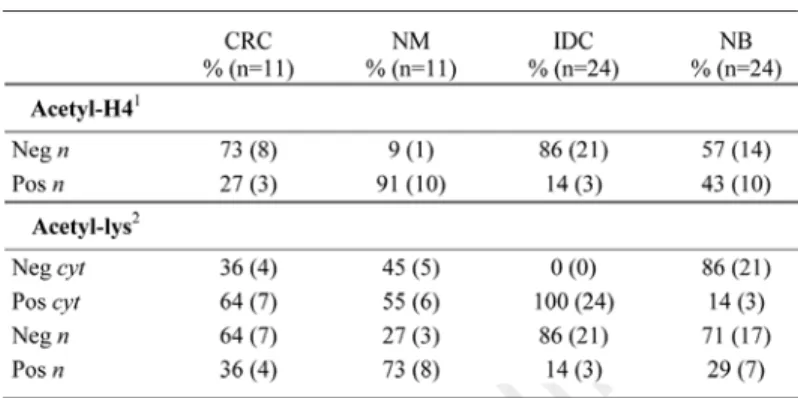
protein was significantly increased in the cytoplasm of the malignant tumors analyzed (tumors vs controls: p = 0.003) (Table 1). Only 13% of tumors (5/35) displayed absence or weak staining in the cytoplasm of neoplastic cells, versus 80% of controls (28/35). Further, strong (2+) specific staining was detected in the 43% of positive tumors (13/30) (Table 1). In particular, the 91% of colon carcinomas (10/11) displayed positive staining (1+, 2+), whereas almost the 100% of normal cases (10/11) displayed absence of specific staining (NM vs CRC: p = 0.003) (Fig. 1C–F).
Expression of CPT1 in human tumors by IHC. The same tumors
were also evaluated for CPT1 protein expression. Corresponding sections of distant normal mucosa (n = 11) were used as controls for the colorectal patients, whereas non neoplastic breast samples (n = 24) were examined for the other cases.
The 63% of the tumoral tissues analyzed (22/35) displayed absent or weak cytoplasmic staining for CPT1, whereas normal samples displayed moderate(1)-to-strong(2) cytoplasmic staining in 72% of cases (tumors vs controls: p < 0.04) (Table 1). The cytoplasmic staining showed the typical granular pattern due to the mitochon-drial localization of CPT1. In particular, the colorectal carcinomas showed a significant decrease of cytoplasmic expression compared with both healthy colonic mucosae and adenomas analyzed (CRC vs NM: p = 0.001 and vs ALGD: p = 0.04). Colon adenomas displayed a protein localization and expression comparable to their normal counterparts (ALGD vs NM: p = 0.06).
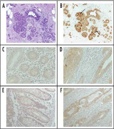
Besides, 8/11 colorectal cancers (73%) and 22/24 breast cancers (92%) showed a speckled nuclear staining, with percentage >20% of positive cells in 7/30 cases (Table 1). In Figure 2, ductal infiltrating breast carcinoma and CRC tissues were compared to matched controls. The specific nuclear staining was evident in the cancerous samples (Fig. 2A–H).
In vitro experiments. CPT1A protein expression. Detection of
CPT1A by immunocytochemical analysis (ICC) showed a marked nuclear staining beside to the cytoplasmic staining in epithelial
Figure 1. Immunohistochemical FAS protein expression in representative human breast (A and B) and colon (C–F) cancer cases. (A) shows the H&E staining in a breast ductal carcinoma with ‘lobular cancerization’. FAS pro‑ tein is strongly (2+) increased in the cancerous part (see arrows), compared with the weak/moderate (0, 1+) expression evident in the nontransformed section (B). FAS expression increases also in two colon carcinomas (D and F), compared with the corresponding distant normal mucosae (C and E) (20X original magnification).
©2008 LANDES BIOSCIENCE
. DO NO
T DISTRIBUTE
.
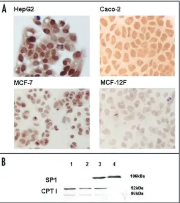
neoplastic cell lines (MCF-7, Caco-2, HepG2) (Fig. 3A). CPT1A nuclear signal varied in intensity through the cell cycle, showing the highest level in condensing chromosomes, as cells progress through mitosis (not shown). Conversely, MCF-12F no tumoral cells displayed a barely detectable staining in the cytoplasm (Fig. 3A).
Cytoplasmic and nuclear protein extracts from MCF-7 and MCF-12F cells were analyzed for CPT1A expression by Western blot analysis. The CPT1A cytoplasmic expression was detected both in MCF-7 and in MCF-12F cells, whereas the nuclear expression was specific of neoplastic cells (Fig. 3B).
Histone deacetylase (HDAC) activity. We also tested HDAC activity on nuclear extracts from neoplastic (MCF-7, Caco-2, HepG2) and control cells (MCF-12F). The HDAC activity was significantly higher in neoplastic cells, than in MCF-12F cells (Fig. 4). Two different HDACI, such as sodium-butyrate and trichostatin A (TSA)21 was effective to inhibit HDAC activity in these cells (not
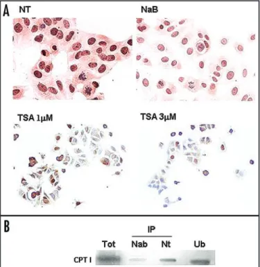
shown). Besides, both treatments with HDAC inhibitors TSA and sodium-butyrate were effective to strongly decrease CPT1 nuclear staining in neoplastic cells (Fig. 5A).
Co‑immunoprecipitation experiments. Tumoral cells (MCF-7)
were treated or not with sodium-butyrate for 24 hours at the concentration 5 mM, able to inhibit HDAC1 activity. The nuclear proteins were extracted, immunoprecipitated with anti-HDAC1 antibody and loaded onto a denaturing poliacrylamide gel. The filter, immunoprobed with anti-CPT1A antibody, showed a coimmuno-precipitation band in nuclear extract from untreated MCF-7 cells. This specific band was strongly reduced after sodium-butyrate treat-ment (Fig. 5B). The immunoprecipitation bands were normalized, reprobing the filter with anti-HDAC1 antibody (not shown).
Figure 3. CPT1A protein expression in no cancerous (MCF‑12F) and cancerous (HepG2, Caco‑2, MCF‑7) cells, by ICC (A) and Western blot analysis (B). The nuclear staining for CPT1A protein is clearly evident in neopla‑ stic cells (panel A, 40X original magnification). (B) shows the Western blot analysis in cytoplasmic (lanes 1,2) and nuclear extracts (lanes 3 and 4) from MCF‑7 (lanes 1,3) and MCF‑12F cells (lanes 2,4). Both the two isoforms of CPT1 (92 kDa and 86 kDa molecular weight) are detected in cytoplasmic extracts from neoplastic and normal cells. CPT1 is undetectable in nuclear extracts from MCF‑12F cells (lane 4). The immunoblotting for the nuclear protein Sp1 is also shown, as a control of extract purity.
Figure 2. Immunohistochemical CPT1A (A and H) and acetyl‑lysine (I and L) protein expression in representative normal (N) and cancerous (K) cases from human breast (A–D) and colon tissues (E–L). The magnification 20X allows to discriminate the cytoplasmic localization of CPT1A in the no cancerous breast (A and C) and colon tissues (E and G) and the specific nuclear staining in breast IDCs (B and D) and colorectal cancer cases (F and H). The decrease of CPT1A cytoplasmic staining is evident in the colon carcinoma (H), compared with the corresponding distant normal mucosa (G). (I and L) show the acetyl‑lysine expression (informative for the global acetylation status of the tissue) on normal mucosa (N) and colorectal cancer tissue (K). The absence of nuclear staining is evident in the colon cancer tissue, compared with the normal control (40X original magnification).
©2008 LANDES BIOSCIENCE
. DO NO
T DISTRIBUTE
.
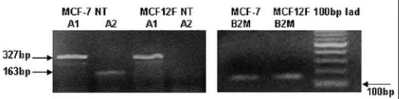
CPT1A transcript variant expression. Two transcript variants
have been described for CPT1-L (CPT1A) isoform, named variant 1 and 2, corresponding to the mRNA sequences NM_001876 and NM_001031847 respectively.19,20 To investigate whether the
different localization of the CPT1A protein found in breast epithelial cells corresponded to a differential expression of the two transcripts, we performed an RT-PCR with primers specific for the two mRNA sequences (see Material and Methods).
As shown in Figure 6, the transcript variant 1 was expressed in the same manner both in MCF-7 and MCF-12F cells, whereas the cDNA of transcript variant 2 was amplified just in the neoplastic cells. A semi-quantitative analysis of PCR reaction was feasible by additional amplification of cDNA for b2 microglobulin (B2M) housekeeping gene (Fig. 6).
Expression of acetyl‑lysine and acetyl‑histone H4 in human tissues. The same colorectal tumors (n = 11) and invasive ductal
breast carcinomas (n = 24) investigated for CPT1 expression, together with corresponding normal tissues were also analyzed for expression of acetyl-histone H4 protein and whole cell acetylation status, by IHC. A significant decrease of acetyl-histone H4 protein expression was evident in the tumoral tissues, as compared with normal counterparts (CRC vs NM: p = 0.02; IDC vs NB: p = 0.03, Table 2). The hypoacetylation at histone level could represent a known mechanism for transcriptional repression of tumor suppressor genes during carcinogenesis.29
The staining with the anti-acetyl-lysine antibody (able to recog-nize the whole cell acetylation status) confirmed a decreased nuclear expression in the same samples (CRC vs NM: p = 0.03; IDC vs NB: p = 0.05), but also showed an increased cytoplasmic staining in tumors as compared with controls (IDC vs NB: p = 0.03) (Table 2, Fig. 2I–L).
diSCuSSion
This study shows, for the first time, the expression of CPT1A in nuclei of human tumors and cancerous cells. We demonstrate a link between nuclear expression of CPT1, uniquely found in human tumors and neoplastic cells and histone hypoacetylation.
Histone deacetylation is associated with transcriptional repression of genes, as the removal of acetyl groups from lysine residues limits accessibility of the DNA for transcription.29 The discovery that
certain HDAC complexes contained known transcriptional repres-sors further strengthened the correlations between hypoacetylation and repression.30 We show that HDAC1 protein interacts with
nuclear CPT1 and that treatment with HDAC inhibitors such as sodium-butyrate and TSA affects the nuclear expression of CPT1 in the tumoral cell. Mammalian HDAC1 exists in large multiprotein complex, Sin3. The Sin3 complex contains mammalian (m)Sin3, Sin3-associated proteins of 18 and 30 kDa and retinoblastoma-asso-ciated proteins (RbAp)46 and -48. HDAC multiprotein complexes are recruited to specific genomic sites by regulatory proteins.31 Figure 4. Histone deacetylase (HDAC) activity on nuclear extracts from neo‑
plastic and control cell lines. We tested HDAC activity on nuclear extracts from neoplastic (Caco‑2, HepG2, MCF‑7) and control cells (MCF‑12F). Optical densities (O.D.) are the mean values ± standard deviations (SD) from three independent experiments. The nuclear activity of histone deacetylase was significantly higher in neoplastic, than in control cells (*O.D. mean values in neoplastic versus control cells, p = 0.03; see also Materials and methods section).
Figure 5. CPT1A protein expression in MCF‑7 cells untreated (NT) and treated with sodium‑butyrate (NaB) or Trichostatin A (TSA) by ICC (A), and western blot on HDAC1‑IP nuclear extracts (B). Cells were treated with sodium‑butyrate at a concentration of 5 mM and with trichostatin A at a concentration of 1 mM and 3 mM for 24 h, as described in Materials and methods. The nuclear staining for CPT1 is strongly reduced after HDAC inhibitors treatments (A). (B) shows the immunoprecipitation experiment with anti‑HDAC1 antibody, in nuclear extracts from MCF‑7 cells treated (NaB) or not (NT) with sodium‑butyrate. Non‑IP nuclear extracts from untreated MCF‑7 (Tot), HDAC1‑IP nuclear extracts from treated MCF‑7 (IP‑NaB), HDAC1‑IP from untreated cells (IP‑NT) and unbound fraction from IP untreated cells (Ub) were electrophoresed on a 10% SDS‑PAGE. The membrane was immunoblotted with CPT1A antibody. CPT1 coimmunoprecipitates with HDAC1 in nuclear extracts from MCF‑7 cells. The treatment with NaB decreases the coimmuno‑ precipitation band. An unbound fraction of CPT1 which does not bind to HDAC1 remains in nuclei from untreated cells (Ub).
©2008 LANDES BIOSCIENCE
. DO NO
T DISTRIBUTE
.
In the nucleus of neoplastic cell, CPT1 could function as a regula-tory protein, recruiting HDAC complexes at acetylated histones of specific promoters, to silence genes involved in the control of cancer development by hypoacetylation.
HDAC inhibitors (HDACI) from a number of chemical classes (e.g., short-chain fatty acids like butyrate, isothiocyanates, hydroxamic acid, etc.) have shown promise as anti-cancer agents in animal studies and early clinical trials.32-35 In fact, HDACI maintain histones in an
acetylated state, and through the resulting alterations in gene regula-tion, inhibit cell cycle progression, differentiation and in some cases induce apoptosis.35,36 Exposure to HDAC inhibitors may also allow
reactivation of tumor suppressor genes, silenced by hypoacetylation during tumorigenesis.36 The use of butyrate to inhibit HDAC1
activity, surprisingly induces, in our model, a decrease of both CPT1 nuclear expression and HDAC1 protein bond to CPT1. Thus, the inhibition of HDAC activity by TSA and butyrate could require the removal of CPT1 from the nucleus of neoplastic cell.
We were able to identify the presence of CPT1A mRNA transcript variant 2 in MCF-7, beside to the classical isoform 1. The mRNA variant 2 codifies for a CPT1A product with a shorter C-terminus. We hypothesize that the shorter CPT1A product could be the one detected in the nucleus of neoplastic cells.
On the other hand, our experiments performed with anti- acetyl-lysine antibody show an hyper-acetylation (most likely of non histonic proteins) in the cytoplasm of human tumors, beside a nuclear histone hypo-acetylation. We can speculate that different mechanisms of post-translational regulation of target proteins could be involved in cell growth/death regulation.36,37
According to previous data, we confirmed an increase of Fatty Acid Synthase expression in human carcinomas14,15 with a corresponding
down-regulation of mitochondrial CPT1A. CPT1A inhibition would prevent oxidation of newly synthesized fatty acids.
Nonetheless, the nuclear CPT1 and not the enzyme as one could represent an highly specific target for new more selective and more effective anti-cancer therapies. In fact, drugs suppressing specifically the CPT1 nuclear activity might allow reactivation of tumor suppres-sors silenced by hypoacetylation, alike molecules who directly inhibit HDAC activity.
References
1. Helmlinger G, Yuan F, Dellian M, Jain RK. Interstitial pH and pO2 gradients in solid tumors in vivo: High-resolution measurements reveal a lack of correlation. Nat Med 1997; 3:177-82.
2. Sutherland RM. Cell and environment interactions in tumor microregions: The multicell spheroid model. Science 1988; 240(4849):177-84.
3. Wang GL, Jiang BH, Rue EA, Semenza GL. Hypoxia-inducible factor 1 is a basic-he-lix-loop-helix-PAS heterodimer regulated by cellular O2 tension. Proc Natl Acad Sci USA 1995; 92:5510-14.
4. Garber K. Energy deregulation: Licensing tumors to grow. Science 2006; 312:1158-9. 5. Shaw RJ. Glucose metabolism and cancer. Curr Opin Cell Biol 2006; 18(6):598-608. 6. Tomiyama A, Serizawa S, Tachibana K, Sakurada K, Samejima H, Kuchino Y, Kitanaka C.
Critical role for mitochondrial oxidative phosphorylation in the activation of tumor sup-pressors Bax and Bak. J Natl Cancer Inst 2006; 98(20):1462-73.
7. Weis BC, Esser V, Foster DW, McGarry JD. Rat heart expresses two forms of mitochondrial carnitine palmitoyltransferase I: The minor component is identical to the liver enzyme. J Biol Chem 1994; 269:18712-15.
8. Zhou W, Simpson PJ, McFadden JM, Townsend CA, Medghalchi SM, Vadlamudi A, Pinn ML, Ronnett GV, Kuhajda FP. Fatty acid synthase inhibition triggers apoptosis during S phase in human cancer cells. Cancer Res 2003; 63:7330-7.
9. Paumen MB, Ishida Y, Muramatsu M, Yamamoto M, Honjo T. Direct interaction of the mitochondrial membrane protein carnitine palmitoyltransferase I with Bcl-2. J Biol Chem 1997; 272:3324-9.
10. Reed JC. Bcl-2 and the regulation of programmed cell death. J Cell Biol 1994; 124:1-6. 11. Nunez G, Clarke MF. The Bcl-2 family of proteins: Regulators of cell death and survival.
Trends Cell Biol 1994; 4:399-403.
12. Pizer ES, Lax SF, Kuhajda FP, Pasternack GR, Kurman RJ. Fatty acid synthase expression in endometrial carcinoma: Correlation with cell proliferation and hormone receptors. Cancer 1998; 83:528-37.
13. Alo’ PL, Visca P, Marci A, Mangoni A, Botti C, Di Tondo U. Expression of fatty acid syn-thase (FAS) as a predictor of recurrence in stage I breast carcinoma patients. Cancer 1996; 77:474-82.
14. Visca P, Alo’ PL, Del Nonno F, Botti C, Trombetta G, Marandino F, Filippi S, Di Tondo U, Donnorso RP. Immunohistochemical expression of fatty acid synthase, apoptotic-regulating genes, proliferating factors, and ras protein product in colorectal adenomas, carcinomas, and adjacent nonneoplastic mucosa. Clin Cancer Res 1999; 5:4111-18.
15. Rashid A, Pizer ES, Moga M, Milgraum LZ, Zahurak M, Pasternack GR, Kuhajda FP, Hamilton SR. Elevated expression of fatty acid synthase and fatty acid synthetic activity in colorectal neoplasia. Am J Pathol 1997; 150:201-8.
16. Menendez J, Decker JP, Lupu R. In support of fatty acid synthase (FAS) as a metabolic oncogene: Extracellular acidosis acts in an epigenetic fashion activating FAS gene expression in cancer cells. J Cell Biochem 2005; 94:1-4.
17. Bremer J. The role of carnitine in cell metabolism. In: De Simone C, Famularo G, eds. Carnitine today. Heidelberg: Springer-Verlag Publishing, 1997:1-37.
18. Murthy MS, Pande SV. A stress-regulated protein, GRP58, a member of thioredoxin super-family, is a carnitine palmitoyltransferase isoenzyme. Biochem J 1994; 304(Pt 1):31-34. 19. Britton CH, Schultz RA, Zhang B, Esser V, Foster DW, McGarry JD. Human liver
mito-chondrial carnitine palmitoyltransferase I: Characterization of its cDNA and chromosomal localization and partial analysis of the gene. Proc Natl Acad Sci USA 1995; 92:1984-8. 20. Gerhard DS, Wagner L, Feingold EA, Shenmen CM, Grouse LH, Schuler G, et al. The
status, quality, and expansion of the NIH full-length cDNA project: The Mammalian Gene Collection (MGC). Genome Res 2004; 14:2121-7.
21. Dashwood RH, Myzak MC, Ho E. Dietary HDAC inhibitors: Time to rethink weak ligands in cancer chemoprevention? Carcinogenesis 2006; 27:344-9.
22. Davis T, Kennedy C, Chiew YE, Clarke CL, deFazio A. Histone deacetylase inhibitors decrease proliferation and modulate cell cycle gene expression in mammary epithelial cells. Clin Cancer Res 2000; 6:4334-42.
23. O’Brien MJ, Winawer SJ, Zauber AG, Gottlieb LS, Sternberg SS, Diaz B, Dickersin GR, Ewing S, Geller S, Kasimian D. The National Polyp Study: Patient and polyp character-istics associated with high-grade dysplasia in colorectal adenomas. Gastroenterology 1990; 98:371-9.
24. Jass JR, Sobin LH. Histological typing of intestinal tumours. World Health Organization International Histological Classification of Tumours. Berlin: Springer-Verlag Publishing, 1989.
25. Rampaul RS, Pinder SE, Elston CW, Ellis IO, On behalf of the Nottingham Breast Team. Prognostic and predictive factors in primary breast cancer and their role in patient manage-ment: The Nottingham Breast Team. EJSO 2001; 27:229-38.
26. Hsu SM, Raine L, Fanger H. The use of avidin-biotin peroxidase complex (ABC) in immunoperoxidase technique: A comparison between ABC and unlabelled antibody (PAP) procedures. J Histochem Cytochem 1981; 29:577-80.
27. Dignam JD, Lebovitz RM, Roeder RG. Accurate transcription initiation by RNA poly-merase II in a soluble extract from isolated mammalian nuclei. Nucleic Acids Res 1983; 1:1475-89.
28. Pucci S, Mazzarelli P, Rabitti C, Giai M, Gallucci M, Flammia G, Alcini A, Altomare V, Fazio VM. Tumor specific modulation of Ku70/80 DNA binding activity in breast and bladder human tumor biopsies. Oncogene 2001; 20:739-47.
29. Kim DH, Kim M, Kwon HJ. Histone deacetylase in carcinogenesis and its inhibitors as anti-cancer agents. J Biochem Mol Biol 2003; 36:110-19.
Figure 6. CPT1A transcript variants expression in normal and cancerous breast epithelial cells. RT‑PCR analysis was performed as described in the Methods section. Both MCF‑7 and MCF‑12F cells express the CPT1A transcript variant 1 (lanes 1, 3), whereas just the neoplastic cells express the CPT1A transcript variant 2 (lanes 2, 4). Additional amplification of b2 microglobulin (B2M) housekeeping gene allows a semi‑quantitative evalua‑ tion of PCR products.
©2008 LANDES BIOSCIENCE
. DO NO
T DISTRIBUTE
.
30. Esteller M, Villar-Garea A. Histone deacetylase inhibitors: Understanding a new wave of anticancer agents. Int J Cancer 2004; 112:171-8.
31. Khochbin S, Verdel A, Lemercier C, Seigneurin-Berny D. Functional significance of histone deacetylase diversity. Curr Opin Genet Dev 2001; 11:162-6.
32. Moe-Behrens GH, Pandolfi PP. Targeting aberrant transcriptional repression in acute myeloid leukemia. Rev Clin Exp Hematol 2003; 7:139-59.
33. Kelly WK, O’Connor OA, Krug LM, Chiao JH, Heaney M, Curley T, MacGregore-Cortelli B, Tong W, Secrist JP, Schwartz L, Richardson S, Chu E, Olgac S, Marks PA, Scher H, Richon VM. Phase I study of an oral histone deacetylase inhibitor, suberoylanilide hydroxamic acid, in patients with advanced cancer. J Clin Oncol 2005; 23:3923-31. 34. Ryan QC, Headlee D, Acharya M, Sparreboom A, Trepel JB, Ye J, Figg WD, Hwang K,
Chung EJ, Murgo A, Melillo G, Elsayed Y, Monga M, Kalnitskiy M, Zwiebel J, Sausville EA. Phase I and pharmacokinetic study of MS-275, a histone deacetylase inhibitor, in patients with advanced and refractory solid tumors or lymphoma. J Clin Oncol 2005; 23:3912-22.
35. Romanski A, Bacic B, Bug G, Pfeifer H, Gul H, Remiszewski S, Hoelzer D Atadja P, Ruthardt M, Ottmann OG. Use of a novel histone deacetylase inhibitor to induce apoptosis in cell lines of acute lymphoblastic leukaemia. Haematologica 2004; 89:419-26. 36. Das C, Kundu TK. Transcriptional regulation by the acetylation of nonhistone proteins in
humans-a new target for therapeutics. IUBMB Life 2005; 57:37-49.
37. Ito A, Kawaguchi Y, Lai CH, Kovacs JJ, Higashimoto Y, Appella E, Yao TP. MDM2-HDAC1-mediated deacetylation of p53 is required for its degradation. EMBO J 2002; 21:6236-45.