A Computational Gene Expression Score for
Predicting Immune Injury in Renal Allografts
Tara K. Sigdel
1☯, Oriol Bestard
1,2☯, Tim Q. Tran
1, Szu-Chuan Hsieh
1, Silke Roedder
1,
Izabella Damm
1, Flavio Vincenti
1, Minnie M. Sarwal
1*
1 Division of Transplant Surgery, Department of Surgery, University of California San Francisco, San Francisco, CA 94017, United States of America, 2 Kidney Transplant Unit, Bellvitge University Hospital, UB, Barcelona, Spain
☯ These authors contributed equally to this work. *[email protected]
Abstract
Background
Whole genome microarray meta-analyses of 1030 kidney, heart, lung and liver allograft
biopsies identified a common immune response module (CRM) of 11 genes that define
acute rejection (AR) across different engrafted tissues. We evaluated if the CRM genes can
provide a molecular microscope to quantify graft injury in acute rejection (AR) and predict
risk of progressive interstitial fibrosis and tubular atrophy (IFTA) in histologically normal
kid-ney biopsies.
Methods
Computational modeling was done on tissue qPCR based gene expression measurements
for the 11 CRM genes in 146 independent renal allografts from 122 unique patients with AR
(n = 54) and no-AR (n = 92). 24 demographically matched patients with no-AR had 6 and 24
month paired protocol biopsies; all had histologically normal 6 month biopsies, and 12 had
evidence of progressive IFTA (pIFTA) on their 24 month biopsies. Results were correlated
with demographic, clinical and pathology variables.
Results
The 11 gene qPCR based tissue CRM score (tCRM) was significantly increased in AR
(5.68
± 0.91) when compared to STA (1.29 ± 0.28; p < 0.001) and pIFTA (7.94 ± 2.278
ver-sus 2.28
± 0.66; p = 0.04), with greatest significance for CXCL9 and CXCL10 in AR (p
<0.001) and CD6 (p<0.01), CXCL9 (p<0.05), and LCK (p<0.01) in pIFTA. tCRM was a
sig-nificant independent correlate of biopsy confirmed AR (p
< 0.001; AUC of 0.900; 95% CI =
0.705
–903). Gene expression modeling of 6 month biopsies across 7/11 genes (CD6,
INPP5D, ISG20, NKG7, PSMB9, RUNX3, and TAP1) significantly (p = 0.037) predicted the
development of pIFTA at 24 months.
a11111
OPEN ACCESS
Citation: Sigdel TK, Bestard O, Tran TQ, Hsieh S-C, Roedder S, Damm I, et al. (2015) A Computational Gene Expression Score for Predicting Immune Injury in Renal Allografts. PLoS ONE 10(9): e0138133. doi:10.1371/journal.pone.0138133
Editor: Niels Olsen Saraiva Câmara, Universidade de Sao Paulo, BRAZIL
Received: June 17, 2015 Accepted: August 25, 2015 Published: September 14, 2015
Copyright: © 2015 Sigdel et al. This is an open access article distributed under the terms of the
Creative Commons Attribution License, which permits unrestricted use, distribution, and reproduction in any medium, provided the original author and source are credited.
Data Availability Statement: All relevant data are within the manuscript text and the figures. Funding: This work was supported by R01 DK083447 (NIDDK) and UCSF internal grant to Minnie Sarwal.
Competing Interests: The authors have declared that no competing interests exist.
Abbreviations: AR, acute rejection; no-AR, no acute rejection; IFTA, interstitial fibrosis tubular atrophy; pIFTA, progressive interstitial fibrosis tubular atrophy; P, progressors; NP, Non-progressors; CRM, common immune response module; tCRM, tissue common
Conclusions
Genome-wide tissue gene expression data mining has supported the development of a
tCRM-qPCR based assay for evaluating graft immune inflammation. The tCRM score
quan-tifies injury in AR and straquan-tifies patients at increased risk of future pIFTA prior to any
pertur-bation of graft function or histology.
Introduction
Kidney transplantation is the preferred modality for treatment of end-stage renal disease by
any cause [
1
] and leads to better outcomes than dialysis [
2
]. However, long-term kidney
allo-graft outcomes have not improved as expected despite a better understanding of the immune
biology of allograft rejection and the advent of novel and more potent immunosuppressive
agents [
3
]. Chronic allograft nephropathy continues to be the main reason for poor outcome
and loss of graft and may be attributed to poor immune-risk assessment of transplant patients
in current clinical practice. The main metrics used for monitoring a renal allograft are the
rela-tively insensitive surrogate markers of allograft dysfunction such as serum creatinine [
4
,
5
] as
well as the use of allograft biopsies to directly diagnose histological lesions that are consistent
with either acute rejection or interstitial fibrosis and tubular atrophy (IFTA). However, the
serum creatinine increases due to many other reasons not related to allograft rejection such as
immunosuppressive drug-related nephrotoxicity, urinary infections, or dehydration. The drift
in serum creatinine is not predictive of tissue injury as the increase is seen late in injury, once
allograft damage is already established; hence it has no utility for modifying treatment for
pre-vention of rejection and/or IFTA. Furthermore, while the use of surveillance biopsies has been
postulated as the gold standard tool for diagnosing allograft lesions, this approach is costly and
invasive, even requiring sedation, particularly among pediatric transplant patients [
6
]. In
addi-tion, we and other have shown that immune injury predates chronic damage [
7
–
10
]. In a
previ-ously published paper we reported a common rejection module (CRM) consisting of 11 genes
that were significantly overexpressed in acute rejection (AR) across all transplanted organs.
The meta-analysis of eight independent transplant datasets from four organs yielded the CRM
genes that could diagnose AR with high specificity and sensitivity in five additional
indepen-dent cohorts [
11
]. In this study have analyzed the 11 CRM genes for their value as biomarker
panel to diagnose AR and predict risk of accelerated or progressive IFTA injury (pIFTA). We
sought out to validate the molecular changes within the allograft before and during acute
rejec-tion injury and evaluated if the combined expression of a finite set of the 11 CRM genes.
Materials and Methods
Study samples
All patients included in the study gave written informed consent to participate in the research,
in full adherence to the Declaration of Helsinki. The study was approved by the institutional
review board at Stanford University and University of California San Francisco. 146 renal
allo-graft biopsies from 122 unique renal transplant patients were collected between 1 month– 10
years post-transplant as protocol biopsies or as indicated by acute graft dysfunction from
pedi-atric and adult renal transplant patients with stable renal function (no-AR), AR, and pIFTA
(for demographics see
Table 1
). Patients with acute rejection had biopsies collected prior to
treatment intensification. Diagnosis of AR and IFTA was made by biopsy histology Banff
immune response module; qPCR, quantitative polymerase chain reaction; cDNA, complementary DNA; Ct, comparative threshold cycle; ROC, Receiver Operating Characteristic; ANOVA, analysis of variance; SEM, standard error of mean.
classification [
12
]. All AR were T-cell mediated or mixed with T cell and antibody mediated
injury (grade IA or higher) and pIFTA samples showed Banff scores grade II or higher (II and
III), without showing any other specific accompanying lesions or AR. 1/3 of a needle biopsy
core was collected in RNAlater solution and stored at -20°C until RNA extraction for the qPCR
studies.
Patient demographics
This study used a total of 146 independent renal allografts collected from 122 unique patients
with biopsy proven AR (n = 54) and no-AR (n = 92). 24 demographically matched patients
with no-AR had 6 and 24 month paired protocol biopsies. Among these 24 patients all of them
had histologically normal biopsies in 6 month post-transplantation time. However, only 12 of
the 24 patients had evidence of progressive IFTA (pIFTA) on their 24 month biopsies. The
remaining 12 patients were normal in their 24 month post-transplantation. The cross sectional
samples were randomly split into two groups of 27 AR and 22 no-AR for the purpose to
deter-mine and validate a tCRM threshold for detection of AR. Of this latter group demographic
var-iables were matched. For the 24 patients with paired samples everyone had normal kidney
function and graft histology on the 6 month biopsies. Twelve patients in this group had pIFTA
injury on their 24 month protocol biopsies (also labeled as progressors or P). The remaining 12
patients had histologically normal 24 month protocol biopsies (also called non-progressors or
NP) (
Table 1
). All the patients received a calcineurin-inhibitor immunosuppressive regimen
based on tacrolimus and induction therapy either with a T-cell depleting agent
(Thymoglobu-lin) or with an anti-IL2 receptor monoclonal antibody (basiliximab). There was no statistical
significance in between the demographical and the clinical parameters.
Total RNA extraction, cDNA synthesis and qPCR
Total RNA was extracted from each biopsy using TRIzol Reagent (Invitrogen, Carlsbad, CA).
RNA integrity was ensured using the RNA 6000 NanoLab Chip Kit (Agilent Technologies).
cDNA synthesis was performed using 25 ng of extracted quality total RNA from the biopsy
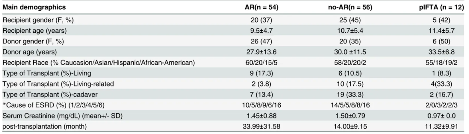
Table 1. Demographic Table.
Main demographics AR(n = 54) no-AR(n = 56) pIFTA (n = 12)
Recipient gender (F, %) 20 (37) 25 (45) 5 (42)
Recipient age (years) 9.5±4.7 10.7±5.4 11.4±5.7
Donor gender (F, %) 26 (47) 20 (35) 6 (50)
Donor age (years) 27.9±13.6 30.0±11.5 33.5±6.8
Recipient Race (% Caucasion/Asian/Hispanic/African-American) 60/20/15/5 58/20/20/2 55/18/19/2
Type of Transplant (%)-Living 9 (17.3) 6 (10.5) 1 (8.3)
Type of Transplant (%)-Living-related 2 (3.8) 10 (17.5) 4(33.3)
Type of Transplant (%)-cadaver 7 (13.4) 19 (33.3) 2 (16.7)
*Cause of ESRD (%) (1/2/3/4/5/6) 10/5/8/9/6/16 14/5/5/8/8/16 2/0/3/2/2/3
Serum Creatinine (mg/dL) (mean+/- SD) 1.45±0.88 1.50±0.79 0.97± 0.0
post-transplantation (month) 33.99±31.58 14.00±9.15 11.32±9.91
*ESRD (1/2/3/4/5/6): 1, glomerulonephritis, 2, polycystic kidney disease, 3, renal dysplasia, 4, reflux nephropathy, 5, obstructive uropathy, 6 = other or unknown. None of these selected patients had delayed graft function. 20% of AR episodes were associated with documentation of non-adherence with medications (self-reported), appointments and/or laboratory measurements. Though the time post-transplant was significantly greater for AR (p<0.05); there was no difference in post-transplant time between no-AR and pIFTA.
samples using SuperScript VILO Master Mix (Invitrogen, Carlsbad, CA) as per the
manufac-turer’s protocol. Briefly, specific target amplification was performed using 3.125 ng relative
amount of cDNA using a pooled individual TaqMan real-time assays for the 11 genes
investi-gated in multiplex with TaqMan PreAmp Master mix Life Technologies) to 10
μl final volume,
for 18 cycles in a thermal cycler (Eppendorf Vapo-Protect, Hamburg, Germany), then diluted
1:20 with sterile water (Gibco, Invitrogen, Carlsbad, CA). The qPCR was performed on the
QuantStudio 6 Flex System (Life Technologies) using 5
μl of the diluted sample from the
spe-cific target amplification, along with the TaqMan Gene Expression Master Mix under standard
conditions (2 min at 50°C, 10 min at 95°C, 40 cycles of 15s at 95°C, 1min at 60°C) using
Taq-Man gene expression assays (Life Technologies) for each of the 11 genes investigated: BASP1,
CD6, CXCL10, CXCL9, INPP5D, ISG20, LCK, NKG7, PSMB9, RUNX3, TAP1. The relative
amount of mRNA expression in each sample was calculated using the comparative threshold
cycle (C
T) method [
13
]. Ribosomal 18S RNA (18S) and Universal RNA (QIAGEN) were used
for normalization of all genes since they showed the least variability in gene expression across
all samples. Final gene expression results were converted to fold change. tCRM score was
calcu-lated by using the geometric mean of the fold changes of the respective genes.
Data analyses
All data are presented as mean ± standard error of the mean (SEM). Groups were compared
using the
χ
2test for categorical variables, the two-way analysis of variance (ANOVA) or t-test
for normally distributed data, and the nonparametric Kruskal–Wallis, Welch’s correction or
Mann
–Whitney U test for non-normally distributed variables. Bivariate correlation analyses
were done using Pearson or Spearman tests for non-parametric variables. Sensitivity/specificity
Receiver Operating Characteristic (ROC) curve analyses were performed to evaluate the most
precise cut-off of the tissue CRM (tCRM) gene scores assessed at the time of biopsy, predicting
the advent of acute rejection (AR). All predictive values were determined by calculating the
area under the curve with SPSS software. Binary linear logistic regression analysis was
per-formed to determine the independent correlation of several independent variables with the
presence of AR. The statistical significance level was defined as p<0.05. Gene expression,
tCRM scores and clinical variables were examined by multivariate analysis to determine
pre-dictive value. Cross sectional analysis was done to determine the significant increase in gene
expression at event. Longitudinal studies were done to determine changes in gene expression
over 2 years in patients with progressive IFTA (pIFTA).
Results and Discussion
The 11-gene intra-allograft common response module (tCRM) score
accurately segregates acute rejection (AR) from stable (no-AR) kidney
transplant patients in kidney tissue biopsies
The individual expressions of all 11 genes of the tCRM score showed a significant increase in
AR, with the most significant increase seen in CXCL9 and CXCL10 (p = 0.0001 in both
com-parisons) between AR and no-AR (
Fig 1
). The tCRM score was also significantly increased in
AR (mean ± SEM = 6.897 ± 1.082, n = 27) when compared to no-AR
(mean ± SEM = 0.8144 ± 0.1374, n = 22, p = 0.000057) (
Fig 1
), and increased for pIFTA cohort
for both the 6 and 24 month biopsies over the biopsies from patients who did not develop any
histological changes (3.33 ± 1.00 versus 1.22 ± 0.2 at 6 months, p = 0.05; 7.94 ± 2.281 versus
2.28 ± 0.66 at 24 months; p = 0.03 at 24 months respectively) (
Fig 1
). Using the first set of 49
biopsies (27AR, 22no-AR), the tCRM score was found to be a significant correlate of Banff
classified AR (AUC of 0.900 (p
<0.001, 95% CI = 0.823–0.976) (
Fig 1
). With the aim of using
the tCRM score as a binary variable to segregate patients at AR risk based on the tCRM score
alone, the data was analyzed to choose a tCRM score of 2.24 which resulted in the greatest
sen-sitivity and specificity for AR prediction (sensen-sitivity = 82.7%, specificity = 82.5%) (
Fig 1
).
When applied to the second independent biopsy set of 49 biopsies (27AR, 22no-AR) set, the
tCRM threshold of 2.24 had a positive predictive value (PPV) for AR of 82.4%.
The tCRM score correlates with the extent of AR lesions
We evaluated whether the tCRM score was associated with the extent of the AR lesions
observed in the matched biopsies. As observed in
Fig 2
, the tCRM score strongly correlated
with the extent of the acute allograft lesions both at the tubular (Banff t score and the
intersti-tum (Banff i score) kidney compartments (R = 0.72, p = 0.001 and R = 0.74, p = 0.001).
Fig 1. Gene expression of tCRM genes in AR. [A] Intra-allograft gene expression for tCRM genes increased in AR when compared to stable with the most significant differences seen between CXCL10 and CXCL9 (p = 0.0001). [B] The tCRM score is significantly increased in AR when compared to Stable (p = 0.0000057). Although there is increased in pIFTA progressors at 6 months, this difference did not meet significance (p = 0.05) until 24 months (p = 0.03). A threshold tCRM score of 2.24 as determined by the discovery set. [C] The tCRM score significantly predicts rejection with an AUC of 0.900 (p = 0.001, 95% CI = 0.823–0.976) with 82.7% sensitivity and 82.5% specificity.
The tCRM score is predictive of chronic allograft nephropathy
Biopsies from patients with chronic allograft changes as defined by the presence of IFTA were
examined at 24 months, and in addition their biopsy pairs were also examined from their 6
month protocol biopsies. Significantly increased fold changes of all genes except BASP1 and
CXLC10 were seen at both 6 and 24 months between pIFTA (P) and no-AR (NP) patients.
While the tCRM score across all 11 genes was significant between pIFTA (P) and no-AR (NP)
(p = 0.05), we found by using an adjusted R score analysis, that among the 11 genes, the
great-est influence on pIFTA was from a subset of 7 genes (CD6, INPP5D, ISG20, NKG7, PSMB9,
RUNX3, and TAP1), with maximal influence from CD6. A modified tCRM score across these
7 genes showed significantly greater scores in the 6 month biopsies of patients destined to
develop pIFTA patients over time (p = 0.037) (
Fig 2
).
Fig 2. tCRM score correlates with AR lesions and chronic allograft nephropathy. [A] The tCRM score positively correlates significantly (p = 0.001) with the degree of infiltrates found on biopsy for the t-score = 0.722 and [B] i score = 0.736. [C] A tCRM score across a subset of 7 of the 11 genes differentiated most samples with pIFTA or progressors (3.29± 0.93) from no-AR patients (1.2 ± 0.18; p = 0.037). Stable/non-progressors (NP) and AR were highly distinguishable (1.198±0.1801 versus 5.582±0.8651; p = 0.0063). pIFTA/Progressors and AR were not different with regards to their tCRM scores (p = 0.16). doi:10.1371/journal.pone.0138133.g002
Discussion
There is an urgent need in transplant medicine for developing reliable and non-invasive
monitoring tools that may help transplant clinicians predict the risk of alloimmune-mediated
allograft injury, preferentially before allograft damage has already been established. While a
number of transcriptional biomarkers have been associated to AR [
14
–
19
], many studies
either have limited sample sizes [
20
–
23
] or have focused on exploratory single biomarkers
that may not necessarily reflect the crux of the molecular complexity in allograft rejection
[
24
,
25
]. Taking advantage of recently reported microarray meta-analyses of eight
indepen-dent transplant datasets by combining effect size and p-values from our group [
11
], showing
a common immune response module (CRM) of gene expression in allograft biopsies during
AR, irrespective of the type of tissue organ, the main goal of this study was to validate these
findings using the more practical method of PCR as well as determine a threshold score. We
found that a qPCR-based intra-allograft tCRM score threshold of
>2.24, can accurately
dis-tinguish AR from no-AR with high sensitivity and specificity. This data allows us to confirm
the association of gene expression across this restricted set of genes with the histological
immune injury of acute rejection, and also supports our earlier findings [
7
] that the
molecu-lar profile of immune injury is a threshold effect, with greater burden of injury in acute
rejec-tion, and similar, but lower injury burden in the subclinical injury of progressive IFTA. The
panel of genes has differential impact on the injury phenotypes such as CXCL9 and CXCL10
are important layers in the injury of AR, and CD6 plays a major role in pIFTA, suggesting
different roles for cell infiltration versus activation in acute and chronic graft injury.
Cyto-kines are involved in all inflammatory responses. Being a class of small cytoCyto-kines, the C-X-C
motif chemokine ligand CXCL9 and CXCL10 play key roles in the initiation and
develop-ment of acute transplant rejection [
26
]. Our observation of CXCL9 and CXCL10 being
important factor in AR corroborates previously reported observations regarding their
involvement in immune mediated graft injury [
14
–
16
,
19
]. Our ongoing studies show the
tCRM score is also elevated in BK viral nephritis in the graft (Sigdel
et al, in submission),
which supports data from previous reports of significant overlap in the molecular
distur-bances from the overlapping intragraft infiltrates in AR and BK viral nephritis [
14
,
27
–
30
],
though in the latter instance the diagnosis of BK viral infection is very evident also by BK
viral urine and blood PCR positivity and tissue SV40 positive stains. Nevertheless, in
histo-logically normal biopsies, and in the absence of AR and confounding intragraft infection, an
elevated tCRM score is a strong harbinger of underlying molecular inflammation and can
provide a very important warning to a clinician to closely monitor this patient for risk of
pIFTA. Recent years have seen an increased effort to develop and validate new assays for
early diagnosis of acute rejection of organ transplantation and transplant monitoring [
31
].
We and others have reported on potential surrogate gene biomarkers for acute rejection
ana-lyzing peripheral blood [
32
,
33
], gene biomarkers using urine [
34
–
36
] and protein
biomark-ers analyzing serum and plasma [
37
], and urine [
10
,
38
–
41
]; many of these are in varying
stages of further validation. We suggest that the inclusion of the tCRM score into the analysis
of the immune profile of protocol biopsies can be very valuable for risk analysis in the context
of clinical trials, as the tCRM score may provide a companion diagnostic for differentiating
patients into high and low immune risk, for stratification into different investigative
treat-ment arms, with an increased margin for patient and graft safety. Because of the restricted
sample size available for this study, we suggest that the value of the tCRM score to
discrimi-nate patients at increased risk of AR or IFTA, prior to any perturbation of graft function or
histology may require further validation in a larger cohort of patients.
Acknowledgments
We would like to acknowledge the assistance of Yolanda Ng in data assimilation, manuscript
preparation and editing, and the patients and their families that contributed to the samples and
clinical data that made this study possible. Some of this work was supported by R01 DK083447
(MMS).
Author Contributions
Conceived and designed the experiments: MMS TKS OB. Performed the experiments: TQT
SCH ID. Analyzed the data: TKS OB MMS SR. Contributed reagents/materials/analysis tools:
FV. Wrote the paper: TKS OB MMS.
References
1. Wolfe RA, Ashby VB, Milford EL, Ojo AO, Ettenger RE, Agodoa LY, et al. Comparison of mortality in all patients on dialysis, patients on dialysis awaiting transplantation, and recipients of a first cadaveric transplant. The New England journal of medicine. 1999; 341(23):1725–30. doi:10.1056/
NEJM199912023412303PMID:10580071.
2. Laupacis A, Keown P, Pus N, Krueger H, Ferguson B, Wong C, et al. A study of the quality of life and cost-utility of renal transplantation. Kidney international. 1996; 50(1):235–42. PMID:8807593. 3. Lodhi SA, Lamb KE, Meier-Kriesche HU. Improving long-term outcomes for transplant patients: making
the case for long-term disease-specific and multidisciplinary research. American journal of transplanta-tion: official journal of the American Society of Transplantation and the American Society of Transplant Surgeons. 2011; 11(10):2264–5. doi:10.1111/j.1600-6143.2011.03713.xPMID:21957938.
4. Pape L, Offner G, Ehrich JH, de Boer J, Persijn GG. Renal allograft function in matched pediatric and adult recipient pairs of the same donor. Transplantation. 2004; 77(8):1191–4. PMID:15114083. 5. Provoost AP, Wolff ED, de Keijzer MH, Molenaar JC. Influence of the recipient's size upon renal
func-tion following kidney transplantafunc-tion. An experimental and clinical investigafunc-tion. Journal of pediatric sur-gery. 1984; 19(1):63–7. PMID:6366181.
6. Davis ID, Oehlenschlager W, O'Riordan MA, Avner ED. Pediatric renal biopsy: should this procedure be performed in an outpatient setting? Pediatric nephrology. 1998; 12(2):96–100. PMID:9543363. 7. Naesens M, Khatri P, Li L, Sigdel TK, Vitalone MJ, Chen R, et al. Progressive histological damage in
renal allografts is associated with expression of innate and adaptive immunity genes. Kidney interna-tional. 2011; 80(12):1364–76. doi:10.1038/ki.2011.245PMID:21881554.
8. Roedder S, Sigdel T, Salomonis N, Hsieh S, Dai H, Bestard O, et al. The kSORT assay to detect renal transplant patients at high risk for acute rejection: results of the multicenter AART study. PLoS medi-cine. 2014; 11(11):e1001759. doi:10.1371/journal.pmed.1001759PMID:25386950; PubMed Central PMCID: PMC4227654.
9. Sigdel TK, Sarwal MM. Recent advances in biomarker discovery in solid organ transplant by proteo-mics. Expert review of proteoproteo-mics. 2011; 8(6):705–15. doi:10.1586/epr.11.66PMID:22087656; PubMed Central PMCID: PMC3282122.
10. Sigdel TK, Salomonis N, Nicora CD, Ryu S, He J, Dinh V, et al. The identification of novel potential injury mechanisms and candidate biomarkers in renal allograft rejection by quantitative proteomics. Molecular & cellular proteomics: MCP. 2014; 13(2):621–31. doi:10.1074/mcp.M113.030577PMID:
24335474; PubMed Central PMCID: PMC3916658.
11. Khatri P, Roedder S, Kimura N, De Vusser K, Morgan AA, Gong Y, et al. A common rejection module (CRM) for acute rejection across multiple organs identifies novel therapeutics for organ transplantation. The Journal of experimental medicine. 2013; 210(11):2205–21. doi:10.1084/jem.20122709PMID:
24127489; PubMed Central PMCID: PMC3804941.
12. Sis B, Mengel M, Haas M, Colvin RB, Halloran PF, Racusen LC, et al. Banff '09 meeting report: anti-body mediated graft deterioration and implementation of Banff working groups. American journal of transplantation: official journal of the American Society of Transplantation and the American Society of Transplant Surgeons. 2010; 10(3):464–71. doi:10.1111/j.1600-6143.2009.02987.xPMID:20121738. 13. Livak KJ, Schmittgen TD. Analysis of relative gene expression data using real-time quantitative PCR
and the 2(-Delta Delta C(T)) Method. Methods. 2001; 25(4):402–8. doi:10.1006/meth.2001.1262
PMID:11846609.
14. Jackson JA, Kim EJ, Begley B, Cheeseman J, Harden T, Perez SD, et al. Urinary chemokines CXCL9 and CXCL10 are noninvasive markers of renal allograft rejection and BK viral infection. American
journal of transplantation: official journal of the American Society of Transplantation and the American Society of Transplant Surgeons. 2011; 11(10):2228–34. doi:10.1111/j.1600-6143.2011.03680.xPMID:
21812928; PubMed Central PMCID: PMC3184377.
15. Hu H, Kwun J, Aizenstein BD, Knechtle SJ. Noninvasive detection of acute and chronic injuries in human renal transplant by elevation of multiple cytokines/chemokines in urine. Transplantation. 2009; 87(12):1814–20. PMID:19543058.
16. Schaub S, Nickerson P, Rush D, Mayr M, Hess C, Golian M, et al. Urinary CXCL9 and CXCL10 levels correlate with the extent of subclinical tubulitis. American journal of transplantation: official journal of the American Society of Transplantation and the American Society of Transplant Surgeons. 2009; 9 (6):1347–53. doi:10.1111/j.1600-6143.2009.02645.xPMID:19459809.
17. Tatapudi RR, Muthukumar T, Dadhania D, Ding R, Li B, Sharma VK, et al. Noninvasive detection of renal allograft inflammation by measurements of mRNA for IP-10 and CXCR3 in urine. Kidney interna-tional. 2004; 65(6):2390–7. doi:10.1111/j.1523-1755.2004.00663.xPMID:15149352.
18. Segerer S, Cui Y, Eitner F, Goodpaster T, Hudkins KL, Mack M, et al. Expression of chemokines and chemokine receptors during human renal transplant rejection. American journal of kidney diseases: the official journal of the National Kidney Foundation. 2001; 37(3):518–31. PMID:11228176.
19. Hauser IA, Spiegler S, Kiss E, Gauer S, Sichler O, Scheuermann EH, et al. Prediction of acute renal allograft rejection by urinary monokine induced by IFN-gamma (MIG). Journal of the American Society of Nephrology: JASN. 2005; 16(6):1849–58. doi:10.1681/ASN.2004100836PMID:15857922. 20. Shi S, Blumenthal A, Hickey CM, Gandotra S, Levy D, Ehrt S. Expression of many immunologically
important genes in Mycobacterium tuberculosis-infected macrophages is independent of both TLR2 and TLR4 but dependent on IFN-alphabeta receptor and STAT1. Journal of immunology. 2005; 175 (5):3318–28. PMID:16116224.
21. Robertson G, Hirst M, Bainbridge M, Bilenky M, Zhao Y, Zeng T, et al. Genome-wide profiles of STAT1 DNA association using chromatin immunoprecipitation and massively parallel sequencing. Nature methods. 2007; 4(8):651–7. doi:10.1038/nmeth1068PMID:17558387.
22. Kuznetsov VA, Orlov YL, Wei CL, Ruan Y. Computational analysis and modeling of genome-scale avid-ity distribution of transcription factor binding sites in chip-pet experiments. Genome informatics Interna-tional Conference on Genome Informatics. 2007; 19:83–94. PMID:18546507.
23. Ellis SL, Gysbers V, Manders PM, Li W, Hofer MJ, Muller M, et al. The cell-specific induction of CXC chemokine ligand 9 mediated by IFN-gamma in microglia of the central nervous system is determined by the myeloid transcription factor PU.1. Journal of immunology. 2010; 185(3):1864–77. doi:10.4049/ jimmunol.1000900PMID:20585034; PubMed Central PMCID: PMC2925661.
24. Sarwal M, Chua MS, Kambham N, Hsieh SC, Satterwhite T, Masek M, et al. Molecular heterogeneity in acute renal allograft rejection identified by DNA microarray profiling. The New England journal of medi-cine. 2003; 349(2):125–38. doi:10.1056/NEJMoa035588PMID:12853585.
25. Der SD, Zhou A, Williams BR, Silverman RH. Identification of genes differentially regulated by inter-feron alpha, beta, or gamma using oligonucleotide arrays. Proceedings of the National Academy of Sci-ences of the United States of America. 1998; 95(26):15623–8. PMID:9861020; PubMed Central PMCID: PMC28094.
26. Zhuang J, Shan Z, Ma T, Li C, Qiu S, Zhou X, et al. CXCL9 and CXCL10 accelerate acute transplant rejection mediated by alloreactive memory T cells in a mouse retransplantation model. Experimental and therapeutic medicine. 2014; 8(1):237–42. doi:10.3892/etm.2014.1714PMID:24944628; PubMed Central PMCID: PMC4061216.
27. Mannon RB, Hoffmann SC, Kampen RL, Cheng OC, Kleiner DE, Ryschkewitsch C, et al. Molecular evaluation of BK polyomavirus nephropathy. American journal of transplantation: official journal of the American Society of Transplantation and the American Society of Transplant Surgeons. 2005; 5 (12):2883–93. doi:10.1111/j.1600-6143.2005.01096.xPMID:16303001.
28. Girmanova E, Brabcova I, Klema J, Hribova P, Wohlfartova M, Skibova J, et al. Molecular networks involved in the immune control of BK polyomavirus. Clinical & developmental immunology. 2012; 2012:972102. doi:10.1155/2012/972102PMID:23251224; PubMed Central PMCID: PMC3521483. 29. Dadhania D, Snopkowski C, Ding R, Muthukumar T, Lee J, Bang H, et al. Validation of noninvasive
diagnosis of BK virus nephropathy and identification of prognostic biomarkers. Transplantation. 2010; 90(2):189–97. PMID:20526237; PubMed Central PMCID: PMC2989149.
30. Lubetzky M, Bao Y, P OB, Marfo K, Ajaimy M, Aljanabi A, et al. Genomics of BK viremia in kidney trans-plant recipients. Transtrans-plantation. 2014; 97(4):451–6. PMID:24310299.
31. Lo DJ, Kaplan B, Kirk AD. Biomarkers for kidney transplant rejection. Nature reviews Nephrology. 2014; 10(4):215–25. doi:10.1038/nrneph.2013.281PMID:24445740.
32. Li L, Khatri P, Sigdel TK, Tran T, Ying L, Vitalone MJ, et al. A peripheral blood diagnostic test for acute rejection in renal transplantation. American journal of transplantation: official journal of the American
Society of Transplantation and the American Society of Transplant Surgeons. 2012; 12(10):2710–8. doi:10.1111/j.1600-6143.2012.04253.xPMID:23009139; PubMed Central PMCID: PMC4148014. 33. Roedder S, Sigdel T, Salomonis N, Hsieh S, Dai H, Bestard O, et al. The kSORT assay to detect renal
transplant patients at high risk for acute rejection: results of the multicenter AART study. PLoS medi-cine. 2014; 11(11):e1001759. doi:10.1371/journal.pmed.1001759PMID:25386950; PubMed Central PMCID: PMC4227654.
34. Fairchild RL, Suthanthiran M. Urine CXCL10/IP-10 Fingers Ongoing Antibody-Mediated Kidney Graft Rejection. Journal of the American Society of Nephrology: JASN. 2015. doi:10.1681/ASN.2015040353
PMID:25948874.
35. Muthukumar T, Lee JR, Dadhania DM, Ding R, Sharma VK, Schwartz JE, et al. Allograft rejection and tubulointerstitial fibrosis in human kidney allografts: interrogation by urinary cell mRNA profiling. Trans-plantation reviews. 2014; 28(3):145–54. doi:10.1016/j.trre.2014.05.003PMID:24929703; PubMed Central PMCID: PMC4118424.
36. Lee JR, Muthukumar T, Dadhania D, Ding R, Sharma VK, Schwartz JE, et al. Urinary cell mRNA pro-files predictive of human kidney allograft status. Immunological reviews. 2014; 258(1):218–40. doi:10. 1111/imr.12159PMID:24517436; PubMed Central PMCID: PMC3947569.
37. Chen R, Sigdel TK, Li L, Kambham N, Dudley JT, Hsieh SC, et al. Differentially expressed RNA from public microarray data identifies serum protein biomarkers for cross-organ transplant rejection and other conditions. PLoS computational biology. 2010; 6(9). doi:10.1371/journal.pcbi.1000940PMID:
20885780; PubMed Central PMCID: PMC2944782.
38. Schaub S, Rush D, Wilkins J, Gibson IW, Weiler T, Sangster K, et al. Proteomic-based detection of urine proteins associated with acute renal allograft rejection. Journal of the American Society of Nephrology: JASN. 2004; 15(1):219–27. PMID:14694176.
39. Sigdel TK, Kaushal A, Gritsenko M, Norbeck AD, Qian WJ, Xiao W, et al. Shotgun proteomics identifies proteins specific for acute renal transplant rejection. Proteomics Clinical applications. 2010; 4(1):32– 47. doi:10.1002/prca.200900124PMID:20543976; PubMed Central PMCID: PMC2883247.
40. Ling XB, Sigdel TK, Lau K, Ying L, Lau I, Schilling J, et al. Integrative urinary peptidomics in renal trans-plantation identifies biomarkers for acute rejection. Journal of the American Society of Nephrology: JASN. 2010; 21(4):646–53. doi:10.1681/ASN.2009080876PMID:20150539; PubMed Central PMCID: PMC2844301.
41. Sigdel TK, Ng YW, Lee S, Nicora CD, Qian WJ, Smith RD, et al. Perturbations in the urinary exosome in transplant rejection. Frontiers in medicine. 2014; 1:57. doi:10.3389/fmed.2014.00057PMID:
![Fig 1. Gene expression of tCRM genes in AR. [A] Intra-allograft gene expression for tCRM genes increased in AR when compared to stable with the most significant differences seen between CXCL10 and CXCL9 (p = 0.0001)](https://thumb-eu.123doks.com/thumbv2/123dokorg/2973567.27650/5.918.60.785.117.678/expression-allograft-expression-increased-compared-stable-significant-differences.webp)
![Fig 2. tCRM score correlates with AR lesions and chronic allograft nephropathy. [A] The tCRM score positively correlates significantly (p = 0.001) with the degree of infiltrates found on biopsy for the t-score = 0.722 and [B] i score = 0.736](https://thumb-eu.123doks.com/thumbv2/123dokorg/2973567.27650/6.918.65.817.119.711/correlates-lesions-allograft-nephropathy-positively-correlates-significantly-infiltrates.webp)
