871
Research Article
Introduction
Nanos is an evolutionarily conserved protein that is essential for the survival of primordial germ cells (PGCs). It has been shown to act as a putative RNA-binding protein involved in germ cell development in both vertebrates (Koprunner et al., 2001; Tsuda et al., 2003) and invertebrates (Fujii et al., 2006; Lehmann and Nusslein-Volhard, 1991; Pilon and Weisblat, 1997; Subramaniam and Seydoux, 1999). In the mouse, three genes encoding Nanos have been identified (Haraguchi et al., 2003). Although Nanos1 is predominantly expressed in the neural system (Haraguchi et al., 2003), Nanos2 and Nanos3 are exclusively expressed in fetal and postnatal germ cells (Tsuda et al., 2006; Tsuda et al., 2003). The expression pattern of Nanos2 is restricted to gonocytes within fetal testis undergoing mitotic arrest, and is then maintained in spermatogonia after birth. Deletion of Nanos2 results in male sterility, due to germ-cell loss during fetal life (Tsuda et al., 2003). Nanos2-null male fetal germ cells enter meiosis before undergoing apoptosis at around 15.5 days post coitus (d.p.c.) (Suzuki and Saga, 2008). Furthermore, misexpression of Nanos2 in female fetal germ cells prevents meiotic entry, strongly suggesting that Nanos2 is an antimeiotic gene (Suzuki and Saga, 2008).
The mechanisms that trigger meiosis have been a subject of debate, but it is now clear that retinoic acid (RA) produced by the mesonephros induces meiosis in female PGCs (Bowles et al., 2006; Koubova et al., 2006), whereas the same action in the fetal testis is prevented by the presence of the retinoid-degrading enzyme CYP26B1 (Bowles et al., 2006; MacLean et al., 2007). The role of RA as a meiotic inducer is also conserved postnatally, because it is able to induce the meiotic entry of differentiating, KIT-positive spermatogonia (Pellegrini et al., 2008). An inverse correlation
between levels of RA and NANOS2 exists in Cyp26b1-null fetal testis – a condition in which RA is elevated (MacLean et al., 2007), gonocytes enter meiosis and NANOS2 levels are low (Suzuki and Saga, 2008). The crucial role of RA in inducing meiosis in both female and male is played by Stra8 (stimulated by retinoic acid gene 8) (Anderson et al., 2008; Mark et al., 2008); indeed, Stra8-null males and females show impairment of gametogenesis owing to failure of meiotic commitment (Anderson et al., 2008; Mark et al., 2008). However, meiotic commitment is aberrantly induced, not only in Nanos2-null mice, but also in Fgf9 knockouts (Colvin et al., 2001).
Fgf9 is known to be expressed by somatic cells in XX and XY gonads at 11.5 d.p.c., but it becomes XY restricted by 12.5 d.p.c. and is then expressed within the testis cords (Schmahl et al., 2004). Fgf9-null mice die shortly after birth owing to defects in lung formation, but XY embryos show phenotypic male-to-female sex-reversal, and the surviving gonocytes are found in meiosis (Colvin et al., 2001). The consequences of Fgf9 ablation on male germ cells are probably independent of the proliferative and differentiative effect on fetal Sertoli cell, because it has been shown that FGF9 is able to promote survival of male germ cells in vitro (DiNapoli et al., 2006).
We report that NANOS2 is continuously expressed in male germ cells from fetal to postnatal development. In particular, we investigated the role of RA and FGF9 on the regulation of NANOS2 in correlation with their ability to influence meiosis. We found that AtRA represses NANOS2 expression, whereas FGF9 upregulates Nanos2 mRNA levels in female and male germ cells and inhibits meiosis in both sexes. Moreover, we show evidence that NANOS2 associates with ribonucleoparticles (RNPs) and polysomes, both in
Opposing effects of retinoic acid and FGF9 on Nanos2
expression and meiotic entry of mouse germ cells
Florencia Barrios1, Doria Filipponi1, Manuela Pellegrini1, Maria Paola Paronetto1, Sara Di Siena1,
Raffaele Geremia1, Pellegrino Rossi1, Massimo De Felici1, Emmanuele A. Jannini2and Susanna Dolci1,* 1Department of Public Health and Cellular Biology, University of Rome ‘Tor Vergata’, 00133 Rome, Italy
2Department of Experimental Medicine, Università dell’Aquila, 67100 L’Aquila, Italy *Author for correspondence ([email protected])
Accepted 9 December 2009 Journal of Cell Science 123, 871-880
©2010. Published by The Company of Biologists Ltd doi:10.1242/jcs.057968
Summary
In the mouse, three genes that are homologous to the Drosophila Nanos (Nos) gene have been identified. Deletion of one of these genes, Nanos2, results in male sterility, owing to loss of germ cells during fetal life. Before apoptosis, Nanos2-null gonocytes enter meiosis, suggesting that Nanos2 functions as a meiotic repressor. Here, we show that Nanos2 is continuously expressed in male germ cells from fetal gonocytes to postnatal spermatogonial stem cells. We observed that the promeiotic factor AtRA, an analog of retinoic acid (RA), downregulates NANOS2 levels, in both fetal and postnatal gonocytes, while promoting meiosis. Interestingly, FGF9, a growth factor crucial for sex differentiation and survival of fetal gonocytes, upregulates levels of NANOS2 in both male and female primordial germ cells (PGCs) and in premeiotic spermatogonia. This effect was paralleled by an impairment of meiotic entry, suggesting that FGF9 acts as an inhibitor of meiosis through the upregulation of Nanos2. We found that NANOS2 interacts with PUM2, and that these two proteins colocalize in the ribonucleoparticle and polysomal fractions on sucrose gradients, supporting the notion that they bind RNA. Finally, we found that recombinant NANOS2 binds to two spermatogonial mRNAs, Gata2 and Taf7l, which are involved in germ-cell differentiation.
Key words: NANOS2, FGF9, RA, Gata2, Taf7l, Meiosis, Spermatogenesis
Jour
fetal and postnatal germ cells. Finally, by GST-RNA pull-down assay, we found that NANOS2 shows affinity for two RNA transcripts, Gata2 and Taf7l, which are involved in spermatogonial differentiation, strengthening the hypothesis that NANOS2 can act as a meiotic inhibitor through a post-transcriptional regulation mechanism.
Results
Developmental expression of Nanos2 in the male gonad
It is known that Nanos2 mRNA starts to be expressed in male PGCs at around 12.5 d.p.c. (Suzuki et al., 2007). Furthermore, by immunofluorescence analysis, it has been very recently shown that undifferentiated spermatogonia type Asingle (As) and Apaired(Apr) are the testicular cells that express NANOS2 in the adult testis (Suzuki et al., 2009). However, it is not clear whether NANOS2 levels drop before birth, or remain elevated in the early postnatal testis. To clearly address this question, we performed a western blot analysis on male germ cells isolated at various stages of fetal and postnatal development. As shown in Fig. 1A we found that NANOS2 protein levels were high at 15.5 d.p.c. and then decreased up to birth. Since NANOS2 levels were already high at 15.5 d.p.c., to better define the fetal period when they start to increase, we analyzed protein extracts obtained from germ cells isolated at 12.5, 13.5, 15.5 d.p.c. and 1 day post partum (d.p.p.). As shown in Fig. 1B, we found that NANOS2 levels were low at 12.5 d.p.c., but they consistently increased up to 15.5 d.p.c. We also found that NANOS2 was expressed after birth and its levels peaked at 5 d.p.p. and then decreased at 7 d.p.p., when differentiating premeiotic spermatogonia (KIT positive) and preleptotene spermatocytes are found (Fig. 1A,B). Isolated pachytene spermatocytes and spermatids did not express Nanos2, either at the RNA or protein level (not shown). Thus, to determine which spermatogonial cell type expressed
Nanos2 in the prepuberal testis, we isolated pure populations of undifferentiated or differentiating spermatogonia by immunomagnetic cell sorting. KIT-negative (undifferentiated) and KIT-positive (differentiating) spermatogonia were obtained by using anti-CD117 (Kit)-coated beads and analyzed for Nanos2 expression by semiquantitative RT-PCR. As shown in Fig. 1C, Nanos2 amplification was obtained only in KIT-negative spermatogonia, whereas KIT-positive cells did not show any signal. Purity of the cell fractions was assessed by western blot analysis probing extracts for KIT and OCT4 (Fig. 1C) or RT-PCR amplification of PLZF and OCT4 (not shown), as previously reported (Lolicato et al., 2008).
Retinoic acid downregulates Nanos2 expression in fetal gonocytes and postnatal spermatogonia
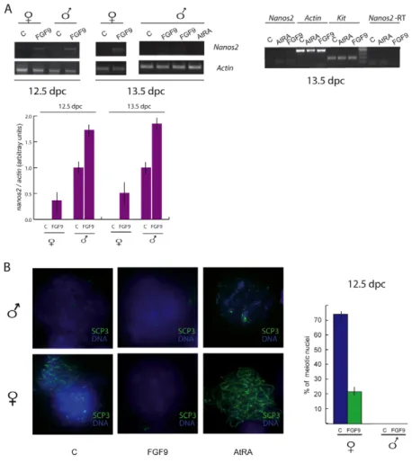
RA has been proposed as a meiosis-inducing substance, both in fetal female germ cells and in postnatal male germ cells (Bowles et al., 2006; Koubova et al., 2006; Pellegrini et al., 2008). In Cyp26b-null fetal testis, gonocytes enter meiosis and Nanos2 mRNA levels are low (Suzuki and Saga, 2008). This suggests that, apart from Stra8 induction, the promeiotic action of RA might also result from the downregulation of Nanos2 expression. To test this hypothesis, we cultured testes from 12.5 d.p.c. embryos, the developmental stage at which Nanos2 starts to be expressed, in the presence of 0.3 M AtRA for 48 hours. As shown in Fig. 2A, AtRA completely abolished the expression of Nanos2 mRNA, whereas it upregulated Stra8 mRNA levels. The same effect was observed when we cultured disaggregated 13.5 d.p.c. testes for 24 hours in the presence of AtRA (Fig. 2A, see also Fig. 3A). Using chromosomal spreads stained for SCP3, we found that about 33% of male germ cells were in the early meiotic leptotene stage in the presence of AtRA whereas no meiotic germ cells were present in the control. Using the same
Fig. 1. Developmental expression of NANOS2.
(A)Western blot analysis of NANOS2 from fetal (15.5 d.p.c.) throughout postnatal life (7 d.p.p.). Male germ cells express NANOS2 at 15.5 d.p.c. and the protein levels decrease around birth, and peak again at 5 d.p.p. to decrease later at 7 d.p.p. Densitometric analysis of western blots from three separate experiments is shown on the right. Bars represent mean ± s.d.; P<0.001. (B)Western blot analysis of NANOS2 expression starting at earlier developmental stages. Densitometric analysis of the western blots from three independent experiments is shown on the right. Bars represent mean ± s.d.; P<0.001. (C)Schematic
representation of spermatogonia development in the prepuberal testis (7 d.p.p.) is shown on the left. Two populations can be distinguished: the undifferentiated spermatogonia, that do not express KIT, and the
differentiated KIT-positive spermatogonia. Adapted from de Rooij and Mizrak (de Rooij and Mizrak, 2008). Nanos2 expression is restricted to the undifferentiated spermatogonia population as shown by semiquantitative RT-PCR (middle panel) or by western blot analysis (right panel).
Jour
culture conditions, in the presence of AtRA about 85% female germ cells were found in leptotene and lepto-zygotene compared with 75% of the control (Fig. 2A, see also Fig. 3B).
We, and others, have previously demonstrated that G0-arrested gonocytes and undifferentiated KIT-negative spermatogonia are not able to enter meiosis in vitro upon stimulation by AtRA (Pellegrini et al., 2008; Trautmann et al., 2008). Since NANOS2 expression is high in these cells (see Fig. 1) we tested whether AtRA was also able to downregulate NANOS2 at these developmental stages. The results showed that 48 hours of AtRA treatment actually downregulated the levels of Nanos2 mRNA and protein in 15.5 d.p.c. and 4 d.p.p. isolated male germ cells (Fig. 2B). This effect was accompanied by upregulation of STRA8 (Fig. 2B) but, as expected, did not result in meiotic induction (Fig. 2C).
FGF9 upregulates Nanos2 expression and represses meiosis in both male and female germ cells
XY Fgf9-null gonocytes enter meiosis before undergoing apoptosis, suggesting that FGF9 prevents meiosis in XY germ cells (DiNapoli et al., 2006). Since this phenotype resembles that of Nanos2-null gonocytes, we hypothesized that FGF9 could directly or indirectly upregulate Nanos2 expression in germ cells. To test this possibility, we chose a developmental stage at which Nanos2 mRNA levels were low in the fetal testis and were obviously absent in the fetal ovary. Thus, 12.5 d.p.c. male or female gonads were cultured for 48 hours in the presence or absence of 25 ng/ml FGF9. By semiquantitative RT-PCR, we found that levels of Nanos2 mRNA were upregulated by treatment with FGF9 in fetal testes and, interestingly, Nanos2 mRNA expression was stimulated also in fetal ovaries (Fig. 3A,B). The same effect was observed when disaggregated 13.5 d.p.c. gonads were cultured for only 24 hours
in the presence of FGF9 (Fig. 3A). A control PCR minus RT was run for Nanos2 mRNA to exclude genomic contamination of the cDNA samples (Fig. 3A) Since the expression of NANOS2 in female germ cells was found to be detrimental for meiotic entry (Suzuki and Saga, 2008), we prepared SCP3-stained nuclear spreads from fetal ovaries after 48 hours of culture with FGF9. The results showed that whereas in the control most of the female germ cells were in leptotene and lepto-zygotene (75%), less than one third of them entered meiosis in the presence of FGF9 (22%; Fig. 3B). The nuclear morphology of the non-meiotic nuclei was similar to that of G0-arrested gonocytes (Fig. 3B, compare also with Fig. 2C) and showed faintly stained chromatin using DAPI stain.
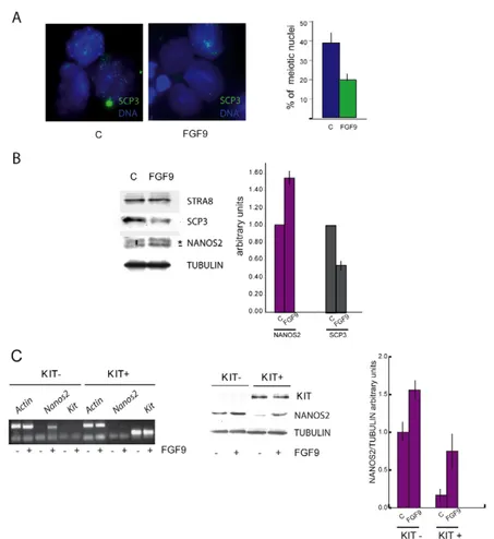
Finally, we investigated whether FGF9 was able to affect meiosis also in 7 d.p.p. spermatogonia. We found that FGF9 reduced the number of meiotic cells after 48 hours of culture, as judged by chromosomal spread counts (40% in the control versus 20% in the FGF9 treated cells, Fig. 4A) and by the decrease of SCP3 levels (Fig. 4B). The role of FGF9 in meiotic inhibition was not mediated by the decrease of STRA8 levels, because they did not change after FGF9 stimulation. To understand which cell population was responsive to FGF9, we then isolated positive and KIT-negative spermatogonia by immunomagnetic cell sorting, and stimulated them with the factor for 24 hours. FGF9 sensitivity was present in both cell types, because it induced upregulation of NANOS2 in KIT-negative and KIT-positive spermatogonia, as shown by RT-PCR and by western blot analyses (Fig. 4C).
NANOS2 associates with RNPs and polysomes and interacts with PUM2
In Drosophila germline progenitor pole cells, NANOS has been shown to interact with pumilio (PUM) to repress Cyclin B (CCNB1)
Fig. 2. AtRA downregulates Nanos2 in both fetal and postnatal male germ cells. (A)Male fetal gonads isolated at 12.5 d.p.c. cultured in vitro for 48 hours in the presence of AtRA. RT-PCR analysis shows that Stra8 and Nanos2 mRNA expression are oppositely modulated by AtRA treatment (top left). Testes from 13.5 d.p.c. embryo were collected, disaggregated, and kept in culture for 24 hours in the presence of AtRA. RT-PCR analysis shows that AtRA strongly reduces Nanos2 expression also at this developmental stage (bottom left). The histogram represents the percentage of meiotic nuclei scored in control or in AtRA-treated cultures of 12.5 d.p.c. gonads. Three independent experiments performed in triplicate are represented. Bars show mean ± s.d.; P<0.001. (B)Isolated male germ cells of 15.5 d.p.c. and 4 d.p.p. were stimulated in vitro with AtRA for 48 hours. Western blots and semiquantitative RT-PCR analyses show that, concomitantly with STRA8 upregulation, NANOS2 is downregulated in AtRA-treated cells. The right panel represents the densitometric analysis of the western blots from three independent experiments performed in triplicate. Bars represent mean ± s.d.; P<0.001. (C)Nuclear spreads from 15.5 d.p.c. (left) or 4 d.p.p. (right) spermatogonia were prepared after 48 hours of culture in the presence or absence of AtRA and probed for SCP3. Merged images of SCP3 (not corresponding to correctly assembled synaptonemal complexes) and DAPI are shown.
Jour
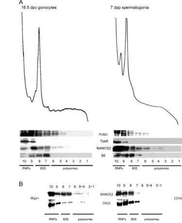
mRNA translation (Kadyrova et al., 2007). To date, none of the mammalian NANOS homologs has been demonstrated to act as a translational repressor, but we have recently shown that NANOS3 is able to interact with PUM2 and associates with the RNPs, in an heterologous system (Lolicato et al., 2008). To test whether NANOS2 might be involved in the control of translation, a Myc-tagged Nanos2-expressing vector was transfected into HEK293T cells. By immunofluorescence analysis using anti-MYC antibodies, we found that NANOS2-MYC was distributed both in the cytoplasmic and the nuclear compartments of transfected cells (Fig. 5A). Cytoplasmic extracts were then fractionated on a sucrose gradient and analyzed by western blot. Fig. 5B shows that NANOS2-MYC was found mostly in the RNP fractions but also in the ribosomal and light polysome fractions. We then followed the distribution pattern of endogenous NANOS2 on sucrose gradients using 16.5 d.p.c. or 7 d.p.p. spermatogonia extracts (Fig. 6A). Similarly to what was observed with transfected cells, NANOS2 colocalized with the RNP fractions, as well as with those containing the ribosomal subunits and light polysomes. The sedimentation pattern of PUM2 on the sucrose gradients mirrored that of NANOS2. However, TIAR, another RNA-binding protein known to be a translational repressor (Mazan-Mamczarz et al., 2006) that is essential for germ-cell survival (Beck et al., 1998), was found to be exclusively present in the RNP fractions (Fig. 6A). The finding that NANOS2 also associates with polysomes was further verified by fractionating 7 d.p.p. spermatogonial extracts on sucrose gradients in the presence of EDTA, to disrupt polysomes. As shown in Fig. 6, association of NANOS2 with the light polysomal fractions
was completely reverted toward the ribosomal subunits and the RNPs in the presence of EDTA, as occurs for DAZL, an RNA-binding protein that has been shown to be present both in the RNP and in the polysomal fractions (Tsui et al., 2000).
NANOS2 interacts with PUM2 and binds to target mRNAs
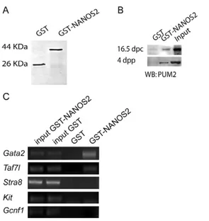
We have previously shown that PUM2 is able to bind not only NANOS3 but also NANOS2 when used as a bait in pull-down experiments (Lolicato et al., 2008). Since the sedimentation profile of NANOS2 and PUM2 suggested that they might associate in vivo (Fig. 6), we tested the ability of NANOS2 to interact with the endogenous PUM2 from germ-cell extracts in a pull-down assay. We prepared protein extracts from fetal gonocytes (16.5 d.p.c.) or (4 d.p.p.) postnatal spermatogonia and incubated them with a GST-or a GST-NANOS2-agarose coupled resin (Fig. 7A). We found that PUM2 from both fetal and postnatal cell extracts was able to interact with GST-NANOS2, whereas a faint background band was observed in the presence of GST (Fig. 7B).
Preliminary data obtained by microarray analysis reported that significant alterations occur in the transcriptome of the developing male gonad of Nanos2-null mice (Saba, 2009). To verify whether murine NANOS2 works as an RNA-binding protein, we undertook a RNA pull-down approach, by probing GST or GST-NANOS2 agarose beads with ribonucleoprotein extracts from postnatal spermatogonia. RNA molecules bound to GST-NANOS2 or to GST were then extracted and reverse transcribed to obtain cDNAs. Using primers that spanned the 3⬘UTR of the putative target genes, we looked for mRNAs whose expression was altered in the
Fig. 3. FGF9 stimulates Nanos2 expression and inhibits meiosis in fetal gonads. (A) Semiquantitative RT-PCR from
control and FGF9-treated cultures of 12.5 d.p.c. whole gonads (48 hours) or disaggregated 13.5 d.p.c. (24 hours) gonads. The expression of Nanos2 is upregulated upon FGF9 stimulation in both sexes, at both stages and in both culture conditions. A control PCR analysis without reverse transcriptase was run with cDNAs from 13.5 d.p.c. male disaggregated gonads cultured with and without FGF9 or AtRA for 24 hours (right). Densitometric analysis of three independent experiments is shown below. Bars represent mean ± s.d.; P<0.001. (B) Nuclear spreads of the corresponding cultures, immunostained for SCP3 to detect the presence of synaptonemal complexes. Graphical representation of meiotic chromosomes counts both in fetal male and female gonads, from three independent experiments, is shown on the right. Bars represent mean ± s.d.; P<0.001.
Jour
Nanos2 knockouts and/or for transcripts that displayed a sexually dimorphic expression pattern during fetal gonad development. Specifically, we screened the cDNAs obtained from the RNA pull-down for Stra8, Taf7l, Kit, Gcnf1 and Gata2, by PCR analysis. We found that Gata2 and Taf7l mRNA was retained specifically by GST-NANOS2 beads (Fig. 7C), whereas we did not observe any specific signal for Kit, Stra8 or Gcnf1 mRNAs (Fig. 7C). We actually found that perfect consensus binding sites for PUM2 [UGUANAUA(A/G)NNNN(C/G/U)(C/G/U)(C/G/U)(C/G/U)(C/G) CC] (White et al., 2001) are located within the 3⬘UTR of both Gata2 and Taf7l mRNAs, supporting the notion that a complex of NANOS-pumilio is essential for mRNA binding. When looking in the Geo Profile database for Taf7l and Gata2 expression in germ cells, we found that these two genes were not only differentially regulated in the developing male and female gonads, but their levels were higher in the differentiating type B compared with type A spermatogonia (GDS2223, GDS2390). Interestingly, Gata2 mRNA levels were found to be significantly increased in the postnatal testis in a period in which high levels of Kit are also found (GDS401). Discussion
NANOS2 is a potential RNA-binding protein that functions cell autonomously to control germ-cell sexual differentiation during fetal development. It has been proposed to act as an antimeiotic gene, because its deletion in mice is associated with abortive meiosis in fetal gonocytes, before they undergo apoptosis. Furthermore, its misexpression in female PGCs prevents meiosis and makes the nuclear morphology of these cells similar to that of G0-arrested gonocytes (Suzuki and Saga, 2008).
In the present paper, we used fetal germ cells and postnatal spermatogonia as models to understand the function of NANOS2 and its relationship with the beginning of meiosis.
NANOS2 expression and meiotic commitment
By analyzing the developmental profile of NANOS2 expression in the testis during the perinatal period, we found that NANOS2 is continuously expressed from the fetal to the postnatal period and it shows two peaks of expression: one at around 15.5 d.p.c. and another one at around 5 d.p.p. The first peak temporally corresponds to the mitotic block period, which male germ cells undergo prenatally. In this period, gonocytes do not enter meiosis and retain stem cell features, as demonstrated by their ability to complete spermatogenesis when transplanted into adult testes (Ohta et al., 2004). The second peak corresponds to a period in which cells expressing neurogenin-3 (NGN3) (a marker for undifferentiated spermatogonia) (Nakagawa et al., 2007; Yoshida et al., 2004) start differentiation into KIT-positive cells (Pellegrini et al., 2008). During this period, the spermatogonial stem cell factor PLZF (promyelocytic leukemia zinc finger) tightly controls Kit mRNA expression in stem cells (Filipponi et al., 2007), and they are completely lost when Kit is not repressed (Buaas et al., 2004; Costoya et al., 2004). By purifying KIT-positive and KIT-negative spermatogonia, we found that NANOS2-expressing cells were present only within the KIT-negative spermatogonia population, which corresponds to PLZF-expressing spermatogonia at 4-5 d.p.n., indicating that Nanos2 is a spermatogonial stem-cell-expressed gene. In agreement with our results, during the revision of this paper, two studies have shown that NANOS2 is mostly expressed in As
Fig. 4. FGF9 stimulates Nanos2 expression and inhibits meiosis in postnatal spermatogonia. Spermatogonia from
prepuberal testes (7 d.p.p.) were cultured in the presence or absence of FGF9 for 48 hours. (A)Nuclear spreads of the corresponding cultures stained for SCP3 and DAPI. The histogram represents the percentage of meiotic nuclei scored in control or in FGF9-treated cultures from three independent experiments. Bars show mean ± s.d.; P<0.001. (B)Western blot analysis for NANOS2 (Abnova antibody), STRA8, SCP3 and tubulin. NANOS2 levels are increased by FGF9 whereas SCP3 levels are decreased; STRA8 is not affected. The bar indicates the specific NANOS2 band, used for densitometry, whereas asterisk denotes a non-specific one. Densitometric analysis of the western blots from three separate experiments is shown on the right. Bars represent mean ± s.d.; P<0.001. (C)Immunomagnetic-sorted KIT-positive and KIT-negative spermatogonia analyzed for FGF9 sensitivity. Semiquantitative RT-PCR on mRNAs (left) or western blot on protein extracts (middle) from positive and KIT-negative spermatogonia were probed for NANOS2, actin and KIT. Densitometric analysis of the western blots from three separate experiments is shown on the right. Bars represent mean ± s.d.; P<0.001.
Jour
and Aprspermatogonia from the adult testis and that its conditional deletion in adulthood leads to spermatogonia stem cell loss (Sada et al., 2009; Suzuki et al., 2009).
Evidence from genetic studies suggests that NANOS2 acts as a suppressor of meiosis in male fetal germ cells (Suzuki and Saga, 2008). We found that AtRA, a derivative of RA and a well-known promeiotic factor (Bowles et al., 2006; Pellegrini et al., 2008), was able to downregulate Nanos2 at both mRNA and protein levels in male PGCs (12.5 d.p.c.) and in undifferentiated spermatogonia. In mitotically arrested gonocytes (15.5 d.p.c.), however, we observed that the strong decrease of Nanos2 mRNA levels induced by AtRA was paralleled only by a less-intense decrease of the protein levels. Since this different behaviour was consistently reproduced, we hypothesize that NANOS2 protein stability is higher in mitotically arrested germ cells than in proliferating germ cells. We found that AtRA increased the percentage of proliferating male PGCs entering meiosis, but it was not effective on mitotically arrested gonocytes or on undifferentiated spermatogonia. For arrested gonocytes, this discrepancy can be explained by the existence of a narrow window for meiotic competence, which corresponds to the period in which NANOS2 starts to be expressed (12.5 d.p.c.) and is needed to repress the meiotic fate (Saga, 2008; Suzuki and Saga, 2008). After this period, high levels of NANOS2, as a result of increased protein stability and/or enhanced synthesis stimulated by factors such as
FGF9 (see below), ‘masculinize’ gonocytes. The evidence that AtRA does not trigger meiosis in undifferentiated spermatogonia, notwithstanding the modulation of NANOS2 and STRA8, suggests that these cells lack other molecular factors required for meiotic entry. One of these factors could be the KIT tyrosine kinase receptor, whose downstream signalling is essential for meiotic entry of differentiating spermatogonia in vitro (Pellegrini et al., 2008). In line with this hypothesis is the observation that Kit is expressed in proliferating fetal germ cells of both sexes up to 12.5-13.5 d.p.c. (Manova and Bachvarova, 1991) when the ‘meiotic window’ is active, and is then re-expressed in the male only postnatally, in differentiating spermatogonia – the meiosis-competent cells of the testis.
In a search for factors that could upregulate NANOS2 levels and negatively influence meiosis, we found that FGF9 was a good candidate. Fibroblast growth factors (FGFs) have a role in the proliferation and survival of many cell types (Ornitz and Itoh, 2001). Fgf9 is expressed in the undifferentiated gonads of both sexes at
Fig. 5. NANOS2 co-sediments with RNPs and polysomes in Hek293T.
(A)Transfection of Myc-tagged Nanos2 in HEK293T cells.
Immunofluorescence analysis with anti-MYC antibodies shows that NANOS2 is distributed mainly in the cytoplasm, but also in the nucleus. (B)RNP-polysome fractionation on sucrose gradients of HEK293T cells transfected with Myc-Nanos2. Western blot analysis of the collected fractions shows that NANOS2 sediments mainly in the light fraction of the gradient (fraction 10, RNPs) but it is also present through the ribosomal subunits (fractions 7-9) and to a lesser extent in the polysomes (fractions 3-6). S6 pattern and UV absorbance profile demonstrate the correct fractionation assay.
Fig. 6. NANOS2 associates with RNPs and polysomes of fetal and postnatal male germ cells. (A)Fetal (16.5 d.p.c.) and postnatal (7 d.p.p.) male germ cells were collected and cytoplasmic extracts were fractionated on sucrose gradients. Western blot analysis of the collected fractions shows that NANOS2 sediments with the RNP fraction, the ribosomal subunits and the polysomal fractions. PUM2 showed a sedimentation pattern similar to NANOS2, suggesting that both proteins can interact in vivo. By contrast, the translational repressor TIAR is found exclusively in the RNPs fraction in both fetal and postnatal stages. S6 and UV absorbance are presented as controls of correct fractionation. (B)Untreated (Mg2+) or EDTA-treated (EDTA) 7 d.p.p. spermatogonial extracts were fractionated on sucrose gradients and analyzed by UV spectrometry (not shown) and western blotting for NANOS2 and DAZL.
Jour
11.5 d.p.c., but it becomes XY restricted by 12.5 d.p.c. and is maintained in the testis cords (Schmahl et al., 2004). Sertoli cell precursors fail to proliferate in Fgf9 mutants and testis differentiation is disrupted (Schmahl et al., 2004). The majority of germ cells are lost at 12.5 d.p.c.; however, the surviving gonocytes within the Fgf9-null gonads are found in meiosis (DiNapoli et al., 2006), suggesting that FGF9 normally promotes survival and prevents meiosis of fetal male germ cells. Indeed, we found that FGF9 upregulated NANOS2 and strongly inhibited meiosis in female PGCs in vitro. This observation is in line with the evidence that Nanos2 misexpression in female germ cells inhibits meiosis and masculinizes germ-cell nuclei (Suzuki and Saga, 2008). FGF9 also upregulated NANOS2 in postnatal spermatogonia, and such an increase was only observed
after a long incubation time (not shown), suggesting that a transcriptional mechanism probably mediates this effect. Sensitivity to FGF9 was present in the undifferentiated and in the differentiating spermatogonia, because both these cell types express an FGF9 receptor, as confirmed by microarray analysis (Rossi et al., 2008). We also observed a negative effect of FGF9 on meiotic entry in differentiating spermatogonia, suggesting a common mechanism shared between female and male germ cells in the control of the mitotic-meiotic switch. It has been hypothesized that a possible role for NANOS2 in the prevention of meiosis is to repress STRA8 expression during the period in which CYP26B1 levels decrease in the male gonad and RA is increased in the mesonephros. In our studies we did not observe STRA8 downregulation after FGF9 stimulation of postnatal spermatogonia in vitro, suggesting that NANOS2 might act independently or downstream of STRA8, even if we cannot exclude that other mechanisms are active in vivo.
NANOS2 subcellular localization and molecular interactions
In contrast to what we observed for NANOS3 (Lolicato et al., 2008), NANOS2 was found to be associated not only with RNPs, but also with the ribosomal and polysomal fractions. This localization is uncommon for translational repressors, but it has been recently shown that translational repression can also occur at the polysomal level, during protein synthesis, through a miRNA repressive mechanism (Cannell et al., 2008; Gebauer and Hentze, 2004; Petersen et al., 2006). Furthermore, the possibility cannot be ruled out that NANOS2, and eventually PUM2, might function both as translational repressors and activators, as has been shown for FMRP (Zalfa et al., 2006); they could also function at other levels of post-transcriptional regulation of mRNA expression, such as the control of mRNA stability.
When transfected in HEK93T cells, NANOS3 associates with PUM2 and is found within the RNPs (Lolicato et al., 2008). Since it has been shown that NANOS2 can complement the NANOS3 defect in Nanos3-null mice, we reasoned that it could also recognize the same partners in male germ cells. Indeed, we found that NANOS2 was able to bind PUM2 expressed either in fetal gonocytes or in postnatal spermatogonia, suggesting that PUM2 can interact in vivo with NANOS2, probably both acting in post-transcriptional mRNA regulation. In the attempt to discover potential NANOS2 targets, we looked for genes showing a sexually
Fig. 7. NANOS2 interacts with PUM2 to regulate target mRNAs. Protein
and RNA pull-downs were performed to investigate the interaction between NANOS2 and PUM2. (A)Coomassie blue staining of GST and GST-coupled NANOS2 used for pull-down experiments. (B)Western blot of cell extracts from fetal (16.5 d.p.c.) and postnatal (4 d.p.n.) male germ cells loaded onto GST or GST-NANOS2-agarose beads and probed for PUM2. A specific band for PUM2 is observed in the presence of GST-NANOS2 in both fetal and postnatal cell extracts. (C) RT-PCR for potential target mRNAs of NANOS2.
Fig. 8. Opposing effects of RA and FGF9 on Nanos2 expression and meiotic entry of mouse germ cells. The
niche between Sertoli cell precursors and fetal gonocytes might be equivalent to the stem cell niche of the postnatal testis. Fetal gonocytes and spermatogonia stem cells could have an equivalent cell fate signed by the expression of
Nanos2. Such fate would be lost when spermatogonia are
committed to meiosis. In the fetal testis, Sertoli cell precursors secrete FGF9 to induce Nanos2 expression and masculinization of PGCs (that become gonocytes). FGF9 upregulates Nanos2 and prevents meiosis. Postnatally, the balance between FGF9 and RA secreted by Sertoli cells regulates the levels of Nanos2 expression in undifferentiated spermatogonia (NANOS2-positive cells) and in
differentiating spermatogonia (STRA8- and KIT-positive cells). In the fetal ovary, FGF9 secretion from somatic cells drops early during gonadogenesis and as a consequence PGCs are not masculinized. High RA levels ensure meiotic entry through induction of Stra8 and possibly Kit.
Jour
dimorphic expression pattern during gonadal differentiation and/or that were more abundantly expressed in differentiating spermatogonia. Among several genes tested (Kit, Taf7l, Gata2, Stra8 and Gcnf1), Gata2 and Taf7l mRNAs were found to interact with GST-NANOS2. GATA2 is a zinc finger transcription factor expressed in fetal oocytes up to 15.5 d.p.c., but not in fetal gonocytes (Siggers et al., 2002). There are no studies on Gata2 expression in postnatal gonads; however, its mRNA levels are strongly increased in differentiating spermatogonia, according to the microarray analysis database (Geo Profiles) at NCBI. Although the role of GATA2 in germ cells is unknown, it is well established that in hematopoietic stem cells, this factor controls Kit expression at the transcriptional level (Orkin, 1992). Taf7l is a component of the transcriptional machinery specific to preleptotene spermatocytes and haploid spermatids (Pointud et al., 2003) and its deletion leads to reduced male fertility (Cheng et al., 2007). Thus, the identification of two transcription factors involved in the differentiation of spermatogonia as targets of post-transcriptional control by NANOS2 is in line with the evidence that NANOS2 negatively regulates differentiation of spermatogonia stem cells (Sada et al., 2009). Studies to identify other NANOS2 mRNA targets, whose post-transcriptional regulation is determinant for spermatogonia differentiation, will be important to understand how commitment of stem cell occurs in the initial phase of spermatogenesis.
In conclusion, our results can be interpreted as schematically depicted in Fig. 8. RA and FGF9 produced locally by the somatic cells of the gonads act in opposite ways to regulate Nanos2 expression and meiotic entry both in fetal and postnatal germ cells. In the fetal ovary, FGF9 levels sharply decrease at 11.5 d.p.c. in parallel with a rise of circulating levels of RA, which stimulates Stra8 expression in PGCs and their commitment to meiosis. In the fetal testis, FGF9 levels are kept high, and circulating RA is degraded by CYP26B1 (not depicted) in somatic cells. High levels of FGF9 without RA signalling in male PGCs upregulate Nanos2, which determines their transition into mitotically arrested gonocytes. In the postnatal testis, Nanos2 is expressed exclusively in SSCs and Sertoli cells to safeguard the delicate balance between stem cell self-renewal and meiotic commitment of spermatogonia by modulating the levels of FGF9 and RA.
Materials and Methods
Cell isolation and culture
Postnatal male germ cells (0-7 d.p.p.) were obtained as previously reported, by sequential enzymatic digestion of testes from CD1 albino mice (Pellegrini et al., 2008). After preplating of cell suspension, germ cells were cultured in modified Earle’s medium (MEM, Gibco) with 20 mM glutamine (Gibco), 2 mM pyruvic acid (Sigma), 1 mM lactic acid (Sigma), non-essential amino acids (Gibco), 100 U/ml penicillin and 100 g/ml streptomycin (Gibco), without serum supplementation.
To obtain enriched fetal germ-cell suspensions, fetal testes and ovaries were collected at different developmental stages and digested with trypsin (Gibco) and DNaseI (Sigma). Cell suspensions were pre-plated in the same medium as postnatal germ cells supplemented with 10% fetal bovine serum, (FBS) and recovered after 3 hours of culture, to allow most of the somatic cells to adhere to the plastic dishes. In the case of organ culture, fetal gonads were cultured for 48 hours onto 0.8% agarose blocks pre-equilibrated with MEM and 10% FBS.
All-trans-retinoic acid (AtRA, Sigma) was dissolved at a concentration of 10 mM in ethanol and diluted to a final concentration of 0.3 M (Pellegrini et al., 2008) in culture medium. Control cultures received the corresponding doses of diluted ethanol. FGF9 (from Società Italiana Chimici) was dissolved in PBS 1 mg/ml BSA and used to a final concentration of 25 ng/ml. All the factors were replaced in the culture medium after 24 hours. Separation of KIT-positive spermatogonia from KIT-negative spermatogonia was performed by magnetic-activated cell sorting (MACS) with CD117 conjugated microbeads (Miltenyi Biotech, Germany) as previously described (Pellegrini et al., 2008).
Definition of the germ-cell stages
Fetal male and female germ cells are defined as PGCs as long as they reach the gonads and proliferate. After gonadal colonization, female PGCs start entering meiosis (at around 13.5 d.p.c.) and are then defined as oocytes. Male PGCs, however, enter mitotic block after gonadal colonization and are then defined as gonocytes. This definition is retained up to birth, when gonocytes resume mitosis and spermatogonial stem cells can be identified. KIT-negative (OCT4- and PLZF-positive) spermatogonia represent the stem cell population of the testis whereas KIT-positive spermatogonia are the differentiating mitotic germ cells which are committed to enter meiosis. Polysome-RNP fractionation
Prepuberal spermatogonia and fetal gonocytes were subjected to polysome-RNP fractionation on sucrose gradients as previously described (Lolicato et al., 2008). In brief, 600 g cytoplasmic extracts in polysome buffer were loaded onto 15-50% sucrose gradients. Ten fractions were collected and protein precipitation from each fraction was performed with 72% trichloroacetic acid (TCA) and subsequently resuspended in 2⫻ sample buffer and processed for SDS-PAGE. Absorbance at 260 nm was continuously recorded while collecting the fractions. EDTA treatment was performed as previously described (Grivna et al., 2006).
Recombinant proteins, transfection, GST protein and RNA pull-down
Nanos2-Myc was produced by RT-PCR using primers using a proofreading polymerase
(Pfu, Stratagene) containing BamHI-EcoRI sites at the extremities. The amplified band was cloned in pcDNA3-Myc (Invitrogen, Milan, Italy) or in pGEX4T1 (GE Healthcare, Milan, Italy). For transient transfections, HEK293T cells were transfected with pcDNA3Myc-Nanos2 by Lipofectamine 2000 (Invitrogen) according to the manufacturer’s instructions.
Pull-down experiments were performed using GST fusion proteins purified from bacterial lysates on glutathione-agarose (Sigma). In the case of protein pull-down, cell extracts (600 g total proteins) of 16.5 d.p.c. gonocytes or 4 d.p.p. spermatogonia, precleared for 1 hour with glutathione-agarose in lysis buffer, were added to 2 g GST protein or GST-NANOS2 fusion protein absorbed on glutathione-agarose in lysis buffer supplemented with 0.05% BSA. After incubation for 90 minutes at 4°C under constant shaking, beads were washed three times with the same buffer, absorbed proteins were eluted in SDS sample buffer and resolved on a 12% SDS-PAGE for western blot analysis. Input represents 1/10 of the initial samples.
For RNA pull-down experiments 1 mg cytoplasmic extracts in polysome buffer were obtained from 7 d.p.p. spermatogonia and processed as described (Keene et al., 2006). Extracts containing RNA and protein were incubated with GST or GST-NANOS2 agarose beads for 4 hours at 4°C. RNA-protein complexes were washed four times in NT2 buffer and RNA was eluted from the beads by incubation at 55° for 30 minutes with 30 g proteinase K. After Trizol extraction (Invitrogen) and DNaseI treatment (Zymo Research, Rome, Italy), RNA was reverse transcribed as described in the following section.
RT-PCR
Total RNA from whole fetal gonads and fetal and postnatal germ cells was extracted with Trizol reagent and treated with DNase I to avoid potential contamination by genomic DNA. DNA-free RNA was reverse transcribed using SuperScript First Strand Synthesis kit (Invitrogen) according to the manufacturer’s instructions. Primers used for PCR were as follows: Nanos2 F, AGTGCCATGGACCTACCGCCCTTT and
Nanos2 R, TCTCAATTATCGCTTGACTCTGC; c-Kit F, GCCACGTCT -CAGCCATCTG and c-Kit R, GTCGGGATCAATGCACGTCA; Stra8 F, TAGGATCCATGGCCACCCCTGGAGAAG and Stra8 R, GAATTCTTA -CAGATCGTCAAAGGTCTCCA; Actin F, GGCTGTATTCCCCTCCATCG and
Actin R, CCAGTTGGTAACAATGCCATGT. For semiquantitative RT-PCR 25
cycles were performed for the amplification of Actin and all other genes, 30 cycles for Nanos2 amplification from male germ cells or 40 cycles from female germ cells. RNA from RNA pull-down experiments was reverse transcribed using SuperScript First Strand Synthesis kit (Invitrogen) according to the manufacturer’s instructions. Primers for putative targets were designed, when possible, to span a region between the last two exons. Primers were the following: Gata2 F, CAAGCT -GCACAATGTTAACAGGC and Gata2 R, ATTCACAGTAATGGCGGCACAAGG;
Taf7l F, GGAGGAGGAAGAGACAGACAATTC and Taf7l R, GTAGAGGGA
-CAGAAGTATGTGGTTCCAC; Stra8 F, TGCCGGACCTCATGGAATTT and Stra8 R, GAATTCTTACAGATCGTCAAAGGTCTCCA; Gcnf1 F, GGATGGAGG -TGATTGAACGACT and Gcnf1 R, AGTCTCCATCTTGGTCTCTGGCT. PCR conditions were: 95°C for 20 seconds, 58°C for 30 seconds, 72°C for 30 seconds, for 40 cycles.
Immunofluorescence
Transfected HEK293T cells were adhered onto poly-L-lysine glass slides and fixed for 10 minutes at room temperature in 2% paraformaldehyde. After washing with PBS, cells were permeabilized for 10 minutes with PBS containing 0.1% Triton X-100, and incubated for 30 minutes at room temperature with PBS containing 0.5% BSA. Samples were then incubated overnight at 4°C in a humidified chamber with anti-MYC antibody from Santa Cruz (sc-2048) at a final concentration of 2 g/ml, then for 1 hour at room temperature with cyanin 3 (AP 180 C; Chemicon). Slides were washed and mounted in 50% glycerol in PBS and immediately examined by
Jour
fluorescence microscopy. Nuclei were counterstained with 1 g/ml Hoechst 33342 (Sigma). Control experiments were performed using non-immune immunoglobulins instead of the specific antibody.
Western blotting
For western blot analysis, germ cells were harvested and washed three times with ice-cold PBS. Cell lysis was performed with 10 mM HEPES, pH 7.9, 1% Triton X-100, 10 mM KCl, 1.5 mM MgCl2, 0.1 mM EGTA, 0.5 mM dithiothreitol (DTT), 10
mM -glycerophosphate, 0.1 mM sodium vanadate and protease inhibitor cocktail (Sigma). 40 g protein extracts were separated by SDS-PAGE in 4-20% gradient gels (Serva, Milan, Italy) or uniform gels of 15% polyacrylamide and transferred to nitrocellulose membrane (Amersham, Piscataway, NJ). The membrane was blocked in phosphate saline buffer with 0.1% Triton (PBST) and 5% fat-free milk powder for 1 hour at room temperature. Incubation with primary antibodies was carried out at 4°C overnight in PBST-5% BSA. Appropriate horseradish-peroxidase-conjugated secondary antibody (Santa Cruz Biotechnology) was used at a 1:5000 dilution in PBST for 1 hour at room temperature. Anti-NANOS2 serum (in this study we used both one obtained from Abnova, Taipei, Taiwan, and a gift from Yumiko Saga, Sokendai, Shizuoka, Japan; in the figure legends we indicated when the commercial antibody was used) was used at a 1:300 dilution in PBST with 5% BSA. Anti-KIT rabbit polyclonal antibody (Albanesi et al., 1996) was diluted at 1:1000. Anti-PUM2 rabbit polyclonal (Abcam) was diluted at 1:3000. Anti-TIAR mouse monoclonal antibody (a kind gift from P. Anderson, Harvard, MA) was diluted at 1:1000. Anti-S6 rabbit polyclonal antibody (Cell Signaling, Beverly, MA) was diluted 1:1000. Anti-Actin rabbit polyclonal (A2066, Sigma) was diluted 1:1000. Anti-MYC antibody was from Santa Cruz (sc-2048) and diluted 1:1000. Mouse monoclonal antibody anti human DAZL was from AbD Serotec, Milan Italy and used at a 1:1000 dilution. Anti-STRA8 rabbit polyclonal from Abcam (Ab 49602) was used at a 1:1000 dilution. Anti-SCP3 rabbit polyclonal (1:1000) was from Novus. Anti-OCT4 mouse monoclonal (1:1000) was from Santa Cruz (sc-5279). The horseradish peroxidase conjugate was detected by chemioluminescence with an ECL Kit (Santa Cruz) and autofluorography. Densitometric analysis was performed by ImageQuantTM TL
software (GE Healthcare). Spreads
Cultured spermatogonia or single cell suspensions from cultured fetal gonads were prepared and stained essentially as previously described (Romanienko and Camerini-Otero, 2000). Slides were washed twice in PBS, and incubated overnight at 4°C with anti-SCP3 rabbit polyclonal antibody (Novus) diluted in blocking solution (1:100 in 10% goat serum, 3% BSA, 0.5% Triton X-100 in PBS). After washing, secondary antibody was added for 1 hour at 37°C. The slides were washed and allowed to dry. Vectashield Mounting Medium with DAPI (Vector Laboratories, Burlingame, CA) was added and the slides were viewed using a Leica microscope. Spreads analysis was performed in three independent experiments.
Statistical analysis
Continuous variables were summarized as means ± s.d. All tests were two-sided and were determined by Monte Carlo’s significance. A significance value threshold of 0.05 was used for the current analysis. Student’s t-test was used to test for differences between two independent groups, whereas one-way ANOVA was used to test for differences among three or more independent groups. For multiple comparisons the Tukey’s HSD test was carried out. All statistical tests were carried out using the SPSS statistical analysis software package, version 10.0.
We thank Yumiko Saga (Sokendai, Japan) for anti-NANOS2 antibody, Paul Anderson (Brigham and Women’s Hospital, MA) for providing anti-TIAR antibody; S. Pedrotti for technical help; C. Sette and P. Grimaldi for critically reading the manuscript and helpful suggestions. This work was supported by grants from the Italian Ministry of University (Prin 2005059793_005, 200734H7WW_004, and 200788TPYE_002).
References
Albanesi, C., Geremia, R., Giorgio, M., Dolci, S., Sette, C. and Rossi, P. (1996). A
cell-and developmental stage-specific promoter drives the expression of a truncated c-kit protein during mouse spermatid elongation. Development 122, 1291-1302.
Anderson, E. L., Baltus, A. E., Roepers-Gajadien, H. L., Hassold, T. J., de Rooij, D. G., van Pelt, A. M. and Page, D. C. (2008). Stra8 and its inducer, retinoic acid, regulate
meiotic initiation in both spermatogenesis and oogenesis in mice. Proc. Natl. Acad. Sci.
USA 105, 14976-14980.
Beck, A. R., Miller, I. J., Anderson, P. and Streuli, M. (1998). RNA-binding protein
TIAR is essential for primordial germ cell development. Proc. Natl. Acad. Sci. USA 95, 2331-2336.
Bowles, J., Knight, D., Smith, C., Wilhelm, D., Richman, J., Mamiya, S., Yashiro, K., Chawengsaksophak, K., Wilson, M. J., Rossant, J. et al. (2006). Retinoid signaling
determines germ cell fate in mice. Science 312, 596-600.
Buaas, F. W., Kirsh, A. L., Sharma, M., McLean, D. J., Morris, J. L., Griswold, M. D., de Rooij, D. G. and Braun, R. E. (2004). Plzf is required in adult male germ cells
for stem cell self-renewal. Nat. Genet. 36, 647-652.
Cannell, I. G., Kong, Y. W. and Bushell, M. (2008). How do microRNAs regulate gene
expression? Biochem. Soc. Trans. 36, 1224-1231.
Cheng, Y., Buffone, M. G., Kouadio, M., Goodheart, M., Page, D. C., Gerton, G. L., Davidson, I. and Wang, P. J. (2007). Abnormal sperm in mice lacking the Taf7l gene. Mol. Cell. Biol. 27, 2582-2589.
Colvin, J. S., Green, R. P., Schmahl, J., Capel, B. and Ornitz, D. M. (2001).
Male-to-female sex reversal in mice lacking fibroblast growth factor 9. Cell 104, 875-889.
Costoya, J. A., Hobbs, R. M., Barna, M., Cattoretti, G., Manova, K., Sukhwani, M., Orwig, K. E., Wolgemuth, D. J. and Pandolfi, P. P. (2004). Essential role of Plzf in
maintenance of spermatogonial stem cells. Nat. Genet. 36, 653-659.
de Rooij, D. G. and Mizrak, S. C. (2008). Deriving multipotent stem cells from mouse
spermatogonial stem cells: a new tool for developmental and clinical research.
Development 135, 2207-2213.
DiNapoli, L., Batchvarov, J. and Capel, B. (2006). FGF9 promotes survival of germ cells
in the fetal testis. Development 133, 1519-1527.
Filipponi, D., Hobbs, R. M., Ottolenghi, S., Rossi, P., Jannini, E. A., Pandolfi, P. P. and Dolci, S. (2007). Repression of kit expression by Plzf in germ cells. Mol. Cell. Biol. 27, 6770-6781.
Fujii, T., Mitsunaga-Nakatsubo, K., Saito, I., Iida, H., Sakamoto, N., Akasaka, K. and Yamamoto, T. (2006). Developmental expression of HpNanos, the Hemicentrotus
pulcherrimus homologue of nanos. Gene Expr. Patterns 6, 572-577.
Gebauer, F. and Hentze, M. W. (2004). Molecular mechanisms of translational control. Nat. Rev. Mol. Cell. Biol. 5, 827-835.
Grivna, S. T., Pyhtila, B. and Lin, H. (2006). MIWI associates with translational machinery
and PIWI-interacting RNAs (piRNAs) in regulating spermatogenesis. Proc. Natl. Acad.
Sci. USA 103, 13415-13420.
Haraguchi, S., Tsuda, M., Kitajima, S., Sasaoka, Y., Nomura-Kitabayashid, A., Kurokawa, K. and Saga, Y. (2003). nanos1: a mouse nanos gene expressed in the
central nervous system is dispensable for normal development. Mech. Dev. 120, 721-731.
Kadyrova, L. Y., Habara, Y., Lee, T. H. and Wharton, R. P. (2007). Translational control
of maternal Cyclin B mRNA by Nanos in the Drosophila germline. Development 134, 1519-1527.
Keene, J. D., Komisarow, J. M. and Friedersdorf, M. B. (2006). RIP-Chip: the isolation
and identification of mRNAs, microRNAs and protein components of ribonucleoprotein complexes from cell extracts. Nat. Protoc. 1, 302-307.
Koprunner, M., Thisse, C., Thisse, B. and Raz, E. (2001). A zebrafish nanos-related
gene is essential for the development of primordial germ cells. Genes Dev. 15, 2877-2885.
Koubova, J., Menke, D. B., Zhou, Q., Capel, B., Griswold, M. D. and Page, D. C.
(2006). Retinoic acid regulates sex-specific timing of meiotic initiation in mice. Proc.
Natl. Acad. Sci. USA 103, 2474-2479.
Lehmann, R. and Nusslein-Volhard, C. (1991). The maternal gene nanos has a central
role in posterior pattern formation of the Drosophila embryo. Development 112, 679-691.
Lolicato, F., Marino, R., Paronetto, M. P., Pellegrini, M., Dolci, S., Geremia, R. and Grimaldi, P. (2008). Potential role of Nanos3 in maintaining the undifferentiated
spermatogonia population. Dev. Biol. 313, 725-738.
MacLean, G., Li, H., Metzger, D., Chambon, P. and Petkovich, M. (2007). Apoptotic
extinction of germ cells in testes of Cyp26b1 knockout mice. Endocrinology 148, 4560-4567.
Manova, K. and Bachvarova, R. F. (1991). Expression of c-kit encoded at the W locus
of mice in developing embryonic germ cells and presumptive melanoblasts. Dev. Biol.
146, 312-324.
Mark, M., Jacobs, H., Oulad-Abdelghani, M., Dennefeld, C., Feret, B., Vernet, N., Codreanu, C. A., Chambon, P. and Ghyselinck, N. B. (2008). STRA8-deficient
spermatocytes initiate, but fail to complete, meiosis and undergo premature chromosome condensation. J. Cell Sci. 121, 3233-3242.
Mazan-Mamczarz, K., Lal, A., Martindale, J. L., Kawai, T. and Gorospe, M. (2006).
Translational repression by RNA-binding protein TIAR. Mol. Cell. Biol. 26, 2716-2727.
Nakagawa, T., Nabeshima, Y. and Yoshida, S. (2007). Functional identification of the
actual and potential stem cell compartments in mouse spermatogenesis. Dev. Cell 12, 195-206.
Ohta, H., Wakayama, T. and Nishimune, Y. (2004). Commitment of fetal male germ
cells to spermatogonial stem cells during mouse embryonic development. Biol. Reprod.
70, 1286-1291.
Orkin, S. H. (1992). GATA-binding transcription factors in hematopoietic cells. Blood 80,
575-581.
Ornitz, D. M. and Itoh, N. (2001). Fibroblast growth factors. Genome Biol. 2,
REVIEWS3005.
Pellegrini, M., Filipponi, D., Gori, M., Barrios, F., Lolicato, F., Grimaldi, P., Rossi, P., Jannini, E. A., Geremia, R. and Dolci, S. (2008). ATRA and KL promote differentiation
toward the meiotic program of male germ cells. Cell Cycle 7, 3878-3888.
Petersen, C. P., Bordeleau, M. E., Pelletier, J. and Sharp, P. A. (2006). Short RNAs
repress translation after initiation in mammalian cells. Mol. Cell 21, 533-542.
Pilon, M. and Weisblat, D. A. (1997). A nanos homolog in leech. Development 124,
1771-1780.
Pointud, J. C., Mengus, G., Brancorsini, S., Monaco, L., Parvinen, M., Sassone-Corsi, P. and Davidson, I. (2003). The intracellular localisation of TAF7L, a paralogue of
Jour
transcription factor TFIID subunit TAF7, is developmentally regulated during male germ-cell differentiation. J. Cell Sci. 116, 1847-1858.
Romanienko, P. J. and Camerini-Otero, R. D. (2000). The mouse Spo11 gene is required
for meiotic chromosome synapsis. Mol. Cell 6, 975-987.
Rossi, P., Lolicato, F., Grimaldi, P., Dolci, S., Di Sauro, A., Filipponi, D. and Geremia, R. (2008). Transcriptome analysis of differentiating spermatogonia stimulated with kit
ligand. Gene Expr. Patterns 8, 58-70.
Saba, R., Suzuki, A., Suzuki, H., Sada, A. and Saga, Y. (2009). 17-P034 Nanos2 regulates
the transcriptome in the embryonic male germ cells. Mech. Dev. 126, S280.
Sada, A., Suzuki, A., Suzuki, H. and Saga, Y. (2009). The RNA-binding protein NANOS2
is required to maintain murine spermatogonial stem cells. Science 325, 1394-1398.
Saga, Y. (2008). Sexual development of mouse germ cells: Nanos2 promotes the male
germ cell fate by suppressing the female pathway. Dev. Growth Differ. 50, S141-S147.
Schmahl, J., Kim, Y., Colvin, J. S., Ornitz, D. M. and Capel, B. (2004). Fgf9 induces
proliferation and nuclear localization of FGFR2 in Sertoli precursors during male sex determination. Development 131, 3627-3636.
Siggers, P., Smith, L. and Greenfield, A. (2002). Sexually dimorphic expression of
Gata-2 during mouse gonad development. Mech. Dev. 111, 159-16Gata-2.
Subramaniam, K. and Seydoux, G. (1999). nos-1 and nos-2, two genes related to
Drosophila nanos, regulate primordial germ cell development and survival in Caenorhabditis elegans. Development 126, 4861-4871.
Suzuki, A. and Saga, Y. (2008). Nanos2 suppresses meiosis and promotes male germ cell
differentiation. Genes Dev. 22, 430-435.
Suzuki, A., Tsuda, M. and Saga, Y. (2007). Functional redundancy among Nanos proteins
and a distinct role of Nanos2 during male germ cell development. Development 134, 77-83.
Suzuki, H., Sada, A., Yoshida, S. and Saga, Y. (2009). The heterogeneity of spermatogonia
is revealed by their topology and expression of marker proteins including the germ cell-specific proteins Nanos2 and Nanos3. Dev. Biol. 336, 222-231.
Trautmann, E., Guerquin, M. J., Duquenne, C., Lahaye, J. B., Habert, R. and Livera, G. (2008). Retinoic acid prevents germ cell mitotic arrest in mouse fetal testes. Cell Cycle 7, 656-664.
Tsuda, M., Sasaoka, Y., Kiso, M., Abe, K., Haraguchi, S., Kobayashi, S. and Saga, Y.
(2003). Conserved role of nanos proteins in germ cell development. Science 301, 1239-1241.
Tsuda, M., Kiso, M. and Saga, Y. (2006). Implication of nanos2-3⬘UTR in the expression and function of nanos2. Mech. Dev. 123, 440-449.
Tsui, S., Dai, T., Warren, S. T., Salido, E. C. and Yen, P. H. (2000). Association of the
mouse infertility factor DAZL1 with actively translating polyribosomes. Biol. Reprod.
62, 1655-1660.
White, E. K., Moore-Jarrett, T. and Ruley, H. E. (2001). PUM2, a novel murine puf
protein, and its consensus RNA-binding site. RNA 7, 1855-1866.
Yoshida, S., Takakura, A., Ohbo, K., Abe, K., Wakabayashi, J., Yamamoto, M., Suda, T. and Nabeshima, Y. (2004). Neurogenin3 delineates the earliest stages of
spermatogenesis in the mouse testis. Dev. Biol. 269, 447-458.
Zalfa, F., Achsel, T. and Bagni, C. (2006). mRNPs, polysomes or granules: FMRP in
neuronal protein synthesis. Curr. Opin. Neurobiol. 16, 265-269.