R
ESEARCHP
ROJECT2010-2012
Investigating the molecular basis of Xotx1,
Xotx2 and Xotx5 differential actions during
Xenopus laevis development
Doctorate Course:Molecular Biotechnology Cycle: XXV
SSD BIO06
Laboratory: Unità di Biologia Cellulare e dello Sviluppo, SS12 Abetone e Brennero 4, Pisa
Supervisor/ Tutor: Prof. Robert Vignali
Student: Pamela Mancini
“Noi stiamo in piedi e camminiamo con parti del nostro corpo che sarebbero servite per pensare se si fossero sviluppate in un’altra parte dell’embrione.”
“We are standing and walking with parts of our body which could have been used for thinking had they developed in another part of the embryo.”
I
Index
Abstract Pag. 1
1- Introduction Pag. 3
1.1- The orthodenticle gene of Drosophila melanogaster Pag. 3
1.2- Vertebrate Otx genes Pag. 6
1.2.1- Paired-like K50 homeobox genes Pag. 6 1.2.2- Otx genes in mammalian anterior development Pag. 7
1.3- Otx genes and evolution Pag. 12
1.3.1- Otx genes in the animal kingdom Pag. 12 1.3.2- Otd/Otx: Insect and Vertebrate nervous system
evolution
Pag. 15
1.4- Xenopus laevis Pag. 17
1.5- Otx genes in Xenopus laevis Pag. 18
1.5.1- Xotx1 (and Xotx4) expression profile Pag. 18 1.5.2- Xotx2 expression profile Pag. 20 1.5.3- Xotx5/5b expression profile Pag. 22 1.5.4- Xotx2 and Xotx5 expression profile in the developing
eye
Pag. 24
1.6- Xotx genes and the Organizer Pag. 25
1.7- Xotx genes in the developing retina: bipolar versus photoreceptor fate
Pag. 30
1.8- Xotx genes and cement gland induction Pag. 32
2- Aim Pag. 37
3- Materials and methods Pag. 39
3.1- DNA constructs section I Pag. 39
3.2- DNA constructs section II Pag. 41
II
3.4- In situ hybridization Pag. 45
3.5- RNAs methods, embryo microinjections and animal cap assays
Pag. 45
3.6- Oligo antisense Morpholino Pag. 46
3.7- RT-PCR Pag. 47
3.8- Two-hybrid screening Pag. 48
3.9- GST-pull down assay Pag. 48
3.10- Western blotting Pag. 49
3.11- XOP-GFP reporter assay Pag. 49
3.12- Immunostaining on sections Pag. 50
3.13- 5’ RACE Pag. 50
3.14- Bioinformatics tools Pag. 50
4- Results section I Pag. 51
4.1- Cement gland induction Pag. 51
4.1 a- Statistical analysis: χ2 homogeneity test Pag. 61
4.2- Convergent extension inhibition Pag. 64
4.3- Neural tissue induction Pag. 66
5- Results section II Pag. 70
5.1- XOTX2 and XOTX5 transactivation domain Pag. 70 5.2- Two-hybrid screen for XOTX2 and XOTX5 potential
interactors
Pag. 71
5.3- Potential interactor database search Pag. 72 5.4- DNA binding ability of potential interactors Pag. 75 5.5- Expression profiles of potential interactors Pag. 75
5.5.1- RT-PCR Pag. 75
5.5.2- In situ hybridization Pag. 77
5.5.2.1- Early developmental stages Pag. 77 5.5.2.2- Later developmental stages Pag. 79 5.6- XOTX interaction domain(s) identification Pag. 81
III 5.7- XOTX2 and XOTX5 potential cofactors interaction with
XOTX1
Pag. 82
5.8- c29 Pag. 83
5.8.1- C29 and XOTX2/XOTX5 in vitro interaction Pag. 83 5.8.2- c29 localization in the X. tropicalis genome Pag. 84 5.8.3- C29 in silico secondary structure prediction Pag. 86 5.8.4- C29 in silico sub-cellular localization prediction Pag. 87 5.8.5- C29 sub-cellular localization Pag. 88 5.8.6- c29 functional analysis: preliminary data Pag. 90
5.9- Xusf2 Pag. 94
5.9.1- XUSF2 and XOTX2/XOTX5 in vitro interaction Pag. 94 5.9.2- XUSF2 and XOTX2/XOTX5 antagonistic action on
Rhodopsin promoter
Pag. 95
5.9.3- XUSF2 and XOTX5 microinjection experiments: preliminary data
Pag. 98
6- Discussion section I: Cement gland, convergent extension and neural tissue
Pag. 101
7- Discussion section II: XOTX potential interactors Pag. 108
8- Conclusions Pag. 113
9- Bibliography Pag. 115
1
Abstract
Otx genes are a class of Vertebrates homeobox genes homologous to the orthodenticle gene of Drosophila melanogaster. In this study we focus on
three members of the Otx class in Xenopus laevis: Xotx1, Xotx2 and Xotx5. These three homeoproteins show a high level of homology and exploit both common and differential actions during Xenopus laevis development. During retinal histogenesis, Xotx2 drives progenitor cells to a bipolar fate, while
Xotx5 guides retinal precursors toward a photoreceptor fate; analogously, Xotx2 and Xotx5 play a similar role in cement gland induction, while Xotx1 is
unable to induce this structure; all three transcription factors seem to be involved in regulating the head organizer activity and convergent extension gastrulation movements.
It has been demonstrated that Xotx2 and Xotx5 specific action in frog retina is due to a small amino acid stretch, highly divergent between the two transcription factors and localized downstream of the homeodomain, named retinal specificity box (RS box). Since the specific actions of different transcription factors can be due to their interaction with different cofactors, we have hypothesized that the RS box specific sequences could make XOTX2 and XOTX5 able to interact with different cofactors, thereby leading to the activation of different specific downstream differentiation pathways. To investigate this, we performed two parallel two-hybrid screens, to search for XOTX2 and XOTX5 specific interactors, in order to clarify their divergent action during Xenopus retinogenesis. Several candidate interactors of the two homeoproteins have thus been isolated, but all these potential cofactors were found able to interact in vitro with both XOTX, and also with XOTX1. However, since XOTX proteins exploit also common actions during Xenopus development, the existence of common XOTX interactors is also feasible; besides, a protein that is able to interact in vitro with several partners, may interact in vivo with only one or few of them simply because it colocalize with them, but not with the others. Thus, we decided to go further with our investigation about identified XOTX hypothetical partners. We performed an
2 extensive in silico-analysis, to find out any homologies with described sequences and we thus selected some of the clones for further analysis:
Xusf1, Xusf2, Xgrn1, Xgrn2 and a hypothetical peptide named c29.
Furthermore, we mapped the specific domain(s) involved in the interaction with each selected cofactor to XOTX N-terminus. An almost partial co-localization of hypothetical partners and Xotx has been found by comparing their expression profiles. After deeply analyzing the data base search results and the expression profiles, we decided to focus our attention on two XOTX hypothetic interactors: USF2, a described transcription factor of bHLH type and C29, a hypothetical so far uncharacterized peptide. We decided to better characterize their molecular interaction with XOTX transcription factors in vitro by GST-pull down assays, as well as their in vivo possible function by performing gain- and loss-of-function experiments. We have predicted in silico the secondary structure of C29 and its subcellular localization; we have demonstrated C29 capability to localize into the nucleus, and we have obtained preliminary data about C29 potential role in
vivo. Besides, we here describe a possible antagonistic action of
XOTX2/XOTX5 and USF2 both in vitro and in vivo. Moreover, it is known that Xotx2 and Xotx5 induce cement gland in Xenopus laevis ectoderm, while Xotx1 does not. Different transcription factors can exert differential actions also on the basis of sequence divergence. Sequence analysis shows the presence of histidine rich and serine rich regions in XOTX1, that are absent in both XOTX2 and XOTX5. We have investigated the molecular basis of XOTX2/5 and XOTX1 differential action in cement gland formation, and we have demonstrated that it is due to the presence/absence of those XOTX1 specific regions. Besides, we have characterized XOTX molecular domains involved in cement gland promoting action, and we have gained some preliminary data concerning XOTX domain(s) involved in neural tissue induction and in regulation of gastrulation movements.
3
1- Introduction
1.1- The orthodenticle gene of Drosophila melanogaster
The Drosophila melanogaster orthodenticle gene was originally isolated in a large scale screen for loci that affect development of the larval cuticle. In otd mutants, differently from wild-type flies, all abdomen cuticles point in the same direction, hence the name: orthodenticle (Wieschaus et al., 1984). In this study it was demonstrated that otd mutant embryos have defects in denticle belt formation, as well as in head development. Besides, it has been shown that otd mutant embryos lack antennal (olfactory fruit fly sensory organ) and pre-antennal structures (Cohen and Jurgens, 1990), and that the ocellar region (ocelli: three simple light sensitive lenses on the dorsal midline at the top of adult head) is sensitive to otd dosage (Wieschaus et al., 1992). The OTD protein contains multiple repeats consisting of single amino acids residues (glycine, serine and glutamine) and pairs of amino acids (i.e. alternating glycine and valine residues). A number of these repeats are the result of the high content of CAG/A sequence in various regions of the coding sequence; the presence of this nucleotide sequence motif has been noted in a number of other developmentally important Drosophila proteins, including Notch (Wharton et al., 1985a; 1985b) and single-minded (Crews et al., 1988). In addition to these repeats, OTD contains a stretch of 19 amino acids precisely repeated in tandem, whose functional role is unknown. Besides, OTD contains several candidate PEST sequences, hypothesized to act as a tag for rapid protein degradation (Finkelstein et al., 1990). Most importantly, OTD protein contains a homeodomain of the paired class K50
4
maagflksgd lgphphsygg phphhsvphg plppgmpmps lgpfglphgl eavgfsqgmw gdlcypgvnt rkqrrerttf traqldvlea lfgktrypdi fmreevalki nlpesrvqvw fknrrakcrq qlqqqqqsns lsssknasgg gsgnscssss ansrsnsnnn gsssnnntqs sggnnsnkss qkqgnsqssq qgggssggnn snnnsaaaaa saaaavaaaq sikthhssfl saaaaasaqs ikthhssfls aaaaasggtn qsannnsnnn nqgnstpnss ssgggsqagg hlsaaaaaaa lnvtaahqns spllptpats vspvsivckk ehlsggygss vggggggggg gassgglnlg vgvgvgvgvg vgvsqdllrs pydqlkdagg digagvhhhh siygsaagsn prllqpggni tpmdssssit tpsppitpms pqsapqrpmp pnrpspptil ppirppicpi miritsgtis tsniritmpr rpatthrwst lairirsttt wairatrppi lvcrhrhpsr apcprrpspr tawitcrrri striwcriys sntaavaatt tvqrgqvvrv rvrvrvrvlv lvvdlvlvlv lvldrgaivl pswsstiiss tstsyssisi triitrintr ittaiiiiss ntimmmnsdr i
Otd transcripts appear at cellular blastoderm stage, when expression is
confined to a broad circumferential stripe at the anterior end of the embryo; this portion of the blastoderm will give rise to many of the structures of the larval head (Jurgens et al., 1986). In otd embryos, a number of structures derived from this region are absent or defective (Finkelstein and Perrimon, 1990). Following gastrulation, expression persists in the procephalic head region. Later, a second domain of expression appears in a longitudinal stripe of cells along the ventral midline of the embryo. These cells will generate mixed population of neurons and glia. As development goes on, expression of otd continues in the ventral nerve cord and in the head region (Finkelstein et al., 1990). The embryonic brain of Drosophila is composed of two supraesophageal ganglia, each subdivided into three neuromeres. The anterior ganglion is subdivided into protocerebral, deuterocerebral and tritocerebral neuromers; otd is expressed mainly in the anteriormost, protocerebral, neuromere, which is deleted almost entirely in otd null embryos (Finkelstein and Perrimon, 1990; Cohen and Jurgens, 1991; Hirth et al., 1995; Younossi-Hartenstein et al., 1997) (Fig. 2). Later on in Fig. 1. Drosophila melanogaster OTD sequence. GenBank accession
number: CAA41732.1. Dark grey: homeodomain; light grey: 19 amino acids repeated sequences; green: candidate PEST sequences (tags for rapid protein degradation).
5 development, otd expression is evident in the eye and antennal primordia and then it covers the vertex primordium (the vertex is the region comprised between Drosophila compound eyes, containing the ocelli and associated cuticles) and it extends along the edge of the antennal disc. Besides, Otd plays a crucial role in Drosophila photoreceptor development (Vandendries et al., 1996) by regulating the expression of opsin genes (Tahayato et al., 2003) Fig. 2. Expression of otd transcripts during Drosophila embryogenesis. (A-D) Anterior is to the left: (A) dorsal view; (B and D) lateral view; (C) ventral view. (A) A cellular blastoderm-stage embryo in which otd expression is
confined to a circumferential stripe extending from 70% to 90% of egg length (arrows). (B, C) Germ-band-extended embryos showing otd transcription in the mesectoderm (me, small arrows) and procephalic head region (pl, large arrow). (D) A germ-band-retracted embryo showing otd expression in the ventral nerve cord (vnc, small arrows) and in a localized region of the head that includes the supraesophageal ganglion (spg, large arrow). (Figure and caption from Finkelstein et al., 1990).
6
1.2- Vertebrate Otx genes
1.2.1- Paired-like K50 homeobox genes
Otx genes encode homeodomain transcription factors of the paired-like
class. The homeodomain is a stretch of 60 amino acid residues and represents a variant of the helix-turn-helix motif found in procariotic transcriptional repressors. It is a DNA binding domain formed by three -helices separated from coiled regions of protein backbone. Helix 3 (recognition helix) binds the DNA major groove, while helix 1 and helix 2 lie outside the DNA double helix. The recognition helix makes contact with both sugar-phosphate backbone and specific bases. An amino-terminus arm makes contact with the DNA minor groove (Lewin, 2003).
Genes belonging to the paired class exert primary developmental functions. They are characterized by six invariant amino acid residues in the homeodomain. The residue at position 50 can be a serine (Pax-type), a glutamine (Q50 paired-like) or a lysine (K50 paired-like); the last is the case of
Otx genes. Only proteins of the first sub-class contain a second DNA binding
domain: a paired (prd) domain (Galliot et al., 1999). This K50 lysine residue
has been reported to confer DNA binding specificity (XOTX2: Pannese et al., 1995).
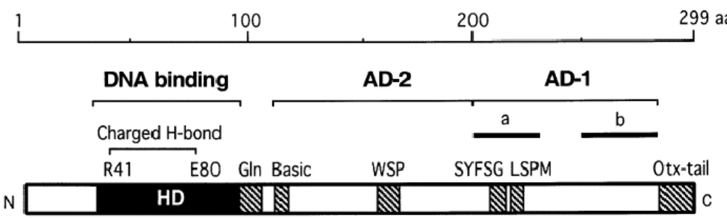
In OTX proteins, the homeodomain is followed by a glutamine rich region, a WSP domain, and, at the C-terminus, by a characteristic region called OTX-tail, generally repeated in tandem, first identified in CRX (Furukawa et al., 1997) (Fig. 3).
The homeodomain of OTX proteins is also involved in their nuclear localization: CRX nuclear localization signal (NLS) is localized in the homeodomain (Fei and Hughes, 2000), as well as, OTX2 NLS (Chatelain et al., 2006). Moreover, it has been demonstrated that the homeodomain is also involved in protein-protein interactions, as in the case of NRL-CRX cooperation (Mitton et al., 2000).
7 Fig. 3. Scheme of human CRX. HD: homeodomain; Gln: glutamine rich region; Basic: basic
region; AD-2/AD-1: transcriptional activation domains; for AD-1, sub-domains “a” and “b” are also shown. Filled boxes represent protein domains shared with OTX1 and OTX2. (Modified from Chen et al., 2002).
1.2.2- Otx genes in mammalian anterior development
The study of Drosophila gene homologues in Vertebrates has provided a large part of the knowledge of development regulating systems. Hox genes are Vertebrates homologues of Drosophila homeotic genes; they control Vertebrates axis specification and provide positional cues in the developing neural tube from hindbrain to tail (Hunt et al., 1991). The Drosophila homeobox gene orthodenticle is involved in fly anterior development (see above), and Drosophila otd sequence has been used to identify and clone
otd Vertebrate homologues Otx1 and Otx2 (Simeone et al., 1992; 1993).
The degree of similarity of mouse and fly homologous homeodomains is striking: mouse OTX1 and OTX2 homeodomains differ for 3 and 2 amino acid residues from OTD homeodomain, respectively. OTX1 and OTX2 homeodomains belong to paired-like K50 class, as well as OTD (see above),
in sharing lysine residue in position 50 (Boncinelli et al., 1993). Murine OTX1 and OTX2 share extensive sequence similarities, even though in OTX1, downstream of the homeodomain, these regions of homology to OTX2 are separated by stretches of additional amino acids containing repetitions of alanine and histidine (Simeone et al., 1993). Nevertheless, OTD and OTX
8 proteins are highly conserved only in the homeodomain; outside the homeodomain, homology is restricted to few short sequences. OTD lacks also the so-called OTX-tail, which is tandemly duplicated in all Vertebrates OTX (Williams and Holland, 1998).
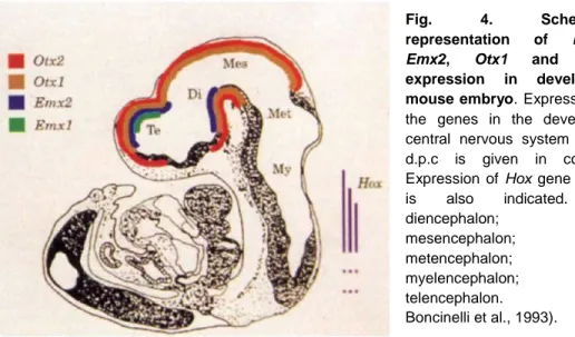
Together with two other Vertebrates homeobox genes, Emx1 and Emx2, the Vertebrate homologues of Drosophila empty spiracles, Otx genes are expressed in restricted regions of the developing mouse brain, including the cerebral cortex and olfactory bulbs (Boncinelli et al., 1993) (Fig. 4). These four genes have a role in establishing the limits and the identity of different brain regions of mouse, resembling, at a more anterior level, the functions of
Hox genes in the embryo posterior part. Otx genes are also expressed in
sense organ primordia, such as the olfactory epithelium, the developing inner ear and the developing eye, and they exploit a major role in the development of these structures (Boncinelli et al., 1993).
In mouse, Otx2 null embryos die early in embryogenesis, lack the rostral neuroectoderm fated to become forebrain, midbrain and rostral hindbrain, and show heavy abnormalities in their body plan (Acampora et al., 1995; Ang et al., 1996, Matsuo et al., 1995). Heterozygous Otx2 +/- embryos, into an appropriate genetic background, show defects in head structures, such as serious brain abnormalities and craniofacial malformations (Matsuo et al., 1995).
Otx1 null mice suffer from spontaneous epileptic seizures and exhibit
abnormalities that affect primarily the entire dorsal telencephalic cortex with a more pronounced effect in the temporal and perirhinal areas (Acampora et al., 1996; Weimann et al., 1999). The development of the visual and acoustic sense organs is also impaired, as the ciliary process in the eye and the lateral semicircular duct in the inner ear are lost (Acampora et al., 1996; Morsli et al., 1999).
Rescue experiments replacing lacking one Otx gene with the other (Acampora et al., 1998; Acampora and Simeone, 1999; Morsli et al., 1999), have shown an extended functional homology between OTX1 and OTX2,
9 and lead to argue that the most of the difference between the two Otx null mutants phenotypes stems from differences in the expression patterns of the two genes (Acampora et al., 1999a). The clearest exception to the overall
Otx functional equivalence is provided by the lateral semicircular canal of the
inner ear, that is never restored in mice replacing mutant Otx1 with human
Otx2 (Morsli et al., 1999). The same phenomenon is observed in mice
replacing Otx1 with otd; these findings suggest that the ability to specify lateral semicircular canal of the inner ear may be an Otx1 specific property (Acampora and Simeone, 1999).
A fundamental step of brain development involving Otx genes is the position of the isthmic organizer (IsO), a signaling center located at the mid-hindbrain boundary (Martinez et al., 1991) that expresses signaling molecules that refine and polarize neighbouring neural tissues (Meinhardt, 1983; Rubenstein et al., 1998). Otx2 plays a crucial role in the IsO positioning together with another homeobox gene, Gbx2. Otx2 defines the anterior fate of the neural plate, while Gbx2 appears to be the major molecular determinant of metencephalic identity (Bouillet et al., 1995; Chapman and Fig. 4. Schematic representation of Emx1,
Emx2, Otx1 and Otx2
expression in developing mouse embryo. Expression of
the genes in the developing central nervous system at 10 d.p.c is given in colours. Expression of Hox gene family
is also indicated. Di:
diencephalon; Mes: mesencephalon; Met: metencephalon; My: myelencephalon; Te: telencephalon. (From Boncinelli et al., 1993).
10 Rathjen, 1995; von Bubnoff et al., 1995; Wassarman et al., 1997). These two genes are essential for correct positioning of the IsO and they exploit this function through mutual repression (Broccoli et al., 1999; Millet et al., 1999; Simeone et al., 2000).
Otx1 and Otx2 are also required in a dose dependent manner for the normal
development of mouse eye. Both Otx1 and Otx2 mutant mice display consistent and profound ocular malformations, including lens, pigmented epithelium, neural retina and optic stalk defects; cell proliferation, differentiation and apoptotic death are severely affected (Martinez-Morales et al., 2001). Otx2 is essential for the development and maintenance of retinal pigmented epithelium (Martinez-Morales et al., 2001; 2003), and is also expressed in post-mitotic retinal neuroblast cells that have the potential to develop into various cell types, including ganglion cells, bipolar cells and photoreceptors (Bovolenta et al., 1997; Baas et al., 2000).
Another Otx-like homeobox gene has been isolated from mouse retina and named Crx: cone-rod homeobox containing gene. CRX is a photoreceptor specific transcription factor, playing a crucial role in their differentiation; its expression is restricted to the developing and mature photoreceptor cells. CRX binds and transactivates a specific sequence found upstream of several photoreceptor-specific genes, including the opsin genes of many species, and is essential for differentiation and maintenance of photoreceptor cells (Freund et al., 1997). Crx overexpression (obtained by retina retroviral transfection) causes an increase in the frequency of clones containing exclusively rods and a reduction of the frequency of clones containing other retinal cell types (amacrine and Müller glia cells). In addition, photoreceptor cells expressing a dominant negative form of Crx failed to form proper photoreceptors outer segments and terminals (Furukawa et al., 1997). Homozygous Crx knockout mice are blind at birth without any detectable photoreceptor function; their photoreceptors never develop the outer segment critical for phototransduction, and subsequently degenerate (Furukawa et al., 1999). Heterozygous Crx+/- mice have
11 normally functioning photoreceptors, but their development is delayed (Furukawa et al., 1999). In humans, Crx is expressed, together with Otx2, in all photoreceptors, from early specification trough adulthood and are important for regulating a wide range of photoreceptors specific genes (Chen et al., 1997; Furukawa et al., 1997; Nishida et al., 2003; Koike et al., 2007; Henning et al., 2008; Corbo et al., 2010; Omori et al., 2011). Mutations in
Crx, as well as in Otx2, are associated with several photoreceptor-specific
retinopathies: mutations in Otx2 or Crx can lead to Leber’s Congenital Amaurosis (LCA) (Freund et al., 1998; Jacobson et al., 1998; Sohocki et al., 1998; Swaroop et al., 1999; Rivolta et al., 2001; den Hollander et al., 2008; Henderson et al., 2009; Nicols et al., 2010). Mutations in Crx are also linked to progressive vision lost in Cone-Rod dystrophy (CORD) and Retinis Pigmentosa (RP) (Freund et al., 1997; 1998; Swain et al., 1997; Sohoki et al., 1998; Swaroop et al., 1999; Rivolta et al., 2001), whereas LCA-associated alleles of Otx2 are also LCA-associated with more severe diseases (Henderson et al., 2009). It has been demonstrated that Crx is a target of
Otx2, together with other Crx direct targets (Nishida et al., 2003; Henning et
al., 2007).
In Drosophila, the single otd gene plays multiple roles in photoreceptor morphogenesis and opsin gene regulation during eye development. OTX1, OTX2 and CRX have been tested for their ability to rescue otd function in fly rhabdomeric eye development. Each mammalian gene has been demonstrated to mediate a defined subset of otd-dependent functions, with
Otx2 and Crx mediating unique cell-specific functions, demonstrating that
during evolution OTX proteins have sub-functionalized (Terrell et al., 2012).
Crx is also expressed in the pineal gland and it is involved in regulating
pineal gene expression trough the interaction with a specific pineal regulatory element located upstream of pineal-specific genes, and it is important for circadian rhythm regulation (Li et al., 1998).
12
1.3- Otx genes and evolution
1.3.1- Otx genes in the animal kingdom
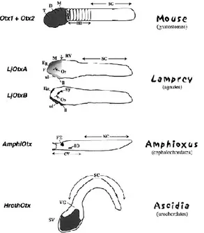
Otd/Otx related genes have been isolated from a wide range of organisms;
most of them, up to the Chordates, have only one Otx member, with few exceptions of duplications in independent lineages (Li et al., 1996; Umesono et al., 1999) (Fig. 5). An Otx related gene is present already in Cnidarians, primitive Metazoans with radial symmetry. In these organisms Otx function is associated with cell movements involved in axes formation rather than with head development (Smith et al., 1999). Rising up the evolutionary scale, Otx genes have been found in animals with primitive bilateral symmetry such as planarians (Stornaiuolo et al., 1998; Umesono et al., 1999). In these organisms Otx expression has been found in regenerating blastemas after transverse sectioning, with an asymmetric distribution: more abundant in regenerating head structures (Stornaiuolo et al., 1998). In planarians, Otx expression starts to be related with anterior patterning. Although not directly correlated with a defined anterior structure, the ancient function of Otx genes seems to deal with body axis patterning and with making tissues competent to respond to anteriorizing signals (Smith et al., 1999). In the nematode
C.elegans three members of the Otx class have been identified. These three
genes are involved in the development of thermo- and chemo-sensory neurons and, as well as Otx genes in mouse, their ablation gives rise to different mutant phenotypes affected in these neuronal populations. This variety of phenotypes could be caused by both divergent expression patterns and divergent protein functions (Lanjuin et al., 2003). The first case of head-associated Otx expression is found in Annelids, in the leech
Holobdella triserialis (Bruce and Shankland, 1998). Otx related genes have
been found in all Chordates, including Urochordates (Wada et al., 1996), Cephalochordates (Williams and Holland, 1996) and Agnathans (Ueki et al., 1998); in all these organisms they are expressed, as in flies, in the anterior
13 rostralmost part of the body and specifically in the CNS, independently from the complexity of these structures (Fig. 5). In Urochordates and Cephalochordates only one member of the Otx family has been isolated, and it is thought to correlate with Vertebrate Otx2 (Wada et al., 1996; Williams and Holland, 1998); indeed, in addition to amino acid sequence homology and similarity in expression patterns, those genes are expressed in endoderm cells during gastrulation, similar to Vertebrate Otx2. This suggests a primitive role of Otx2 in anterior endoderm to elicit signals specifying anterior neuroectoderm.
Another ancient Otx2 function has been proposed: a role in cell movements regulation. This idea is consistent with functional data in frog (Blitz and Cho, 1995; Pannese et al., 1995; Andreazzoli et al., 1997; Vignali et al., 2000) and mouse (Acampora et al., 1995; Matsuo et al., 1995; Ang et al., 1996) that suggest Otx2 involvement in cell movements occurring during gastrulation.
Fig. 5. Schematic representation of Otx-related gene expression (grey) in some representative Protochordates (Ascidia and Amphioxus) and Vertebrates (Lamprey and mouse). D: diencephalon; Ep: epiphysis; Ey: eye; F: forebrain; FE: frontal eye; HB: hindbrain; IO: infundibular organ; ll: lower lip; M: mid brain; MHB: mid-hindbrain boundary; NC:
nerve cord; Oe: olfactory
epithelium; Os: optic stalk; RV: rhomboencephalic vesicle; SC: spinal cord; SV: sensory vesicle; T: telencephalon; ul: upper lip; VG: visceral ganglion (Figure and caption from Acampora et al., 2001).
14
Otx2 sequences and expression patterns are quite conserved during
evolution from low Chordates to Vertebrates (Acampora et al., 2001 and references therein). The duplication event generating Otx1 branch from the ancestor Otx2 gene has occurred in gnathostome Vertebrates (Williams and Holland, 1998). This is coherent with Otx1 new function in specifying Gnathostomes specific structures (i.e. lateral semicircular canal of the inner ear). Otx1 genes evolve more rapidly than Otx2, as also shown by further duplications events occurred in both Xenopus (Kablar et al., 1996) and
zebrafish (Mori et al., 1994) and by the ratio of sequence divergence higher
than in Otx2 genes (Williams and Holland, 1998). These data are reinforced by notable changes in Otx1 expression patterns in different Vertebrates (Simeone et al., 1993; Mori et al., 1994), that underlie a rapid evolution of the regulatory elements as well. A particular case is that of lamprey: the lamprey genome has two Otx cognates LjOtxA and LjOtxB (Fig. 5). Phylogenetic analyses suggest that LjOtxA clusters with Gnathostomes Otx2 gene, while LjOtxB does not belong to either Otx1 or Otx2 lineages. Beside,
LjOtxB is not expressed in lamprey brain, but only in olfactory placode,
epiphysis, optic stalks and lower and upper lips, together with LjOtxA; moreover, LjOtxB is expressed in the eyes where LjOtxA is not detected (Fig. 5). Thus, Otx1 and Otx2 functions for the development of forebrain and midbrain in Gnathostomes appear to be shouldered by LjOtxA alone in lamprey. LjOtxB may have diverged from the stem of the Otx1 and Otx2 and it may have evolved independently (Ueki et al., 1998), with some weak similarity to Otx5/Crx lineage (see below).
Another Gnathostome Otx orthology class comprises Xenopus Otx5/5b, fish
Xotx5/Crx and the higly divergent Crx gene characterized in Mammals
(Plouhinec et al., 2003; Germot et al., 2001). Otx5 and Crx share highly specific expression domains: developing eye and epiphysis (Furukawa et al., 1997; Vignali et al., 2000). Such expression patterns substantially differ from the broad Otx1 and Otx2 expression areas, spanning the whole
15 prosencephalon and diencephalon. Genes of Crx orthology class may have been recruited for specific roles in photoreceptors development.
1.3.2- Otd/Otx: Insect and Vertebrate nervous system evolution
Until a few years ago it was widely assumed that Insects and Vertebrates nervous system had evolved independently (Garstand, 1928; Lacalli, 1994). This was due to their position at opposite side of the dorso-ventral body axis. Nevertheless, nowadays a common evolutionary origin is supported by several evidences. Two groups of homologues genes, Hom/Hox and
Otd/Otx, play crucial roles in the regional specification of the neuroectoderm
fated to form nerve cord/posterior brain and anterior brain, respectively. Many studies carried out in Drosophila have shown that Mammalian Hox genes could either partially rescue phenotypes due to mutation of their fly orthologues, or elicit responses similar to those of their endogenous counterparts when transiently overexpressed (Bachiller et al., 1994; Malicki et al., 1990; Zhao et al., 1993). On the other side Drosophila otd has been used to rescue either mouse Otx1 or Otx2 gene. Mice in which a full-coding
otd was introduced to replace Otx1 showed the rescue of several
abnormalities: brain size, as well as the thickness and cell number of the temporal and perirhinal cortices, that are reduced in Otx1 -/- mice, are very similar to wild-type (Acampora, 1998). Moreover, replacement of Otx1-/- by
otd leads to rescue of some sensory and sensory associated structures,
such as iris and ciliary process; on the contrary, lateral semicircular canal of the inner ear is never rescued (Fritzch et al., 1986; Torres and Giraldez, 1998), as it is the case when Otx1 is rescued with Otx2. This observation leads to the conclusion that the development of the lateral semicircular canal requires newly established properties specific of Otx1. Also mice in which
Otx2 has been substituted with otd show an almost partial rescue, providing
16 and OTX proteins are able to drive cephalic development possibly through the activation of genetic pathways conserved between Insects and Vertebrates, reinforcing the idea that the nervous systems of these two taxa are homologous structures sharing a common ancestor (Acampora and Simeone, 1999; Reichert and Simeone, 1999; Sharman and Brand, 1998).
Otd and Otx functions have been established in a common ancestor of fly
and mouse and retained during evolution; at the same time, copy number of
Otx genes and transcriptional/translational regulation have been modified by
evolution, leading to the specification of the more complex Vertebrates brain (Acampora and Simeone, 1999; Reichert and Simeone, 1999; Sharman and Brand, 1998). Otx gene duplication and modification of sequences and regulatory control may have contributed, from this point of view, to mammalian brain evolution (Boyl et al., 2001). Additional properties may have been acquired also by sequence divergences that endow the proteins with new specific abilities, as may be the case of Otx1 in the inner ear. As previously mentioned, sequence similarities between Otx and Otd genes are restricted to the homeodomain; it is possible that while the ability to recognize the same DNA targets by the homeodomain might be evolutionary conserved, beside this, murine Otx genes have acquired, outside the homeodomain, additional functional features that are different from those of
otd (Acampora et al., 2001). As mentioned above, rescue experiments on otd mutant flies using Vertebrate Otx genes, have shown that Otx1, Otx2
and Crx each mediate a defined subset of otd-dependent function in the fly eye, showing how OTX proteins have sub-functionalized during evolution (Terrell et al., 2012).
17
1.4- Xenopus laevis
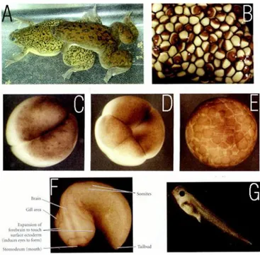
Xenopus laevis (Fig. 6) is a species of the genus Xenopus, that belongs to
the Pipidae family. It is widespread in sub-Saharan Africa. It is commonly known as South Africa clawed frog (or toad). It is an acquatic animal living in stagnant waters where it eats almost every kind of food, directing it into its toungless mouth using its front limbs. Its body is flat, with lidless eyes on the top of a small head. The hind legs have webbed feet with small claws on three toes, since the greek name Xenopus that means “strange foot”.
Fig. 6. Xenopus laevis. A: Mating frogs, the male
grasping the female around the belly and fertilizing the eggs as they are released. B: newly laid clutch of eggs. The brown area of each egg is the pigmented animal cap. The white spot in the middle of the pigment is where the egg nucleus resides. C: 2-cell embryo near the end of its first cleavage. D: An 8-cell embryo. E: Early blastula; the cells get smaller, but the volume of the egg remains the same. F: pre-hatching tadpole, as the protrusions of the forebrain begin to induce eyes to form. G: mature tadpole, having swum away from the egg mass and feeding independently. (modified from Gilbert, 2000)
18 Many years ago it was discovered that it could be used for human pregnancy tests; this led to the worldwide distribution of Xenopus laevis which turned out to be an ideal laboratory animal and from the ‘60 it is commonly used as a model system in many laboratories all over the world. As a model system it presents several advantages. For instance, eggs are large and embryos are suitable for microsurgical dissections; its development is rapid and in vitro fertilization is quite simple. However, there are some drawbacks: it is pseudotetraploid and it requires about 3 years to reach sexual maturity.
1.5- Otx genes in Xenopus laevis
In Xenopus laevis four members of the Otx class have been isolated and characterized: Xotx1 (Kablar et al., 1996), Xotx2 (Pannese et al., 1995; Blitz and Cho, 1995), Xotx4 (Kablar et al., 1996), and Xotx5/5b (Kuroda et al., 2000; Vignali et al., 2000). Xotx1 is a homologue of mouse Otx1, Xotx2 is a homologue of mouse Otx2, Xotx5/5b are homologues of mouse Crx; Xotx4 may be a derived copy of Xotx1.
1.5.1- Xotx1 (and Xotx4) expression profile
Xotx1 and Xotx4 have both been isolated during a Xenopus cDNA library
screening using murine Otx1 as probe (Kablar et al., 1996). The peptide sequence of the homeodomain is fully conserved between Xotx1, Xotx4 and mouse Otx1, but for a single amino acid change at position 18. Similarity between Xotx1 and Otx1 extends outside the homeodomain, where they share serine and histidine rich regions, Otx1 diagnostic characters, while
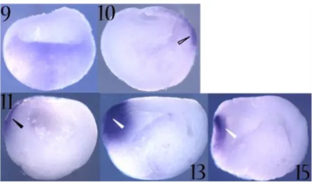
19 Fig. 8. Xotx1 expression as detected by in situ hybridization on whole embryos.
Embryos stages are
indicated. Up is
animal/dorsal; down is vegetative/ventral; right is posterior; left is anterior. Empty black arrowhead: dorsal ectoderm; white empty arrowhead: notoplate (anterior part); black arrow:
cement gland
anlage/cement gland; white arrow: anterior brain; grey arrow: otic vesicle; grey empty arrowhead: optic vesicle.
Xotx1 transcripts become visible by in situ hybridization (Fig. 7 and Fig. 8) at
stage 10 in the outer layer of the dorsal mesoderm; then, at stage 11, Xotx1 is detectable in the anterior neural plate; at early neurula stage (13/14) a strong expression is observed in the anterior neuroectoderm, within this area no labeling is detectable in the midline region, putatively corresponding to the anterior part of the notoplate. In the neuroectoderm, transcripts are Fig. 7. Xotx1 expression as detected by in situ hybridization on bisected embryos. Embryos
stages are indicated. Up is
animal/dorsal; down is
vegetative/ventral; right is
posterior; left is anterior. Empty black arrowhead: dorsal ectoderm;
black arrowhead: presumptive
anterior neuroectoderm; white
20 present in both the epithelial and sensorial layer. Moreover, transcripts are also detectable in the sensorial layer of the ectoderm at the level of the mesoderm-free zone where the cement gland and the stomodeal-hypophyseal anlagen will appear. At stage 18, Xotx1 is expressed in the prospective brain regions, but it never reaches the very anterior tip of the brain. This anterior expression persists till tailbud stage (st. 23). Weak labeling is also detectable in the prospective pigmented layer of the eye vescicle. At stage 33 brain expression persists, as well as the exclusion from the most anterior part; Xotx1 is also expressed in pigmented retinal layer, otic vesicles, olfactory placodes and, at a lower level, in a thin stripe of cells in the cement gland region. At a later stage (st. 37) cephalic expression persists (Kablar et al., 1996).
Xotx4 display a similar, although not perfectly superimposable expression
profile (Kablar et al., 1996).
1.5.2- Xotx2 expression profile
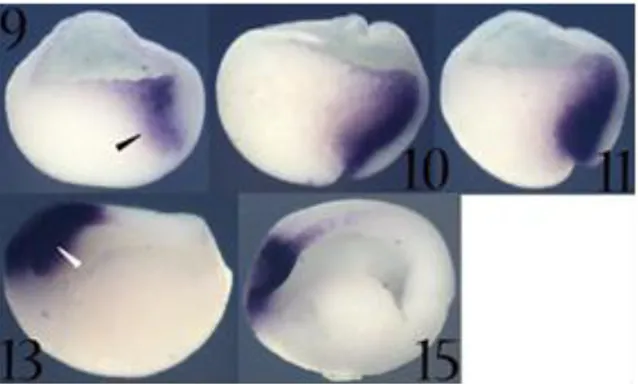
Xotx2 transcripts are primarily detected at stage 9 in the internal region of
the dorsal marginal zone, the future Spemann’s organizer region. At stage 10, the major expression site is in the migratory deep zone cells that are fated to give rise to prechordal mesendoderm (Keller et al., 1992). In addition it is also expressed in dorsal bottle cells. At stage 10.5, Xotx2 expression persists in these cell types and posteriorly, above the dorsal blastopore lip, this expression clearly respects the boundary between internal deep zone cells and external cell layer (boundary known as Brachet’s cleft) (Keller et al., 1992). Conversely, in the anterior region, Xotx2 expression extends to cells of the presumptive anterior neuroectoderm (Fig. 9 and Fig. 10).
21 Fig. 10. Xotx2 expression as detected by in situ hybridization on whole embryos.
Embryos stages are
indicated. Up is animal/dorsal; down is vegetative/ventral; right is posterior; left is anterior. Empty black arrowhead: dorsal migrating zone; white empty arrowhead: presumptive anterior neuroectoderm; black arrow: cement gland anlage/cement gland; white arrow: optic chiasma; grey empty arrowhead: optic vesicle; grey arrowhead: anterior neural tube/brain.
At neurula stage (st.14), expression is confined to mesoderm and ectoderm cells of anterior/dorsal regions, and to stomodeal-hypophyseal and cement gland anlagen. Xotx2 is not expressed in the region corresponding to the optic chiasma (Eagleson and Harris, 1990; Pannese et al., 1995). This expression profile persists till stage 18. At tailbud stage (st. 23) Xotx2 transcripts are present in the anterior part of the brain, excluding optic chiasma area, in whole eye vesicles, in cement gland and in forming
Fig. 9. Xotx2 expression as detected by in situ hybridization on bisected embryos. Embryos
stages are indicated. Up is
animal/dorsal; down is
vegetative/ventral; right is
posterior; left is anterior. Black arrowhead: migratory deep zone; white arrowhead: anterior neural plate.
22 olfactory placodes, where they persist during later phases of olfactory organ development. At stage 33 brain expression is still present, with the exception of chiasmatic region, and Xotx2 becomes detectable also in otic vesicle. This expression profile persists with no substantial variations till stage 37 (Kablar et al., 1996). (Fig. 9 and Fig. 10)
1.5.3- Xotx5/5b expression profile
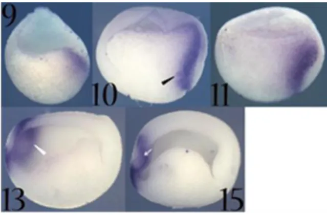
Xotx5 is initially transcribed at early gastrula stage (st. 10) in the dorsal
blastopore lip (Spemann’s organizer region). During subsequent gastrula stages this expression persists and intensifies; the major expression site corresponds to the migratory deep zone cells fated to give rise to prechordal mesoderm. At stage 10.5 Xotx5 expression clearly respects the boundary represented by the Brachet’s cleft; at this stage Xotx5 is also strongly transcribed in the dorsal ectoderm. During later gastrula stages expression disappears from the dorsal blastopore lip, while Xotx5 transcripts are still detectable in the anterior neuroectoderm, including the whole presumptive anterior neural plate. At early neurula stages (st. 13-14) Xotx5 expression intensifies in the anterior neural plate, but disappears from a central area corresponding to the presumptive retina and optic chiasma territories. At these stages Xotx5 transcripts are also detectable in a ventral anterior area corresponding to the cement gland anlage, where it persists until tailbud stage. At stage 17 a new expression site appears, corresponding to the epiphyseal anlage; this expression persists during epiphysis development. From stage 22 the expression in the cement gland anlage becomes weaker and disappears during subsequent developmental stages. Starting from stage 24, a new expression site appears in the eye region, where Xotx5 is transcribed in a small cluster of cells corresponding to the presumptive neural retina. At stage 27, Xotx5 is expressed in a broad dorsal region of the neural retina, where it persists till stage 30. At tadpole stage (st. 35) Xotx5
23 expression covers the entire eye region, except for the lens territories (Vignali e al., 2000) (Fig. 11 and Fig. 12).
Fig. 12. Xotx5
expression as detected by in situ hybridization on whole embryos.
Embryos stages are
indicated. Up is
animal/dorsal; down is vegetative/ventral; right is posterior; left is anterior. Empty black arrowhead: dorsal migrating zone; grey arrowhead: dorsal ectoderm; white arrow: optic chiasma; white empty arrowhead: presumptive anterior neuroectoderm; black arrow: cement gland anlage/cement gland; grey empty arrowhead: optic vesicle; black empty arrowhead: pineal gland.
Fig. 11. Xotx5 expression as detected by in situ
hybridization on bisected embryos. Embryos stages are
indicated. Up is animal/dorsal; down is vegetative/ventral; right is posterior; left is anterior.
Black arrowhead: migratory
deep zone; white arrowhead: anterior neural plate; white
arrow: presumptive optic
24
1.5.4- Xotx2 and Xotx5 expression profile in the developing eye
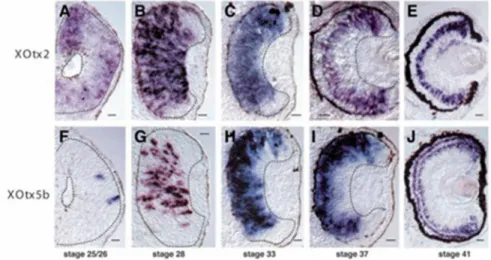
In Xenopus, Xotx2 and Xotx5 are expressed in different patterns during retinal histogenesis (Fig. 13). At stage 20, Xotx2 is expressed in the optic vesicles, while Xotx5 expression in the eye starts only at stage 25, when it is expressed in a small cluster of cells of the presumptive neural retina; at this stage Xotx2 is expressed throughout the presumptive retinal pigmented epithelium (RPE) and neural retina. A few hours later, at stage 28, Xotx2 expression has narrowed to the central retina and RPE, while Xotx5 has expanded. Starting from stage 31, during retinal cells differentiation, the two genes expression patterns seem almost superimposable and they are transcribed in a diffuse fashion throughout all retinal thickness.
At stage 33, the two retinal expression profiles are indistinguishable, in that they are distributed throughout the developing retina except in the most peripheral region, corresponding to the ciliary marginal zone (CMZ). Starting from stage 37 the two gene expression patterns become progressively restricted; at the level of the mature retina (stage 41) Xotx2 mRNA is
Fig. 13. In situ hybridisation on sections of XOtx2 (A-E) and XOtx5b (Xotx5) (F-J). Stages are indicated. Bars: 20 m; dashed lines indicate the extent of developing neural retina in A-D,F-I. (Figure and caption modified from Viczian et al., 2003)
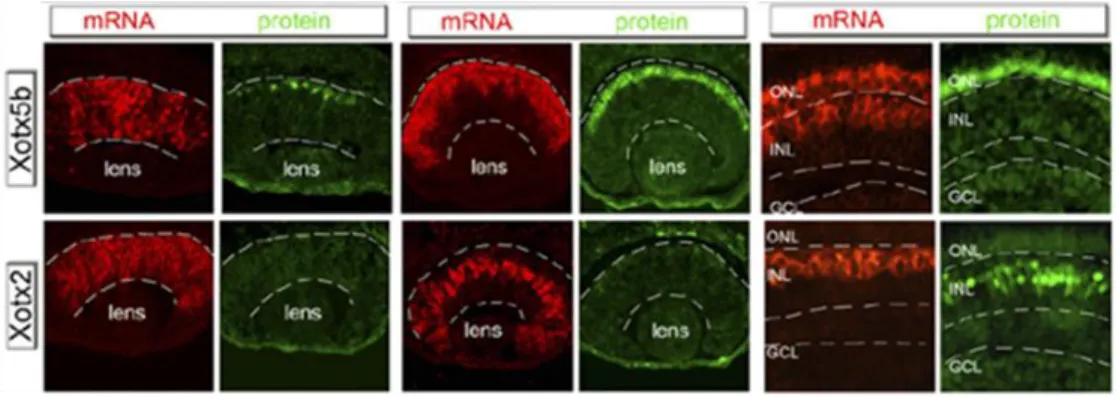
25 localized only in bipolar cells, while Xotx5 is transcribed only in photoreceptors and in a subset of bipolar cells (Viczian et al., 2003). Even more dramatic is the difference in the protein expression patterns: XOTX2 protein is detected only in bipolar cells, while XOTX5 is produced only in photoreceptors, due to precise translational control through the 3’UTR untranslated regions of their mRNA (Fig. 14) (Decembrini et al., 2006).
1.6- Xotx genes and the Organizer
Neural axial patterning in Vertebrates is the result of inductive events, in which dorsal mesoderm plays a crucial role (Spemann, 1938). Dorsal mesoderm, forming blastopore lip, is called the Organizer because of its ability to recruit cells to form axial structures. The observation that early dorsal lips are able to induce secondary heads, while late dorsal lip transplantations give rise to ectopic posterior structures, led to a distinction between a head and a trunk organizer (Spemman, 1938; Hamburger, 1988). The Organizer itself is induced from signals coming from embryo
dorso-Fig. 14. Translation of the Xenopus Homeobox Xotx5b (Xotx5), and Xotx2 mRNAs.
In situ hybridization of Xotx2 and Xotx5b (Xotx5) compared to immunostaining of the
corresponding proteins on embryonic retinas sections at st. 34 (mid-neurogenesis), st. 37 (late-neurogenesis), and st. 42 (mature embryonic retina). Magnification of central retinal aspect); GCL: ganglion cell layer, INL: inner nuclear layer, ONL: outer nuclear layer. (Figure and caption adapted from Decembrini et al., 2006)
26 vegetal cells forming the Nieuwkoop center (Nieuwkoop, 1973), and becomes able to induce animal pole ectoderm toward a neural fate, contemporarily establishing its antero-posterior pattern. The neuroectoderm is initially specified as anterior (activation step) and only later posterior neural structures are specified from anterior neuroectoderm (transformation step) (Nieuwkoop, 1952). Regions that will give rise to the head do not undergo convergent extention movements typical of the trunk- and tail-forming regions that are responsible for their elongation.
A number of evidences suggest that Xotx genes are involved in head-organizing activity (Pannese et al., 1995; Blitz and Cho, 1995; Kablar et al., 1996; Andreazzoli et al., 1997; Morgan et al., 1999; Vignali et al., 2000). First of all a common feature of Xotx genes is that their early expression patterns correspond to presumptive head regions (Kablar et al., 1996; Pannese et al., 1995; Vignali et al., 2000) that do not undergo convergent extention movements (see above: Xotx expression patterns); a number of evidences demonstrated that XOTX proteins play a role in specifying anterior structures, rather than being mere positional markers.
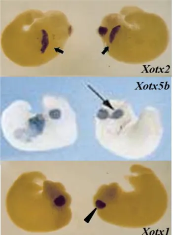
Gain of function experiments have shown that Xotx2 microinjection results in a delay in gastrulation movements and in a failure of blastopore lip closure. In embryos showing these gastrulation alterations trunk and tail fail to develop properly, the size of these structures is considerably reduced and the embryonic axis is bent dorsally (Pannese et al., 1995). In addition, several embryos show ectopic cement gland formation, as well as the presence of neural tissue in ectopic positions (Fig. 15) (Pannese et al., 1995).
In embryos treated with UV light or retinoic acid (RA) Xotx2 expression is strongly inhibited, as well as the development of anterior structures, suggesting a direct correlation between this two phenomena and implicating a fundamental role for Xotx2 in anterior structures development. This also suggests a role of Xotx2 as an important intermediary between the first positional specification mediated by the cortical rotation originated by sperm
27 entry (Gerhart et al., 1989) and the establishment of anteroposterior axis (Pannese et al., 1995).
Xotx2 microinjection significantly increases goosecoid expression, suggesting that the regulation of goosecoid expression is among the functions of Xotx2 regulatory action during early development, and
goosecoid has been suggested to play a role in executing Spemann’s
organizer phenomenon (Cho et al., 1991), thus suggesting a crucial role for
Xotx2 in mediating dorsal blastopore lip activities (Pannese et al., 1995).
It has been shown that Xotx2 prevents cells that express it from participating in the convergent extention movements that shape the posterior part of the body. Xotx2 exerts this function by directly activating XclpH3, Xenopus
Fig. 15. Xenopus laevis embryos injected with
Xotx2, Xotx1 and Xotx5b
(Xotx5) mRNA. Xotx1, Xotx2 and Xotx5b (Xotx5)
mRNA microinjection all lead to embryos with typical posterior defects, but only
Xotx2 and Xotx5b (Xotx5)
can induce ectopic cement gland formation. Black bold
and thin arrow: ectopic
cement gland; black
arrowhead: cement gland.
(Figures adapted from
Andreazzoli et al., 1997; Vignali et al., 2000).
28 homologue of Mammalian calponin; the product of this gene binds both actin and myosin filaments preventing the generation of contractile force and thereby the generation of movements (Morgan et al., 1999).
Phenotypes shown by Xotx1 injected embryos strongly resemble those obtained after Xotx2 microinjection (Fig. 15) and analyses performed on exogastrulae have clearly shown that Xotx1, as well as Xotx2, inhibits convergent extention movements essential for trunk and tail formation (Andreazzoli et al., 1997). It has been hypothesized that Xotx1 might inhibit convergent extention movements acting on cell adhesion molecules, and that this activity could be either direct or mediated by Otx1 repression of other genes like Xbra, Pintavillas and Xnot, that are expressed in trunk and tail cells that undergo mediolateral intercalation movements (Andreazzoli et al., 1997). A major difference between Xotx1 and Xotx2 overexpression phenotypes is the presence of ectopic cement glands that is never detected in Xotx1 injected embryos (Andreazzoli et al., 1997). Moreover, einsteck experiments performed on injected embryos have shown that Xotx2 is able to convert a tail organizer into a head organizer, while Xotx1 seems able only to inhibit the tail-organizing activity of the late blastopore lip, leading to the development of non-posterior bulging structures (swollen vesicles not showing typical tail features), without turning a tail into a head organizer (Andreazzoli et al., 1997). A possible interpretation of these data is that
Xotx1 could be able only to inhibit convergent extension movements, and
this should be sufficient only to repress tail-inducing activity. On the other hand, Xotx2 may also be able to act as specific anteriorizing factor reconstituting a head organizing activity. This hypothesis is consistent with
Xotx2 capability to induce ectopic cement gland, differently from Xotx1
(Andreazzoli et al., 1997).
Both Xotx1 and Xotx2 are activated by the injection of siamois, noggin and
Xwnt-8, factors able to trigger head organizer induction, and are repressed
by posteriorizing agent such as RA, suggesting, again, an involvement of both in anteriorizing activity (Andreazzoli et al., 1997 and reference therein).
29
Xotx5 microinjected embryos fully resemble Xotx2 over-expression
phenotype (Fig. 15): failure of blastopore proper closure, posterior defects and ectopic cement glands formation (Vignali et al., 2000). Moreover, Xotx5, as well as Xotx2, is able to turn a tail organizer into a head organizer (Vignali et al., 2000). Ectopic neural tissue is also detected in injected embryos (Vignali et al., 2000). Further analyses have shown that the induction of ectopic neural tissue may not reflect a direct effect of Xotx5 within the ectoderm, since in animal cap experiments Xotx5 is weakly able, if at all, to trigger neural genes (sox-2, nrp-1) expression (Vignali et al., 2000); however, Xotx5 may play a role in neural induction somehow sensitizing the anterior dorsal ectoderm towards a neural fate, possibly suppressing the ectodermal fate. On the other hand, Xotx2 activates general neural and anterior neural markers in isolated ventral ectoderm (Gammill and Sive, 1997; 2001). These differences may be due to objective differences in proteins functions, or may be due to different experimental approaches or to different dosages used for the two transcripts (see Gammill and Sive, 2001). The induction of ectopic cement gland is instead the result of a direct action of Xotx2, and maybe of Xotx5, within the ectoderm (Vignali et al., 2000). In addition, Xotx2 efficiently prevents the expression of posterior neural and ventral markers, suggesting that part of the mechanism through which it promotes anterior fate is to repress formation of non-anterior positions (Gammill and Sive, 2001). These data suggest that Xotx2 and Xotx5 may perform cooperative roles during early embryogenesis, while they show divergent expression patterns (see above) and different functions (see below) during later developmental stages (Vignali et al., 2000).
Relative size of body regions allocated in early embryogenesis for the development of head and trunk structures are altered in Xotx microinjected embryos. Regions specified for presumptive head structures are slightly expanded at the expense of those giving rise to trunk and tail structures. At the same time, reduced trunk and tail structures result from the interference
30 with convergent extension movements taking place during gastrulation and neurulation and giving rise to more posterior regions (Pannese et al., 1995).
1.7- Xotx genes in the developing retina: bipolar versus photoreceptor fate
Vertebrate retina is a highly specialized sensorial tissue whose correct functions depend on the development of its complex cytoarchitecture. This tissue is made up of six different neuronal cell types: bipolar, horizontal, amacrine and ganglion cells and two types of photoreceptors (cones and rods). Besides, a single type of glia cells (Müller glia cells) is present. All these cell types arise from the same retinal multipotent neuroblast cell population and the specification of different neuronal cell types follows a precise temporary and spatially order. The competence model proposes that progenitor cells pass through a series of competence states, during each of which the progenitors are competent to produce a subset of retinal cell types (Livesey and Cepko, 2001). Competence states seem to be intrinsically defined and thus cell fate choices are intrinsically regulated through the definition of progenitor competence. Within a given competence state, the generation of a particular type of cell is regulated by positive and negative extrinsic signals (Livesey and Cepko, 2001). It has been demonstrated that bHLH factors are essential in promoting retinal neurogenesis, but additional factors, like homeodomain containing transcription factors are also crucial in specifying the different cell types (Hatakeyma and Kageyama, 2004 and references therein).
In Xenopus laevis, it has been demonstrated that Xotx2 and Xotx5 are responsible for bipolar and photoreceptor differentiation respectively (Viczian et al., 2003). Coherently with their expression pattern in the eye (see above), lipofections of multipotent retinal progenitors cells with constitutively expressed Xotx2 and Xotx5 cDNA, lacking 3’ UTR regulating elements,
31 showed dramatically different effects: Xotx2 drives them to a bipolar fate, while Xotx5 promotes a photoreceptor fate (Fig. 16) (Viczian et al., 2003; Wang et al., 2005; Decembrini et al., 2006).
Xotx2 suppresses Xotx5 photoreceptor inducing action, and the
co-lipofection of Xotx2 with Xotx5 overrides the latter’s ability to promote photoreceptor fate and the combination drives cells towards bipolar fates (Viczian et al., 2003). A small divergent region confers to the two homeoprotein their differential activities in Xenopus developing retina. This region has been called retinal specificity box (RS box) and spans amino acid residues 100-109 of XOTX2 and 100-107 of XOTX5 (Fig. 17). The RS box lies directly carboxyterminal to the homeodomain, and the two proteins differ for 6 amino acid residues at this level (Onorati et al., 2007). Swap domain experiments have shown that the RS box is necessary and sufficient to confer to the two transcription factors their specific retinal actions (Onorati et al., 2007). Significantly, deletion of the RS box completely abrogates any cell fate activity of both Xotx, while the insertion of the RS box into Drosophila
otd, which has no cell fate activity in the frog retina, endows it with the retinal
activity of either Xotx2 or Xotx5, suggesting that in the absence of the RS Fig.16. Overexpression of
Xotx5b (Xotx5) or Xotx2 in
developing Xenopus laevis
retinoblast. (A) Colipofection of
GFP and pCS2 vector in the Xenopus retinae. A diversity of
retinal cell types express the fluorescent marker. (B) Retinae co-lipofected with Xotx5b (Xotx5) and
GFP show an increase in
lipofected photoreceptor cells. (C) Retinae co-lipofected with Xotx2 and GFP vector show an increase in the number of lipofected bipolar cells. (D,E) The graphs show an
average of the percentages
obtained (Figure and caption
32 box OTD fails to properly target the gene sets normally activated by either XOTX2 or XOTX5. Moreover the grater ability of XOTX5, compared to XOTX2, to synergize with Xenopus NRL to activate the rhodopsin promoter is also switched depending on this box. OTD protein is also able to interact with NRL, demonstrating that the RSbox is not essential for this interaction, but it may modulate this contact in a way that is consistent with the roles of XOTX2 and XOTX5 in frog retinogenesis (Onorati et al., 2007). These data provide strong evidence on how closely related homeodomain factors differentiate their functions to regulate distinct cell fates. To explain RS box capability to modulate XOTX2 and XOTX5 retinal actions two not mutually exclusive possibilities have been proposed: the box refines the DNA binding ability of XOTX proteins towards different sets of promoters, or it modulates interactions with different specific molecular partners of either XOTX protein.
1.8- Xotx genes and cement gland induction
Cement gland (CG) (or adhesive organ) is a mucus-secreting organ, localized at the extreme anterior of the Xenopus embryo where embryonic ectoderm and endoderm contact each other, without mesoderm interposition (Fig. 18); this region corresponds to the chin primordium of Mammals. In all Deuterostomes this region will give rise to the stomodeum (primitive mouth). The adhesive organ secrets a waterproof glue that attaches the newly hatched embryo to a solid support, in a phase when it can swim only poorly
Fig. 17. XOTX2/5b and XOTX5 RS box. On the left are schematics of the two XOTX; on
the right are their sequences in the region directly downstream of the homeodomain (HD), with different colors shading the XOTX2 (red) and XOTX5b (XOTX5) (yellow) sequences. Lines are introduced for sequence alignment. The divergent region responsible for the different retinal activities of XOTX2 and XOTX5b (XOTX5) (RS box) is shown in the blue frame. (Figure adapted from Onorati et al., 2007)
33 and cannot feed autonomously. It is innervated by the mandibular branch of the trigeminal nerve, whose neurites make the cement gland work as a sensory device that mediates the “stopping response”, to keep the embryo from moving after it is safety attached by glue (Boothby and Roberts, 1992 a, b; Davies et al., 1982; Roberts and Blight, 1975). This mechanism saves energy and makes embryos less obvious to predators.
Fig. 18. The Xenopus cement gland forming region. The Xenopus cement
gland (CG; shaded area) forms from the outer layer of ectoderm that overlies the endoderm, in the mesoderm-free area at the anterior of the embryo. This region lies between the dorsal neural plate and ventral epidermis. Outer ectodermal layer (dark blue), inner ectodermal layer (light blue), mesoderm (red) and endoderm (yellow) are indicated. (Figure and caption adapted fromWardle and Sive, 2003).
CG arises from the outer or epithelial layer of the ectoderm (Drysdale and Elinson, 1992; Nieuwkoop and Faber, 1967); this layer also gives rise to epidermis, hatching gland, certain neurons and to the ependymal lining of the neural tube.
CG primordium becomes morphologically visible at the onset of neurulation, when a patch of cells, lying anterior to the neural folds, becomes more darkly pigmented than the surrounding tissue. As the neural tube closes, this organ is differentiated and begins secreting mucus before the embryo hatches. Completely differentiated adhesive organ consists in a pseudostratificated columnar epithelium, made up by very polarized cells containing mucus and maternal pigment granules (Sive and Bradley, 1996 and reference therein); these cells form a cone shape structure with an oval apex. During successive phases of Xenopus development, CG cells decrease in height and become vacuolated to disappear about at stage 45 (Van Evercooren and Picard, 1978). CG degeneration is coordinated with the opening of the mouth and initiation of feeding.