Alma Mater Studiorum
Alma Mater Studiorum –
– Università di Bologna
Università di Bologna
DOTTORATO DI RICERCA IN
Scienze Mediche Specialistiche
Ciclo XXVIII
Settore Concorsuale di afferenza: 06/D1 Settore Scientifico disciplinare: MED11
TITOLO TESI
EFFECTS OF PERCUTANEOUS MITRAL VALVE REPAIR WITH MITRACLIP ON CLINICAL STATUS, VENTRICULAR REMODELING AND NEUROHORMONAL PROFILE IN PATIENTS WITH ADVANCED HEART FAILURE AND SIGNIFICANT FUNCTIONAL MITRAL REGURGITATION
Presentata da: Dott.ssa Berardini Alessandra
Coordinatore Dottorato
Relatore
Prof. Roberto Di Bartolomeo
Prof. Claudio Rapezzi
Esame finale anno 2016
TABLE OF CONTENTS INTRODUCTION……….………3 AIM OF THE STUDY………10 METHODS……….……10 Patient selection………..………….10 Echocardiographic examination………..…..11 Device and procedure……….………...11 Study objectives and follow-up……….…12 Statistical analysis………..…..13 RESULTS………13 Baseline characteristic of patient population……….…………13 Acute results………..………13 Six-month outcome………..……….14 LV remodeling………..………..………..14 DISCUSSION..………..………15 CONCLUSIONS……….……..17 TABLES………..………..18 FIGURES……….…..22 REFERENCES………..24
INTRODUCTION Mitral valve regurgitation (MR) is the second most frequent valve disease requiring invasive treatment in the Western world1. Significant functional mitral regurgitation (FMR) can be found in around 30% of the patients with previous myocardial infarction (MI) and in 35% of the patients with chronic heart failure (HF). Data from many observational studies showed a twofold mortaliy rate in advanced HF with severe MR2-4. MR can derive from anatomo-structural or functional changes of the components of the valve apparatus. FMR is due to changes of the valve apparatus following left ventricle (LV) remodeling 5-7. Etiopathogenesis of FMR can be classified as “ischemic” or “non ischemic”. In the former, the major determinant of MR is valve tethering, i.e. the downward strecthing of both valve leaflets due to LV dilatation and increase of sfericity index2. These two factors cause the apical displacement of papillary muscles and the lateral displacement of closure forces acting on the leaflets. As a consequence, there is incomplete coaptation of mitral valve leaflets and MR. Chronic LV volume overload causes an increase of LV wall stress, that in turn cause a progressive reduction of ventricular contractility and annular closure forces, further increasing MR volume. The most important mechanism of ischemic FMR is tethering of a single valve leaflet, which is caused by displacement of the papillary muscle located in the infarcted zone, where LV remodeling occurs8. Independently from etiology, LV remodeling secondary to chronic volume overload is directly responsible of FMR and its progression, because of the vicious circle between volume overload and LV remodeling (figure 1).
Figure 1: Etiology of functional mitral regurgitation Surgical therapy of MR in advanced HF is still object of controversy. It has been shown that, when irreversible LV remodeling and pulmonary hypertension occur, reduction of MR grade is not associated with any benefit 6,9. A number of studies identified reduced LV ejection fraction (EF) as an independent negative prognostic factor following mitral valve surgery6,10. On the other hand, there is evidence showing that treatment of MR in patients with severe LV dysfunction is associated with a significative reverse LV remodeling (increase of EF, reduction of telesystolic and telediastolic volumes and sfericity index), improvement of functional class and quality of life6. Yet, patients with advanced HF and extreme LV remodeling exhibit very high peri- and post-operative mortalty and, for many of these patients, surgical risk is deemed prohibitive. Percutaneous mitral valve repair represent a therapeutic option in alternative to conventional cardiac surgery in a selected patient population, comprising mainly subjects who are not candidate to cardiac surgery because of a too high or prohibitive surgical risk, caused by comorbidity, advanced age or severe LV dysfunction. Based on these premises, several devices for transcatheter treatment of MR have been developed and are under investigation. All these devices aim at reproducing conventional surgical techniques for mitral valve repair: edge-to-edge technique, indirect anuloplasty using a device positioned in the coronary sinus, direct anuloplasty, LV remodeling with transventricular or transatrial devices, mitral valve replacement. Presently, the
only percutaneous device with CE market approval is the MitraClip system (Abbott Vascular, Abbott Park, Illinois, USA). The Mitraclip procedure consists in positioning a clip which captures both valve leaflets at the level of mitral regurgitation. This generate a new permanent coaptation point between the two leaflets and valve opening with a double orifice, determining reduction of MR grade similarly with the surgical edge-to-edge technique first described by Alfieri11. Positioning and release of the device is performed, under general anesthesia and transoesophageal echocardiography guide, through a catheter introduced in the right femoral vein and reaching the valve through the interatrial septum. To date, more than 10.000 percutaneous mitral valve repair procedures with the Mitraclip system have been performed worldwide. The first clinical data come from the North-American EVEREST trials 12,13, followed by several clinical European registries that have been launched after CE market approval. Data from EVEREST I and II trials showed feasibility and safety of percutaneous mitral valve repair with the Mitraclip system, which was associated with a low mortality and morbility rate in the perioperative phase and during follow-up12,13. Subsequently, the results of the randomized controlled EVEREST trial have been reported 14. This trial compared in a randomized 2:1 fashion 184 patients treated with the Mitraclip system with 95 patients undergoing surgical MV repair. All patients had MR grade ≥ 3+ and were eligible to both treatments. The study reported a significant reduction of adverse events in patients receiving the MitraClip procedure (15%) vs. those who underwent surgery (48%, p< 0.0001), whereas procedural success and long-term efficacy in terms of reduction of MR grade to a degree ≤ 2+ were more commonly achieved in surgical patients (73%) in comparison with the percutaneous treatment arm (55%). Inverse LV remodeling has been observed in both treatment arms, whilst in the Mitraclip group there was a better recover of functional class and quality of life. Data from the “real world” 15-18 demonstrate that patients treated with the Mitraclip procedure have a higher risk in comparison with patients enrolled in the EVEREST study, showing that this procedure is feasible and safe in patients that would have been excluded based on the strict enrollment criteria of the trial. In all real world series, patients who underwent the Mitraclip procedure had predominantly functional rather than degenerative MR, with reduced LV function and a higher surgical risk profile. Based on available evidence, current indications for Mitraclip MV repair are the presence of symptomatic MR grade ≥3+, of degenerative or functional etiology, in patients with very high of prohibitive surgical risk. Surgical risk assessment is complex, and requires a multidisciplinary
sonographers and interventional cardiologists. The heart team express an opinion on feasibility, risks and expected benefits of both procedures, surgical and percutaneous, in terms of NYHA functional class and quality of life improvement, and prognosis quoad vitam. As far as the Mitraclip procedure is concerned, clinical evaluation and meticolous echocardiographic assessment are key to perform a safe and effective procedure. In particular, trans-esophageal ecocardiography (TEE) is necessary to precisely characterize specific anatomic factors associated with procedural feasibility and technical success (figure 2). Figure 2: Anatomic criteria evaluated during screening trans-esophageal echocardiography in patients who are candidate to percutaneous mitral valve repair with the MitraClip system. A: flail gap (< 10 mm), defined as the maximal distance between the leaflet prolaxing in the left atrium (LA) and the leaflet on the annular plane, measured in 4- or 5-chamber long-axis view, where the gap is larger. B: flail width (< 15 mm), measured in short-axis view where the lesion width is larger. C: coaptation depth (< 11 mm), evaluated in 4-chamber or LV outflow tract view. D: coaptation lenght (≥ 2 mm) evaluated in 4-chamber or LV outflow tract view. Ao, aorta; LA, left atrium; LV, left ventricle. The MitraClip system (figure 3) consists of a triassial delivery catheter and an implantable clip. The clip delivery system has a proximal part with a 24F diameter and e distal 22F portion. The clip has two arms, is made of chromo/cobalt alloy with a polyesther coating, pre-assembled at the distal tip of the catheter. Orientation, positioning, opening, leaflets’ capture and release of the clip are controlled by specific handles located on the external portion of the catheter, which is fixed on an
external sterile metallic support located on a small table at the level of the patient’s legs. Figure 3: MitraClip device. The procedure is performed in a hybrid operative room or in the catheterization laboratory under general anesthesia, with fluoroscopic and TEE guidance. The right femoral vein is cannulated, and a radial artery is commonly used for invasive pressure monitoring. Trans-septal puncture is then performed under TEE guidance to access the left atrium. Trans-septal puncture must be located in the postero-superior portion of the fossa ovalis, to allow the correct positioning of the clip delivery system toward the MV orifice. The punctute must be between 35 and 45 mm above the mital valve annulus, in order to have enough space for the clip to be correctly oriented with respect to the annular plane and the jet of regurgitation (Figure 4).
Figure 4. Trans-esophageal echocardiography monitoring during trans-septal puncture, at the level of the postero-superior portion of the interatrial septum, at a distance from the mitral valve annulus plane around 35-40 mm. A: visualization of the tenting of the interatrial septum caused by the Mullins catheter and the Brockenbrough needle pushed from the right atrium, in medio-esophageal short-axis view (45-60°), for a correct puncture location in the antero-posterior plane. B: tenting visualization in bicaval projection at 90°, for a correct location of the needle in the infero-superior plane. C: measurement of the distance between the tenting on the left portion of the interatrial septum and the mitral valve plane in 4-chamber view at 0°. All these manouvres are visualized and guided by TEE. For example, trans-septal puncture is guided through the visualization of the “tenting” zone, which is the septal deformation caused by the Mullins catheter pushed in the fossa ovalis. TEE also allows the visualization of the guiding catheter, the monitoring of clip insertion in the left atrium, avoiding trauma and perforation of the atrial walls or the appendage, and finally guide the orientation of the clip perpendicularly to the mitral valve annulus, centered in the direction of the regurgitant jet. The right clip trajectory and its perpendicularity with the rim of coaptation of the valve leaflets is constantly verified through a number of projections which are perpendicular to each other. Tridimensional echocardiography, if available, represents a valid support for the operators, mainly for the clip orientation, allowing an immediate visualization of the device in relationship with copatation rim (Figure 5).
Figure 5: Tridimensional, real time trans-esophageal echocardiography images acquired during clip orienting on the mitral valve plane. A: incorrect, too anterior, position of the clip delivery system. B: correct position, central and perpendicular to the coaptation rim. C: clip orientation not perpendicular to the coaptation rim (dotted red line; the dotted green line indicates the correct orientation). D: correct clip orientation on the coaptation plane, along the green line. When the device is correctly oriented, it can be advanced in the left ventricle and retrieved to capture both valve leaflets (grasping). If a double orifice has been created and echocardiography confirms the effective reduction of MR grade and the perfect inserction of the valve leaflets within the clip, clip arms are completely closed and locked and the clip is released. If the position of the clip is not satisfactory or the grasping is not complete or not effective in reducing MR, before the final release the clip can be re-opened and repositioned. When necessary, a second or a third clip can be implanted to optimize procedural results, providing a series of additional echocardiographic criteria are satisfied. In general, if no complications occur, the patient is estubated immediately after the procedure.
AIM OF THE STUDY Significant functional mitral regurgitation (FMR) is common in heart failure (HF) patients with increased LV volumes and depressed LV ejection fraction (EF) and has been associated with a ominous prognosis5,19. Additionally, perioperative mortality after surgery for FMR is not negligible, and a large number of patients with FMR are judged inoperable or at high surgical risk because of severe LV dysfunction and/or comorbidities4,20. Percutaneous mitral valve repair (PMVR) with the MitraClipTM device (Abbott, Abbott Park, IL, USA) has recently evolved as a therapeutic alternative for patients with significant MR, of both degenerative and functional origin, whose surgical risk is considered very high or prohibitive14,16,21. Although less effective than surgery in reducing mitral regurgitation, PMVR has been demonstrated to be safe and capable of leading to significant improvement in clinical outcomes. Additionally, reverse cardiac remodeling at mid-term follow-up has been reported after successful PMVR in patients with left ventricular dysfunction 17,22,23. However, few data are currently available on ventricular and neurohormonal changes in patients with FMR due to severe LV dysfunction and refractory, advanced heart failure, otherwise destined to heart transplant or death 24. The aim of this study was to assess and compare the effects of PMVR therapy on clinical outcomes, LV remodeling and neurohormonal changes in inoperable end-stage patients with chronically symptomatic FMR despite optical medical / electrical therapy and severe LV dysfunction. METHODS Patient selection We analyzed the clinical and echocardiographic data of a consecutive cohort of patients with advanced heart failure and moderate to severe FMR who underwent PMVR therapy for significant symptoms not responding to optimal medical therapy and CRT. All patients had been judged inoperable by the local heart team on the basis of extreme cardiac remodeling and / or the presence of several comorbidities. Candidates for PMVR were selected among patients followed at our advanced heart failure department, within a comprehensive program which includes CRT, left ventricular assist device and heart transplant. When applicable, CRT had to be performed first, and PMVR was considered only after ≥ 6 months of CRT with persisting New York Heart Association
(NYHA) functional class III or IV despite pharmacological optimization22. All potential candidates underwent transthoracic (TTE) and transesophageal Doppler echocardiography (TEE). Anatomic feasibility for PMVR was largely based on the Endovascular Valve Edge-to-Edge REpair Study (EVEREST) criteria for FMR. Surgical risk was estimated using the Society of Thoracic Surgeons (STS) score and the logistic EuroSCORE (European System for Cardiac Operative Risk Evaluation), although additional clinical and instrumental parameter were taken into account. The decision whether to proceed with MitraClip implantation was always taken by a multidisciplinary team composed by clinical cardiologist, interventional cardiologist, expert echocardiographer, cardiac surgeon and cardiac anesthesiologist. In particular, patients with criteria beyond the EVEREST recommendations were the object of an in depth collegial discussion13. Echocardiographic examination The severity of FMR at baseline was graded according to the American Society of Echocardiography guidelines 25 as: mild 1+, moderate 2+, moderate-to-severe 3+ and severe 4+. In addition, the vena contracta width at the narrowest portion of the regurgitant jet was measured 25. Measurement of LV volumes and EF were performed according to the biplane Simpson’s method 26. The mitral valve orifice area, when feasible, was assessed using the pressure half-time method 27. Systolic pulmonary artery pressure was measured using the gradient derived from the maximal velocity of tricuspid regurgitation, adding 5 mmHg if the inferior vena cava had a normal diameter (<21mm), 10 mmHg if the vena cava was dilated with reduced respiratory excursions, 15 mmHg when the vena cava was dilated without respiratory excursions . Right ventricular dimensions and contractile function were also evaluated according to standardized criteria28. Device and procedure The MitraClip system has been previously described in details 13,14. Briefly, it is a catheter based system designed to perform a double orifice repair of the mitral valve. The system includes a clip, a steerable guide catheter, and a clip delivery system that enables positioning and placement of the clip on the mitral valve leaflets, resulting in permanent leaflet approximation. The procedure was performed under general anesthesia under TEE and fluoroscopic guidance. Live real-time 3-D echocardiography was used to improve the conduct of the implantation. When necessary, more than a clip was implanted. Procedural success was defined as the implantation of at least 1 clip
Study objectives and follow-up The aim of this study was to assess the effect of PMVR on cardiac remodeling, clinical status and neurohormonal profile (or BNP plasma levels measurement) at 6 months after procedure in patients with severe FMR and severe LV dysfunction for whom risk of surgical repair/replacement of the mitral valve was prohibitive. Clinical evaluation was performed before MitraClip treatment, after 30 days and 6 months, or more frequently if clinically-indicated. We assessed the incidence of death, myocardial infarction, stroke, conversion to conventional surgical mitral valve replacement and re-admission for heart failure. In-hospital outcomes included also bleeding, need for inotropic drugs and acute kidney injury. Bleeding was classified as life-threatening (fatal bleeding, bleeding in a critical organ, bleeding with severe hemodynamic consequences, drop in hemoglobin ≥5 g/dL or need for red blood cells transfusion ≥4 units), major bleeding (overt bleeding with drop in hemoglobin ≥3 g/dL or requiring transfusion or causing hospitalization or permanent injury, or requiring surgery), and minor bleeding (all other bleedings). Acute kidney injury was defined as increase in serum creatinine ≥ 0.3 mg/dL compared with baseline. Trans-thoracic echocardiography was performed at baseline, after PMVR and planned every 6 months thereafter. Main parameters of interest were LVEF, LV end-diastolic volume (EDV), and LV end systolic volume (ESV). Pro-brain-natriuretic-peptide (proBNP) plasma level measurements were performed before the procedure and at 6 months. Statistical analysis Continuous variables were expressed as mean ± standard deviation (SD). Categorical variables were presented as frequencies and percentages. Paired Student t-tests were utilized to assess the differences in the means of continuous variables before and after procedures. Comparison test between proportions were utilized to assess the differences in the categorical variables before and after procedure. The Wilcoxon matched pairs signed-ranks test were utilized for comparison of BNP value before and after procedure. A P value < 0.05 was considered statistically significant. The study investigations were in accordance with the Declaration of Helsinki, the study was approved by our institutional review board and all patients provided written informed consent for participation.
RESULTS Baseline characteristic of patient population Between June 2013 and April 2015, 25 consecutive patients with advanced heart failure and severe FMR (60% ischaemic aetiology; 40% idyopathic dilated cardiomyopathy) were evaluated. Baseline patients’ characteristics are listed in Table 1.The study population included 76% men with a mean age of 64 ± 14 years. All patients were in NYHA functional class III/IV before the procedure. In 3 cases, PMVR had been performed as a bridge to heart transplant because of the very unstable conditions and the low probability to have a heart donation within a short time. Mean LVEF was 31 ± 7 %, mean LVEDV 201 ± 53 ml, mean LVESV was 141 ± 48 ml. Ten patients (40%) had previously undergone implantable cardioverter defibrillator-CRT implantation and had been subsequently judged nonresponders. STS mortality score was 14±12%, logistic EuroSCORE 29±19%, depicting a very high-risk population. More than half of the patients showed ≥moderate renal failure, 5 (20%) chronic obstructive pulmonary disease (COPD), and 2 patients (8%) had a previous cerebrovascular accident. Fourteen patients (56%) had been admitted because of heart failure in the previous 6 months (average number of admissions = 1.7 ±1.5). Ten patients (40%) were dependent from iv diuretics, and six patients (24%) needed iv inotropes infusion. Six patients (24%) had been hospitalized for an average period of 2 months at the time of procedure because they could not be weaned from iv drugs infusion. Average daily furosemide dose among ambulatory patients was 178±64 mg. The preoperative median pro-BNP value was 5291 pg/ml (interquartile range, IQR, 1587-8176 pg/mL). Acute results In 3 patients (12%), coaptation of the mitral leaflets was absent generating massive MR. In order to enhance coaptation and facilitate the leaflets’ grasp with the Mitraclip system, iv inotropes were initiated a few hours before the procedure and in 1 case intra-aortic balloon pump was placed. No intraprocedural complication occurred and no patient was converted to conventional surgical mitral valve replacement. In 16% of the patients 1 clip was implanted, in 76% 2 clips and in 8% 3 clips. Perioperative outcomes, in-hospital and at 30-day, are summarized in Table 2. No case of “afterload mismatch” was observed. No major bleeding occurred, whereas two minor bleedings (asymptomatic microhematuria in both cases) resolved spontaneously before discharge. No
deaths nor adverse events after discharge up to 30 days were reported. Pre-discharge echocardiography showed residual MR ≤ 2+ in 76% of the patients. Six-month outcome One patient died 2 months after PMVR (this patient had a severe LV dysfunction, LVEDV 250 ml, LVEF 18% and residual MR grade 4). One patient underwent heart transplant around 1 month after the index procedure, and another one received a left ventricular assist device on an elective basis (both patients underwent PMVR as a bridge-to-heart transplant). Among the other 22 patients, 15/22 patients (68%) improved from NYHA functional class III or IV at baseline to NYHA ≤ II. No patient was in NYHA functional class IV (vs. 6/22, 27% at baseline) (figure 6). Five patients (23%) were re-admitted for heart failure. In the first one, post-procedural mitral valve stenosis (average gradient 16 mmHg) had developed in combination with known severe COPD. A second patient, who was undergoing screening for heart transplantation, had residual MR grade 3, 22% LVEF and 85 mmHg systolic pulmonary pressure. The third patient, who had been treated as a bridge to heart transplantation, had residual MR grade 3, LVEF 21% and LVEDV 257 ml. The fourth patient, 80 years old with multiple comorbidities, was readmitted to optimize medical treatment. The fifth patient had end-stage hypertrophic cardiomyopathy with severe LV dysfunction and massive MR: in this case intra-aortic balloon pump was placed during procedure in order to enhance coaptation and facilitate the leaflets’ grasp with the Mitraclip system. On average, the number of hospitalizations for heart failure in comparison with the 6 months before PMVR were reduced from 1.05±1.10 to 0.3±0.7 (p=0.0009) (figure 7). Two patients (9%) without indication to CRT received an ICD in primary prevention of sudden death because of LVEF <35%. Both patients showed a worsening of the degree of mitral regurgitation at six months (from 2+ to 3+). LV remodeling Six-month echocardiographic follow-up was available for 22 patients (3 excluded or missing were: 1 death, 1 patient after heart transplant, 1 patient with left ventricular assist device. Changes from baseline to 6 months echocardiographic characteristics are summarized in Table 3. MR ≤ 2+ was reported in 17 patients (77%), whereas MR 3+ and MR 4+ in 3 (14%) and 2 (9%) of the patients respectively (figure 8) . There were no significant changes from baseline in LVEDV (195±53 ml at baseline vs. 193±58 at 6-month, p=0.90) and LVESV (134±49 ml at baseline vs. 134±51 at 6-month,
p=0.54) or LVEF (32±7 % at baseline vs. 32±7 at 6-month, p=0.35). No significant change of these parameters was observed considering only patients with residual MR≤2 (LVEDV 184±41 ml at baseline vs. 182±47 at 6-month, p=0.65; LVESV 123±37 ml at baseline vs. 123±41 at 6-month, p=0.91; LVEF 33±6 % at baseline vs. 33±6 at 6-month, p=0.56). Right ventricular function and systolic pulmonary artery pressure remained also unchanged (table 3). Measurement of pro-BNP plasma levels was performed at baseline and at 6 months. A significant decrease from a median of 4395 pg/mL (IQR, 1606-5884 pg/mL) at baseline to a median of 2594 pg/mL (IQR, 1406-3924 pg/mL, P=0.003) at follow up occurred. DISCUSSION The present study describes the results of PMVR with the MitraClip device in an “extreme” setting of patients with severe FMR. Indeed, all patients enrolled presented chronic advanced heart failure with long lasting symptoms and were non responders to optimal medical therapy and CRT. All patients had been judged inoperable because of severe LV dysfunction and/or comorbidities. Most of them had either massive mitral regurgitation, very unstable hemodynamics and/or extreme LV remodeling. In this end stage setting, the Mitraclip procedure was confirmed to be safe. In fact, no serious complication occurred neither in the periprocedural phase nor after 6-month follow-up. In terms of efficacy, improvement of symptoms , reduction of re-admissions for heart failure and favorable changes of neurohormonal profile were evident despite any reverse LV remodeling. In previous studies, the effects of MitraClip implantation in patients with FMR and severe LV dysfunction and/or heart failure have been evaluated heterogeneously. While reduction of MR severity, of NYHA class and mortality have been considered in all published series, LV remodeling has been studied in detail only in few studies16,22,23,29. Single studies have evaluated
the variations in 6-minute walking test 17,23 and the neurohormonal profile 17,22,23. A single paper
reports the variations in hospital admissions for heart failure after the procedure 24. In our study, the simultaneous availability of all these parametrs (except the 6-minute walking test) allows some pathophysiological and methodological insights as well as clinical considerations. In our series, the absence of cases of acute post-procedural afterload mismatch despite a severe baseline reduction of LV ejection fraction is an encouraging observation. The issue of acute afterload mismatch has been focused essentially by the S. Raffaele, Milan group. In a series of patients comparable to ours for clinical and hemodynamic severity, Melisurgo et al30 report the
possible that pre-treatment with inotropes of the more severe cases in our series, contributed to preventing the phenomenon. Regarding mid-term clinical outcome, the reduction of symptoms and hospital admissions for heart failure represent an important result in a cohort of advanced heart failure patients with a high use of human and economical resources. The observed reduction of NYHA class (at 6-months no patient was in NYHA class IV, and 68% were in NYHA class ≤II vs. none at baseline, p<0.001) is in line with the published data17,22,23. Notably, as in the only other available study to investigate the issue 24, the number of hospitalizations for heart failure decreased significantly in the six months following the procedure (from 1.05±1.10 to 0.3±0.7, p=0.0009, compared to the 6 months preceding PMVR). This information is potentially useful from a pharmacoeconomic standpoint and could contribute to correctly evaluate the net cost of MitraClip implantation in terms of quality of life. An important objective of the study was the evaluation of LV remodeling and of neurohormonal changes during follow-up. While a significant decrease in NT-pro BNP occurred (from a median value of 4395 pg/mL [interquartile range (IQR, 1606-5884 pg/mL] at baseline to 2594 pg/mL (IQR, 1406-3924 pg/mL, P=0.003, at 6 months), LV volumes remains substantially unchanged. This is in contrast with the majority (but not the totality) of previous studies, that showed a significant reduction of both LVEDS and LVESV with increase of LVEF17,22,23,29. The reasons of this discrepancy is not unclear. Reduction of LV volumes depends on correction of volume overload and reverse remodeling is known to occur mainly when MR severity is reduced to either 1+ or 2+29. In the present study MR grades ≤2 persisted in the majority of patients at 6-months. A possible explanation could be a somewhat later stage of the disease in our series, although this was not completely captured by commonly used parameters of MR grade, LV dilatation/function and risk scores. It should be highlighted that all our patients were recruited through an “advanced heart failure and heart transplantation program”; thus really end-stage patients were selected, including subjects already on the waiting list for heart transplantation and patients who could not be weaned from iv diuretics or inotropes. In one out of six cases, coaptation of the mitral valve leaflets was so compromised that initiation of inotropes, vasodilators or IABP a few hours before the procedure was necessary in order to accomplish PMVR. Another hypothesis could be the low number of observations and the relatively short follow-up. However, in our study a “freezing” of LV volumes was confirmed even considering only patients with final MR grade ≤2. Indeed, our results are consistent with other small studies in similar patient groups.24,31.
CONCLUSIONS Despite the lack of reverse LV remodeling, in our study clinical and neurohormonal endpoints were clearly improved, delineating an overall positive clinical impact of PMVR. In particular, the average downgrade of NYHA class and the reduction of hospitalizations for heart failure are noteworthy. In order to impact LV remodeling parameters as well, PMVR should be anticipated in the natural history of FMR disease, as shown by both PMVR and surgical series32. Taken together, these observations suggest that reduction of both NT-pro BNP and NYHA class in conjunction with reversed or at least halted LV remodeling represent a reasonable end point in the evaluation of PVMR treatment in patients with severe HF and functional MR.
TABLES Table 1. Baseline clinical, echocardiographic and procedural characteristics Variable n= 25 Age, y 64± 14 Male, n (%) 19 (76) NYHA functional class, n (%) III 19 (76) IV 6 (24) Etiology Ischaemic cardiomyopathy, n (%) 15 (60) Dilated cardiomyopathy, n (%) 10 (40) Clinical history Prior CABG, n(%) 5 (20) Prior PCI, n(%) 13 (52) Previous ICD without CRT, n(%) 7 (28) Previous CRT-D, n (%) 10 (40) Waiting list for heart transplantation, n (%) 2 (8) Screening for heart transplantation, n (%) 1 (4) Comorbidity Impired renal function ≥ moderate, n(%) 15 (60) COPD, n(%) 5 (20) Previous stroke, n(%) 2 (8) Diabetes mellitus, n (%) 8 (32) Logistic EuroScore (%),mean ± SD 29 ± 19 STS mortality score (%),mean ± SD 14 ± 12
Echocardiography LV ejection fraction, % 31 ± 7 LV end-diastolic volume, ml 201± 53 LV end-systolic volume, ml 141± 48 LV end-systolic diameter, mm 53 ± 7 LV end-systolic diameter > 55 mm, n (%) 13 (52) Systolic pulmonary artery pressure, mmHg 49 ± 14 Right ventricle dilatation grade ≥ moderate, n (%) 7 (28) Right ventricle dysfunction grade ≥ moderate, n (%) 11 (44) Pro-BNP, median [IQR], pg/ml 5291 [1587-8176] Continuous variables are reported as mean ± standard deviation or median ± IQR, categorical variables as number (%). CABG= coronary artery bypass graft; CRT= cardiac resynchronization therapy; IABP= intra-aortic balloon pump; ICD= implantable cardioverter defibrillator; COPD= chronic obstructive pulmonary disease; LV= left ventricular; PCI= percutaneous coronary intervention.
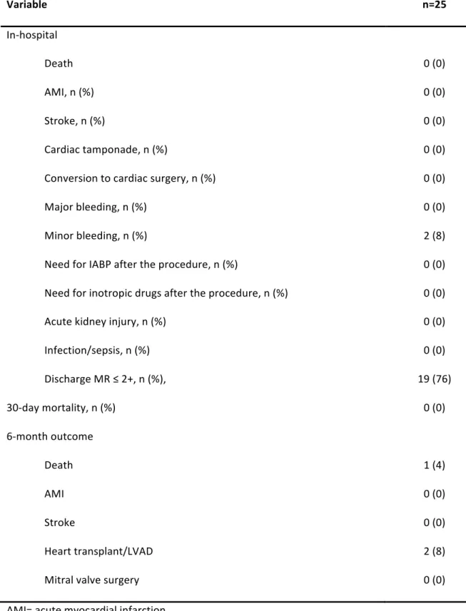
Table 2. Clinical outcome. Variable n=25 In-hospital Death 0 (0) AMI, n (%) 0 (0) Stroke, n (%) 0 (0) Cardiac tamponade, n (%) 0 (0) Conversion to cardiac surgery, n (%) 0 (0) Major bleeding, n (%) 0 (0) Minor bleeding, n (%) 2 (8) Need for IABP after the procedure, n (%) 0 (0) Need for inotropic drugs after the procedure, n (%) 0 (0) Acute kidney injury, n (%) 0 (0) Infection/sepsis, n (%) 0 (0) Discharge MR ≤ 2+, n (%), 19 (76) 30-day mortality, n (%) 0 (0) 6-month outcome Death 1 (4) AMI 0 (0) Stroke 0 (0) Heart transplant/LVAD 2 (8) Mitral valve surgery 0 (0) AMI= acute myocardial infarction
Table.3 Changes from baseline to 6 months in echocardiographic characteristics, neurohormonal profile and hospital admissions Variable Baseline (n=22) 6-month (n=22) P Echocardiography Mitral regurgitation grade ≤ 2+, n (%) 0 (0) 17 (77) 0.00 Mitral regurgitation grade≥ 3+, n (%) 22 (100) 5 (23) 0.00 LV Ejection fraction, mean ±SD ,% 32 ± 7 32 ± 7 0.35 LV end-diastolic volume, mean ± SD, ml 195± 53 193± 58 0.90 LV end-systolic volume, mean ± SD, ml 134± 49 134± 51 0.54 LV end-systolic diameter, mean ± SD, mm 52 ± 8 55± 9 0.12 LV end-systolic diameter > 55 mm, n (%) 11 (50) 12 (55) 0.70 Systolic pulmonary artery pressure mean ± SD 49± 15 43± 12 0.14 Right atrial pressure mean ± DS 9 ± 3 8 ± 2 0.22 Right ventricular dilatation > moderate, n (%) 7 (32) 7 (32) - Hypokinetic right ventricle > moderate, n (%) 9 (41) 9 (41) - Clinical status NYHA functional class ≤ II, n (%) 0 (0) 15 (68) 0.00 NYHA functional class ≥ III, n (%) 22 (100) 7 (32) 0.00 Pro-BNP, median [IQR], pg/ml 4395 [1606-5884] 2594 [1406-3924] 0.04 Hospital admissions Total admissions, n (mean±SD) 1.7 ± 1.5† 0.7 ± 0.8 0.011 Admissions for HF, n (mean±SD) 1.05 ± 1.1† 0.3 ± 0.7 0.0009 † Hospitalizations in the 6 months before percutaneous mitral valve repair HF= heart failure; IQR=interquartile range; NYHA=New York heart association; pro-BNP=pro- brain-natriuretic-peptide; SD= standard deviation.
FIGURES
0% 10% 20% 30% 40% 50% 60% 70% 80% 90% 100% Baseline 6 Months NYHA IV NYHA III NYHA ≤ 2 27% 73% 32% 68% Figure 6. Changes from baseline to 6 months in NYHA functional class. Data refer to 22 patients with paired data at baseline and 6-month follow-up.Figure 7: Changes in hospitalizations in the 6 months before and after percutaneous mitral valve repair. Data refer to 22 patients with paired data at baseline and 6-month follow-up. 0 0,2 0,4 0,6 0,8 1 1,2 1,4 1,6 1,8 Total Admissions Admissions for heart failure Before Auer P= 0.011 P= 0.0009
0% 10% 20% 30% 40% 50% 60% 70% 80% 90% 100%
Baseline Discharge 6 Months
MR 4+ MR 3+ MR ≤ 2+ 86% 14% 9% 14% Figure 8. Changes from baseline to 6 months in mitral regurgitation (MR) grade. Data refer to 22 patients with paired echocardiographic data at baseline and 6-month follow-up. 77% 86% 9% 5%
REFERENCES 1. Enriquez-Sarano M, Akins CW, Vahanian A. Mitral regurgitation. Lancet 2009;373:1382-94. 2. Levine RA, Schwammenthal E. Ischemic mitral regurgitation on the threshold of a solution: from paradoxes to unifying concepts. Circulation 2005;112:745-58. 3. Allen LA, Felker GM. Advances in the surgical treatment of heart failure. Curr Opin Cardiol 2008;23:249-53. 4. Wu AH, Aaronson KD, Bolling SF, Pagani FD, Welch K, Koelling TM. Impact of mitral valve annuloplasty on mortality risk in patients with mitral regurgitation and left ventricular systolic dysfunction. J Am Coll Cardiol 2005;45:381-7. 5. Trichon BH, Felker GM, Shaw LK, Cabell CH, O'Connor CM. Relation of frequency and severity of mitral regurgitation to survival among patients with left ventricular systolic dysfunction and heart failure. Am J Cardiol 2003;91:538-43. 6. Di Salvo TG, Acker MA, Dec GW, Byrne JG. Mitral valve surgery in advanced heart failure. J Am Coll Cardiol 2010;55:271-82. 7. Carabello BA. The current therapy for mitral regurgitation. J Am Coll Cardiol 2008;52:319-26. 8. Kaul S, Spotnitz WD, Glasheen WP, Touchstone DA. Mechanism of ischemic mitral regurgitation. An experimental evaluation. Circulation 1991;84:2167-80. 9. Hill JA, Olson EN. Cardiac plasticity. N Engl J Med 2008;358:1370-80. 10. Bishay ES, McCarthy PM, Cosgrove DM, Hoercher KJ, Smedira NG, Mukherjee D, White J, Blackstone EH. Mitral valve surgery in patients with severe left ventricular dysfunction. Eur J Cardiothorac Surg 2000;17:213-21. 11. Alfieri O, Maisano F, De Bonis M, Stefano PL, Torracca L, Oppizzi M, La Canna G. The double-orifice technique in mitral valve repair: a simple solution for complex problems. J Thorac Cardiovasc Surg 2001;122:674-81. 12. Feldman T, Wasserman HS, Herrmann HC, Gray W, Block PC, Whitlow P, St Goar F, Rodriguez L, Silvestry F, Schwartz A, Sanborn TA, Condado JA, Foster E. Percutaneous mitral valve repair using the edge-to-edge technique: six-month results of the EVEREST Phase I Clinical Trial. J Am Coll Cardiol 2005;46:2134-40. 13. Feldman T, Kar S, Rinaldi M, Fail P, Hermiller J, Smalling R, Whitlow PL, Gray W, Low R, Herrmann HC, Lim S, Foster E, Glower D. Percutaneous mitral repair with the MitraClip system:
Study) cohort. J Am Coll Cardiol 2009;54:686-94. 14. Feldman T, Foster E, Glower DD, Kar S, Rinaldi MJ, Fail PS, Smalling RW, Siegel R, Rose GA, Engeron E, Loghin C, Trento A, Skipper ER, Fudge T, Letsou GV, Massaro JM, Mauri L. Percutaneous repair or surgery for mitral regurgitation. N Engl J Med 2011;364:1395-406. 15. Tamburino C, Ussia GP, Maisano F, Capodanno D, La Canna G, Scandura S, Colombo A, Giacomini A, Michev I, Mangiafico S, Cammalleri V, Barbanti M, Alfieri O. Percutaneous mitral valve repair with the MitraClip system: acute results from a real world setting. Eur Heart J;31:1382-9. 16. Franzen O, Baldus S, Rudolph V, Meyer S, Knap M, Koschyk D, Treede H, Barmeyer A, Schofer J, Costard-Jackle A, Schluter M, Reichenspurner H, Meinertz T. Acute outcomes of MitraClip therapy for mitral regurgitation in high-surgical-risk patients: emphasis on adverse valve morphology and severe left ventricular dysfunction. Eur Heart J 2010;31:1373-81. 17. Franzen O, van der Heyden J, Baldus S, Schluter M, Schillinger W, Butter C, Hoffmann R, Corti R, Pedrazzini G, Swaans MJ, Neuss M, Rudolph V, Surder D, Grunenfelder J, Eulenburg C, Reichenspurner H, Meinertz T, Auricchio A. MitraClip(R) therapy in patients with end-stage systolic heart failure. Eur J Heart Fail 2011;13:569-76. 18. Reichenspurner H, Schillinger W, Baldus S, Hausleiter J, Butter C, Schaefer U, Pedrazzini G, Maisano F. Clinical outcomes through 12 months in patients with degenerative mitral regurgitation treated with the MitraClip(R) device in the ACCESS-EUrope Phase I trial. Eur J Cardiothorac Surg 2013;44:e280-8. 19. Koelling TM, Aaronson KD, Cody RJ, Bach DS, Armstrong WF. Prognostic significance of mitral regurgitation and tricuspid regurgitation in patients with left ventricular systolic dysfunction. Am Heart J 2002;144:524-9. 20. Mirabel M, Iung B, Baron G, Messika-Zeitoun D, Detaint D, Vanoverschelde JL, Butchart EG, Ravaud P, Vahanian A. What are the characteristics of patients with severe, symptomatic, mitral regurgitation who are denied surgery? Eur Heart J 2007;28:1358-65. 21. Grasso C, Capodanno D, Scandura S, Cannata S, Imme S, Mangiafico S, Pistritto A, Ministeri M, Barbanti M, Caggegi A, Chiaranda M, Dipasqua F, Giaquinta S, Occhipinti M, Ussia G, Tamburino C. One- and twelve-month safety and efficacy outcomes of patients undergoing edge-to-edge percutaneous mitral valve repair (from the GRASP Registry). Am J Cardiol 2013;111:1482-7. 22. Auricchio A, Schillinger W, Meyer S, Maisano F, Hoffmann R, Ussia GP, Pedrazzini GB, van der Heyden J, Fratini S, Klersy C, Komtebedde J, Franzen O. Correction of mitral regurgitation in
nonresponders to cardiac resynchronization therapy by MitraClip improves symptoms and promotes reverse remodeling. J Am Coll Cardiol 2011;58:2183-9. 23. Taramasso M, Maisano F, Latib A, Denti P, Buzzatti N, Cioni M, La Canna G, Colombo A, Alfieri O. Clinical outcomes of MitraClip for the treatment of functional mitral regurgitation. EuroIntervention 2014;10:746-52. 24. Adamo M, Barbanti M, Curello S, Fiorina C, Chiari E, Chizzola G, Capodanno D, Tamburino C, Metra M, Ettori F. Effectiveness of MitraClip therapy in patients with refractory heart failure. J Interv Cardiol 2015;28:61-8. 25. Zoghbi WA, Enriquez-Sarano M, Foster E, Grayburn PA, Kraft CD, Levine RA, Nihoyannopoulos P, Otto CM, Quinones MA, Rakowski H, Stewart WJ, Waggoner A, Weissman NJ. Recommendations for evaluation of the severity of native valvular regurgitation with two-dimensional and Doppler echocardiography. J Am Soc Echocardiogr 2003;16:777-802. 26. Lang RM, Bierig M, Devereux RB, Flachskampf FA, Foster E, Pellikka PA, Picard MH, Roman MJ, Seward J, Shanewise JS, Solomon SD, Spencer KT, Sutton MS, Stewart WJ. Recommendations for chamber quantification: a report from the American Society of Echocardiography's Guidelines and Standards Committee and the Chamber Quantification Writing Group, developed in conjunction with the European Association of Echocardiography, a branch of the European Society of Cardiology. J Am Soc Echocardiogr 2005;18:1440-63. 27. Baumgartner H, Hung J, Bermejo J, Chambers JB, Evangelista A, Griffin BP, Iung B, Otto CM, Pellikka PA, Quinones M. Echocardiographic assessment of valve stenosis: EAE/ASE recommendations for clinical practice. J Am Soc Echocardiogr 2009;22:1-23; quiz 101-2. 28. Rudski LG, Lai WW, Afilalo J, Hua L, Handschumacher MD, Chandrasekaran K, Solomon SD, Louie EK, Schiller NB. Guidelines for the echocardiographic assessment of the right heart in adults: a report from the American Society of Echocardiography endorsed by the European Association of Echocardiography, a registered branch of the European Society of Cardiology, and the Canadian Society of Echocardiography. J Am Soc Echocardiogr 2010;23:685-713; quiz 786-8. 29. Grayburn PA, Foster E, Sangli C, Weissman NJ, Massaro J, Glower DG, Feldman T, Mauri L. Relationship between the magnitude of reduction in mitral regurgitation severity and left ventricular and left atrial reverse remodeling after MitraClip therapy. Circulation 2013;128:1667-74. 30. Melisurgo G, Ajello S, Pappalardo F, Guidotti A, Agricola E, Kawaguchi M, Latib A, Covello RD,
functional mitral regurgitation. Am J Cardiol 2014;113:1844-50. 31. Koifman E, Fefer P, Hay I, Feinberg M, Maor E, Guetta V. MitraClip implantation for high risk patients with severe mitral regurgitation: the Sheba experience. Isr Med Assoc J 2014;16:91-5. 32. Braun J, Bax JJ, Versteegh MI, Voigt PG, Holman ER, Klautz RJ, Boersma E, Dion RA. Preoperative left ventricular dimensions predict reverse remodeling following restrictive mitral annuloplasty in ischemic mitral regurgitation. Eur J Cardiothorac Surg 2005;27:847-53.