DIABETICMedicine DOI: 10.1111/j.1464-5491.2008.02575.x
Blackwell Publishing Ltd
Original Article: Metabolism
Relationship between soluble CD40 ligand and
γγγγ
-glutamyltransferase concentrations in non-drinking,
young Type 1 diabetic individuals
G. Zoppini
1, G. Targher
1, M. Trombetta, G. Lippi* and M. Muggeo
Section of Endocrinology, Department of Biomedical and Surgical Sciences and *Section of Clinical Chemistry, Department of Biomedical and Morphological Sciences, University of Verona, Verona, Italy
Accepted 13 August 2008
Abstract
Aims To assess the association between circulating levels of soluble CD40 ligand (sCD40L), an emerging cardiovascular risk factor, and γ-glutamyltransferase (GGT) activity concentrations in Type 1 diabetic subjects.
Methods Plasma concentrations of sCD40L and GGT activity, a marker of liver dysfunction, were measured in 54 non-smoking, non-drinking, young Type 1 diabetic patients, who were free of diagnosed cardiovascular disease. Results When participants were grouped according to tertiles of GGT, plasma sCD40L concentrations steadily increased across GGT tertiles (P= 0.007 for trend). Similarly, plasma sCD40L concentrations were positively correlated with plasma GGT levels in the whole group of participants (r= 0.532; P < 0.0001). In multivariate linear regression analysis, plasma GGT activity levels were positively associated with sCD40L (standardized beta coefficient = 0.342;
P= 0.027) independently of age, gender, diabetes duration, glycated haemoglobin, total cholesterol and systolic blood pressure. Further adjustment for the presence of diabetic retinopathy and microalbuminuria did not appreciably attenuate this association.
Conclusions Our findings suggest that there is a strong, graded, relationship between plasma GGT activity and sCD40L concentrations in non-smoking, non-drinking, young Type 1 diabetic individuals. This association appears to be independent of numerous confounding factors. Further studies are required to confirm the reproducibility of these results. Diabet. Med. 25, 1283–1288 (2008)
Keywords cardiovascular risk factors, γ -glutamyltransferase, liver fat, sCD40 ligand, Type 1 diabetes
Abbreviations ALT, alanine aminotransferase; AST, aspartate aminotransferase; BMI, body mass index; CVD, cardiovascular disease; GGT, γ-glutamyltransferase activity; GSH, glutathione; HbA1c, glycated haemoglobin; NAFLD, non-alcoholic fatty liver disease; sCD40L, soluble CD40 ligand; TNF, tumour necrosis factor
Introduction
The past two decades have been characterized by growing interest in the idea that atherosclerosis is an inflammatory disease [1]. Interaction of the multi-potent immuno-modulator CD40 ligand (CD40L) with its receptor CD40 has emerged as an important contributor to the inflammatory processes that
lead to atherosclerosis and thrombosis. CD40 and CD40L, a member of the tumour necrosis factor (TNF) receptor super-family, are widely expressed in human atheroma and on a range of atheroma-associated cells, including endothelial cells, macrophages, T cells, smooth muscle cells and platelets [1–3]. The CD40–CD40L interaction promotes a broad range of pro-thrombotic and pro-inflammatory effects, including the expres-sion of adheexpres-sion molecules and the secretion of numerous cytokines and matrix metalloproteinases involved in extracellular matrix degradation [2,4–7]. Accordingly, inhibition of CD40 signalling reduces experimental atherosclerosis development [8] as well as the evolution of established atherosclerosis [9]. In addition to the cell-associated form, CD40L also exists in a Correspondence to: Dr. Giovanni Targher, Section of Endocrinology,
Department of Biomedical and Surgical Sciences, University of Verona, Ospedale Civile Maggiore, Piazzale Stefani, 1, 37126 Verona, Italy. E-mail: [email protected]
1G. Zoppini and G. Targher contributed equally to this work. dme(04)_2575.fm Page 1283 Tuesday, October 28, 2008 8:55 AM
DIABETICMedicine sCD40L and γ-glutamyltransferase in Type 1 diabetes • G. Zoppini et al.
soluble form that is fully active biologically, termed soluble CD40L (sCD40L). Interestingly, elevated plasma sCD40L concen-trations are closely associated with an increased risk of future cardiovascular events in healthy women [10], in patients with acute coronary syndromes [11] and in those with end-stage renal disease on haemodialysis [12].
Recent studies suggested that non-alcoholic fatty liver disease (NAFLD), which is the commonest cause of abnormal serum liver enzymes in adults in Western countries [13–15], may be linked to an increased incidence of cardiovascular disease (CVD), especially in diabetic populations [16–20]. Type 2 diabetic patients appear to have a higher risk of NAFLD, especially in its more progressive forms, than non-diabetic subjects [13–16]. Available information on the frequency of NAFLD in people with Type 1 diabetes is quantitatively limited. However, elevated serum alanine aminotransferase (ALT) levels (defined as ALT > 50 U/l) were found in 9.5% of patients with Type 1 diabetes and 12.1% of those with Type 2 diabetes in a cohort of 1353 adult patients (62% of whom had Type 2 diabetes) [21]. In addition, further indirect evidence of a high frequency of NAFLD in Type 1 diabetes might be derived from recent results of large epidemiological studies reporting a higher frequency of the metabolic syndrome (ranging from 20 to 40%) in this patient population than that observed in non-diabetic individuals [22,23].
The exact biological mechanisms by which NAFLD may contribute to the development and progression of CVD have not been completely elucidated. To our knowledge, the relationship between serum liver enzymes, as markers of NAFLD [13,14] and circulating levels of sCD40L in Type 1 diabetes has not yet been investigated.
Thus, the aim of this study was to assess the association between plasma sCD40L and liver enzymes in non-drinking, young Type 1 diabetic individuals without any clinical evidence of CVD.
Methods
After written informed consent was obtained, we enrolled 54 young Type 1 diabetic patients, who were selected from all outpatients with Type 1 diabetes regularly attending our diabetes clinic after excluding (i) subjects who currently smoked or drank alcohol, (ii) those who had a history of recent acute illness or clinical evidence suggestive of CVD, kidney or liver diseases and (iii) those who were taking anti-platelet drugs. Overall, these participants represented approximately 30% of all the Type 1 diabetic patients (n= 202) who regularly attended our clinic. A group of these participants (n= 27) has been previously included in a pilot study assessing plasma sCD40L concentrations in relation to diabetes status [24]. All patients were treated with insulin; none was prescribed lipid-lowering drugs, whereas six patients were taking ramipril. Information on daily alcohol consumption and other lifestyle characteristics was obtained from all participants by a questionnaire. To exclude the presence of clinically manifest CVD, a resting electrocar-diogram, measurement of ankle brachial pressure index and
carotid ultrasonography (to exclude the presence of significant carotid stenoses but to measure carotid intima-media wall thickness) were performed in all participants.
Body mass index (BMI) was calculated by dividing weight in kilograms by height in meters squared. Blood pressure was measured in triplicate with a standard mercury manometer. Venous blood was drawn in the morning (08.00–08.30 h) after an overnight fast in all participants. Serum liver enzymes [ALT, aspartate aminotransferase (AST) and γ-glutamyltransferase (GGT)], lipids and other biochemical blood measurements were determined by standard laboratory procedures (DAX 96; Bayer Diagnostics, Milan, Italy). Reference ranges for ALT, AST and GGT levels, in our laboratory, were 5– 40 and 5–55 U/l for women and 5–45 and 5–55 U/l for men, respectively. Serology for viral hepatitis B and C was assessed in all participants. Glycated haemoglobin (HbA1c) was measured by an automated high-performance liquid chromatography analyser (Bio-Rad Diamat, Milan, Italy); normal range values in our laboratory were 3.0–5.5%. Blood for coagulation analysis was collected in citrated tubes and immediately centrifuged at 2500 g for 15 min at 4°C. Several aliquots of each plasma sample were then quick-frozen and stored at −80°C, in plastic vials, until determinations were performed. Within 6 months, all stored plasma samples were assayed in a single session by an experienced laboratory technician. Determination of plasma sCD40L was performed in duplicate with a commercially available ELISA-kit (Bender Med-Systems Diagnostics, Vienna, Austria) with intra-assay and interassay coefficients of variation of 6.6 and 9.7%, respectively. Plasma concentrations of fibrinogen (IL-test-PT-fibrinogen HS; Instrumentation Laboratory, Lexington, MA, USA), adiponectin (by an ELISA-method; B-Bridge International, San Jose, CA, USA), soluble intercellular adhesion molecule-1 (ICAM-1) (by an ELISA-method; Bender Med-Systems Diagnostics, Vienna, Austria) and soluble P-selectin (by an ELISA-method; Bender Med-Systems Diagnostics, Vienna, Austria) were measured in duplicate. Intra- and interassay coefficients of variation were 6.5 and 7.2%, 3.5 and 7.3%, 3.8 and 9%, 4 and 8.9% for fibrinogen, adiponectin, sICAM-1 and P-selectin, respectively.
In all patients, urinary albumin excretion was measured in an early morning urine sample as the albumin-to-creatinine ratio by an immuno-nephelometric method; micro-albuminuria and macro-albuminuria were present if urinary albumin excretion was 30–299 mg/g creatinine and ≥ 300 mg/g creatinine, respec-tively [25]. No patients had macro-albuminuria. A single ophthalmologist diagnosed retinopathy by fundoscopy after pupillary dilation, according to a clinical disease severity scale [26]. The presence of sensory neuropathy (by biothesiometer) was not extensively recorded in the study participants.
Statistical analysis
Data are presented as means ±SD or proportions. Skewed
variables (sCD40L, adiponectin, sICAM-1, P-selectin, fibrinogen and triglycerides) were logarithmically transformed to improve normality before analysis. The following statistical tests were performed: one-way analysis of variance (for continuous variables), χ2-test (for categorical variables) and univariate linear correlation.
The independent association between sCD40L (logarithmically transformed) and GGT (included as a continuous measure) was assessed by two multivariate linear regression models as
Original article DIABETICMedicine
follows: model 1 including age, gender, diabetes duration, HbA1c, total cholesterol, GGT and systolic blood pressure as covariates; and model 2 including age, gender, diabetes duration, HbA1c, GGT, retinopathy (yes/no) and microalbuminuria (yes/no) as covariates. P < 0.05 were considered statistically significant. Statistical analyses were performed using SPSS 14.0 (SAS Institute, Cary, NC, USA).
Results
Patients enrolled in the study were predominantly males (53.7% of total, n= 29), lean (BMI = 23.8 ± 2.8 kg/m2), normotensive (systolic blood pressure = 126 ± 15 mmHg; diastolic blood pressure = 80 ± 8 mmHg), normolipidaemic (total cholesterol = 4.7 ± 0.7 mmol/l; triglycerides = 1.0 ± 0.6 mmol/l) and had a mean age of 32 ± 9 years, duration of disease of 14.8 ± 8 years (range: 2–40 years) and were in good glycaemic control (HbA1c= 6.7 ± 1.1%). All participants had normal serum liver enzymes (AST = 17 ± 7 U/l, ALT = 19 ± 6 U/l, GGT = 13 ± 8 U/l) and were non-drinkers and non-smokers. Of participants, 18.5% (n= 10) had microalbuminuria and 35.2% (n= 19) had retinopathy (seven of whom had proliferative retinopathy). The prevalence of microvascular complications was similar to that described in other European populations with comparable age, diabetes duration and glycaemic control [27].
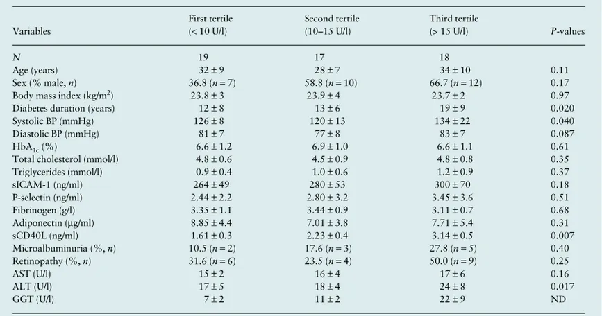
Table 1 shows the clinical and biochemical characteristics of participants grouped according to tertiles of serum GGT activity levels. Diabetes duration, ALT levels and systolic blood pressure increased across GGT tertiles, whereas all other measured variables were not significantly different among the groups.
Notably, plasma sCD40L concentrations steadily increased across GGT tertiles (P= 0.007 for trend). Similar insignificant trends were observed across tertiles of ALT (P= 0.083) or the ALT-to-AST ratio (P= 0.091), respectively (data not shown). Consistently, as shown in Fig. 1, plasma sCD40L concentrations were positively correlated with plasma GGT levels in the whole group of participants (r= 0.532; P < 0.0001). The significant increase in plasma sCD40L concentrations across GGT tertiles remained essentially unchanged after excluding those (n= 6), who were treated with ramipril (P= 0.019 for trend).
In multivariate linear regression analysis, GGT was positively associated with log-transformed sCD40L (standardized beta coefficient = 0.342; P= 0.027) independently of age, gender, diabetes duration, HbA1c, total cholesterol and systolic blood pressure. All other variables included in this model were not independently associated with sCD40L.
Almost identical results were obtained in another multivariate regression model that included age, gender, diabetes duration, HbA1c, presence of retinopathy and microalbuminuria as covariates. Also in this case, GGT was the only variable
Table 1Main clinical and biochemical characteristics of young Type 1 diabetic patients grouped according to tertiles of serum GGT activity concentrations Variables First tertile (< 10 U/l) Second tertile (10–15 U/l) Third tertile (> 15 U/l) P-values N 19 17 18 Age (years) 32 ± 9 28 ± 7 34 ± 10 0.11 Sex (% male, n) 36.8 (n = 7) 58.8 (n = 10) 66.7 (n = 12) 0.17
Body mass index (kg/m2) 23.8 ± 3 23.9 ± 4 23.7 ± 2 0.97
Diabetes duration (years) 12 ± 8 13 ± 6 19 ± 9 0.020
Systolic BP (mmHg) 126 ± 8 120 ± 13 134 ± 22 0.040
Diastolic BP (mmHg) 81 ± 7 77 ± 8 83 ± 7 0.087
HbA1c (%) 6.6 ± 1.2 6.9 ± 1.0 6.6 ± 1.1 0.61
Total cholesterol (mmol/l) 4.8 ± 0.6 4.5 ± 0.9 4.8 ± 0.8 0.35
Triglycerides (mmol/l) 0.9 ± 0.4 1.0 ± 0.6 1.2 ± 0.9 0.37 sICAM-1 (ng/ml) 264 ± 49 280 ± 53 300 ± 70 0.18 P-selectin (ng/ml) 2.44 ± 2.2 2.80 ± 3.2 3.45 ± 3.6 0.51 Fibrinogen (g/l) 3.35 ± 1.1 3.44 ± 0.9 3.11 ± 0.7 0.68 Adiponectin (μg/ml) 8.85 ± 4.4 7.01 ± 3.8 7.71 ± 5.4 0.31 sCD40L (ng/ml) 1.61 ± 0.3 2.23 ± 0.4 3.14 ± 0.5 0.007 Microalbuminuria (%, n) 10.5 (n = 2) 17.6 (n = 3) 27.8 (n = 5) 0.40 Retinopathy (%, n) 31.6 (n = 6) 23.5 (n = 4) 50.0 (n = 9) 0.25 AST (U/l) 15 ± 2 16 ± 4 17 ± 6 0.16 ALT (U/l) 17 ± 5 18 ± 4 24 ± 8 0.017 GGT (U/l) 7 ± 2 11 ± 2 22 ± 9 ND
Data are means ± SD or proportions.
Differences were tested by the one-way ANOVA (for continuous variables) and by the χ2-test (for categorical variables).
ALT, alanine aminotransferase; AST, aspartate aminotransferase; BP, blood pressure; GGT, γ-glutamyltransferase; HbA1c, glycated
haemoglobin; ND, not determined; sCD40L, soluble CD40 ligand; SD, standard deviation; sICAM-1, soluble intercellular adhesion molecule-1. dme(04)_2575.fm Page 1285 Tuesday, October 28, 2008 8:55 AM
DIABETICMedicine sCD40L and γ-glutamyltransferase in Type 1 diabetes • G. Zoppini et al.
independently associated with sCD40L (standardized beta coefficient = 0.324; P = 0.024).
Identical results were observed even when the independent association between sCD40L and GGT was assessed by multi-variate logistic regression analysis. In that analysis, GGT was positively associated with sCD40L (1st tertile vs. 2nd–3rd tertiles combined) independently of age, gender, total cholesterol, systolic blood pressure, HbA1c and duration of diabetes [multiple-adjusted odds ratio 1.31 (95% confidence intervals 1.06–1.49), P = 0.011].
Discussion
NAFLD, in its whole spectrum of disease ranging from pure steatosis to steatohepatitis and cirrhosis, is the commonest chronic liver disease in the developed world and is now regarded as the liver manifestation of the metabolic syndrome [13,14]. NAFLD prevalence has been estimated to range from 15 to 30% in the general population in Western countries [14,15] and is almost certainly increasing. Compared with non-diabetic subjects, people with diabetes appear to be at increased risk of developing NAFLD and certainly have a higher risk of developing fibrosis and cirrhosis [13–16,21].
Mild elevations of serum GGT activity levels and other serum liver enzymes are suggested to have a clinical and epidemiological significance as markers of NAFLD and related liver dysfunction [13,14]. A strong positive association between mildly elevated serum GGT levels and increased incidence of CVD has been shown in several population-based studies [28–32]. For example, in a study of 163 944 middle-aged Austrian men and women, serum GGT levels, even within their reference range, were independently associated with increased CVD mortality in both genders [29].
The major finding of our study is that, in Type 1 diabetic patients, increasing GGT levels are closely associated with elevated plasma sCD40L concentrations independently of a
broad range of confounding factors, including age, gender, systolic blood pressure, total cholesterol, HbA1c, diabetes duration and microvascular complications status. Notably, our patients were young, in good glycaemic control and free of diagnosed CVD. Moreover, all our patients were non-smokers, non-drinkers and had serum liver enzymes within the reference ranges. We believe that all of these conditions would possibly enhance the validity of our findings.
As many patients with NAFLD have normal serum liver enzymes [13–17], there is now increasing consensus among hepatologists supporting the notion that the currently used ‘normal’ reference values for serum liver enzymes for excluding NAFLD need to be revised. Recently, Prati et al. have proposed upper limits of normal range for ALT (i.e. 30 U/l for men and 19 U/l for women) that were substantially lower than the levels that laboratories currently consider to be the upper range of normal (for example, 45 U/l for men and 40 U/l for women in our laboratory) [33]. Using these new definitions of normal, these researchers could more accurately identify the patients with NAFLD and other chronic liver diseases than they could using the old ranges [33]. Notably, when we used these more stringent criteria, nearly half of our patients in the highest GGT tertile had serum ALT levels above the new proposed cut-off values, thus possibly having NAFLD.
Overall, therefore, our findings support a potential link between NAFLD, as reflected by mildly elevated serum GGT levels, and the CD40–CD40L pathway in Type 1 diabetic patients, thus suggesting an additional, underlying, mechanism by which NAFLD might contribute to the development and progression of CVD. The possible molecular mediators linking NAFLD and CVD have been extensively reviewed elsewhere [34], but include the release of pro-atherogenic mediators from the liver, including increased reactive oxygen species, C-reactive protein, fibrinogen, plasminogen activator inhibitor-1, TNF-alpha and other pro-inflammatory cytokines. In this context, it has been experimentally shown that the CD40–CD40L pathway is markedly activated in the liver of patients with chronic liver diseases (especially in those with autoimmune hepatitis and primary biliary cirrhosis) [35–37]. Moreover, in in vitro studies it has been demonstrated that TNF-alpha stimulates CD40 expression on hepatocytes via activation of nuclear factor kappa B (NF-κB) and activator protein-1 (AP-1)/jun N-terminal kinase (JNK), which are two transcription factors that control the expression of several pro-inflammatory cytokines and adhesion molecules and that are implicated in the control of epithelial cell proliferation and apoptosis [36,37].
Another possible explanation for our findings can rely on recent data suggesting that GGT, even within its reference range, is not simply a marker of liver dysfunction or alcohol abuse but may also be a risk factor actively involved in CVD pathogenesis—possibly also through the activation of the CD40–CD40L pathway [38,39]. GGT, which is found on all cell membranes, with the exception of erythrocytes, is the main determinant of extracellular hydrolysis of glutathione (GSH). In this process, GGT releases the dipeptide cysteinyl–glycine,
FIGURE 1Relationship between plasma soluble CD40 ligand (sCD40L) concentrations (logarithmically transformed) and γ-glutamyltransferase (GGT) activity levels in non-smoking, non-drinking, young Type 1 diabetic patients.
Original article DIABETICMedicine which is subsequently cleaved to cysteine and glycine by
plasma membrane dipeptidase activities [38]. Thus, GGT activity provides cells with a means for the recovery of precursors needed to reconstitute intracellular levels of GSH, the main cellular antioxidant. However, recent studies have shown that the reactive thiol of cysteinyl–glycine originated during GGT-mediated cleavage of GSH may cause the reduction of ferric Fe(III) to ferrous iron Fe(II), thus starting a redox-cycling process resulting in increased production of the reactive oxygen species superoxide anion and hydrogen peroxide, both capable of stimulating pro-oxidant reactions [38,39]. In turn, oxidative stress can induce chronic inflam-mation with subsequent activation of the CD40–CD40L pathway. GGT-mediated pro-oxidant/inflammatory effects are likely within atherosclerotic coronary, carotid and cerebral plaques, where catalytically active GGT has been identified histochemically and can be sustained by iron storage proteins such as transferrin and ferritin, or even by free iron, shown to be present within the plaque at sufficient concentrations [38,39].
Our study has some important limitations. First, the cross-sectional design of our study precludes the establishment of causal or temporal relationships between plasma sCD40L and GGT concentrations. Prospective studies will be required to clarify the time sequence of events. Second, NAFLD diagnosis was based on serum liver enzymes and exclusion of other common causes of chronic liver disease (i.e. alcohol abuse, viral hepatitis, use of hepato-toxic medications), but was not confirmed by liver biopsy. However, liver biopsy would be unethical to perform in our patients who had serum liver enzymes within the reference range. Finally, whether these observations can also be extended to non-diabetic individuals remains to be determined.
In conclusion, our findings suggest that there is a strong, graded, relationship between plasma GGT activity and sCD40L concentrations in young Type 1 diabetic patients without any clinical evidence of CVD. This association appears to be independent of numerous confounding factors. Further research is needed to elucidate the underlying molecular mechanisms linking GGT activity and the CD40–CD40L pathway before causality can be firmly established.
Competing interests
Nothing to declare.References
1 Packard RR, Libby P. Inflammation in atherosclerosis: from vascular biology to biomarker discovery and risk prediction. Clin Chem 2008;
54: 24–38.
2 Schonbeck U, Libby P. CD40 signalling and plaque instability. Circ
Res 2001; 89: 1092–1103.
3 Mach F, Schonbeck U, Sukhova GK, Bourcier T, Bonnefoy JY, Pober JS et al. Functional CD40 ligand is expressed on human vascular endothelial cells, smooth muscle cells, and macrophages:
implications for CD40-CD40 ligand signalling in atherosclerosis.
Proc Natl Acad Sci U S A 1997; 94: 1931–1936.
4 Henn V, Slupsky JR, Gräfe M, Anagnostopoulos I, Förster R, Müller-Berghaus G et al. CD40 ligand on activated platelets triggers an inflammatory reaction of endothelial cells. Nature 1998; 391: 591–594.
5 Dechanet J, Grosset C, Taupin JL, Merville P, Banchereau J, Ripoche J
et al. CD40 ligand stimulates proinflammatory cytokine production
by human endothelial cells. J Immunol 1997; 159: 5640 –5647. 6 Slupsky JR, Kalbas M, Willuweit A, Henn V, Kroczek RA,
Müller-Berghaus G. Activated platelets induce tissue factor expression on human umbilical vein endothelial cells by ligation of CD40. Thromb Haemost 1998; 80: 1008 –1014.
7 Zhou L, Stordeur P, de Lavareille A, Thielemans K, Capel P, Goldman M et al. CD40 engagement on endothelial cells promotes tissue factor-dependent procoagulant activity. Thromb Haemost 1998; 79: 1025–1028.
8 Mach F, Schonbeck U, Sukhova GK, Atkinson E, Libby P. Reduction of atherosclerosis in mice by inhibition of CD40 signalling. Nature 1998; 394: 200–203.
9 Schonbeck U, Sukhova GK, Shimizu K, Mach F, Libby P. Inhibition of CD40 signalling limits evolution of established atherosclerosis in mice. Proc Natl Acad Sci U S A 2000; 97: 7458 –7463.
10 Schonbeck U, Varo N, Libby P. Soluble CD40L and cardiovascular risk in women. Circulation 2001; 104: 2266 –2268.
11 Heeschen C, Dimmeler S, Hamm CW, van den Brand MJ, Boersma E, Zeiher AM et al. Soluble CD40 ligand in acute coronary syndromes.
N Engl J Med 2003; 348: 1104 – 1111.
12 Hocher B, Liefeldt L, Quaschning T, Kalk P, Ziebig R, Godes M
et al. Soluble CD154 is a unique predictor of non-fatal and fatal
atherothrombotic events in patients who have end-stage renal disease and are on haemodialysis. J Am Soc Nephrol 2007; 18: 1323 – 1330.
13 Angulo P. Non-alcoholic fatty liver disease. N Engl J Med 2002; 346: 1221– 1231.
14 de Alwis NMW, Day CP. Non-alcoholic fatty liver disease: the mist gradually clears. J Hepatol 2008; 48: S104 –S112.
15 Browning JD, Szczepaniak LS, Dobbins R, Nuremberg P, Horton JD, Cohen JC et al. Prevalence of hepatic steatosis in an urban population in the United States: impact of ethnicity. Hepatology 2004; 40: 1387–1395.
16 Targher G, Bertolini L, Padovani R, Rodella S, Tessari R, Zenari L
et al. Prevalence of non-alcoholic fatty liver disease and its association
with cardiovascular disease among Type 2 diabetic patients. Diabetes
Care 2007; 30: 1212–1218.
17 Targher G, Bertolini L, Poli F, Rodella S, Scala L, Tessari R et al. Non-alcoholic fatty liver disease and risk of future cardiovascular events among Type 2 diabetic patients. Diabetes 2005; 54: 3541–3546. 18 Ekstedt M, Franzen LE, Mathiesen UL, Thorelius L, Holmqvist M, Bodemar G et al. Long-term follow-up of patients with NAFLD and elevated liver enzymes. Hepatology 2006; 44: 865– 873.
19 Targher G, Bertolini L, Rodella S, Tessari R, Zenari L, Lippi G et al. Non-alcoholic fatty liver disease is independently associated with an increased incidence of cardiovascular events in Type 2 diabetic patients. Diabetes Care 2007; 30: 2119– 2121.
20 Hamaguchi M, Kojima T, Takeda N, Nagata C, Takeda J, Sarui H et al. Non-alcoholic fatty liver disease is a novel predictor of cardiovascular disease. World J Gastroenterol 2007; 13: 1579 – 1584.
21 West J, Brousil J, Gazis A, Jackson L, Mansell P, Bennett A et al. Elevated serum alanine transaminase in patients with Type 1 or Type 2 diabetes. Q J Med 2006; 99: 871 – 876.
22 Thorn LM, Forsblom C, Fagerudd J, Thomas MC, Pettersson-Fernholm K, Saraheimo M et al. Metabolic syndrome in Type 1 diabetes. Association with diabetic nephropathy and glycemic control (the FinnDiane study). Diabetes Care 2005; 28: 2019–2024.
DIABETICMedicine sCD40L and γ-glutamyltransferase in Type 1 diabetes • G. Zoppini et al. 23 Pambianco G, Costacou T, Orchard TJ. The prediction of major
outcomes of Type 1 diabetes: a 12-year prospective evaluation of three separate definitions of the metabolic syndrome and their components and estimated glucose disposal rate. The Pittsburgh Epidemiology of Diabetes Complications Study Experience.
Diabetes Care 2007; 30: 1248 –1254.
24 Targher G, Zoppini G. Soluble CD40L in young Type 1 diabetic individuals without clinical microvascular and macrovascular complications. Diabetes Care 2004; 27: 1236–1237.
25 Molitch ME, DeFronzo RA, Franz MJ, Keane WF, Mogensen CE, Parving HH et al. Nephropathy in diabetes. Diabetes Care 2004; 27: S79 – S83.
26 Early Treatment Diabetic Retinopathy Study Research Group. Fundus photographic risk factors for progression of diabetic retinopathy. ETDRS report number 12. Ophthalmology 1991; 98: 823 – 833. 27 Stephenson J, Fuller JH; the EURODIAB Complications Study Group.
Microvascular and acute complications in IDDM patients: the EURODIAB Complications Study. Diabetologia 1994; 37: 278–285. 28 Wannamethee G, Ebrahim S, Shaper AG. Gamma-glutamyltransferase: determinants and association with mortality from ischemic heart disease and all causes. Am J Epidemiol 1995; 142: 699–708. 29 Ruttmann E, Brant LJ, Concin H, Diem G, Rapp K, Ulmer H.
Gamma-glutamyltransferase as a risk factor for cardiovascular disease mortality: an epidemiological investigation in a cohort of 163 944 Austrian adults. Circulation 2005; 112: 2130–2137. 30 Lee DH, Silventoinen K, Hu G, Jacobs DR Jr, Jousilahti P, Sundvall
J et al. Serum γ-glutamyltransferase predicts non-fatal myocardial infarction and fatal coronary heart disease among 28 838 middle-aged men and women. Eur Heart J 2006; 27: 2170–2176.
31 Schindhelm RK, Dekker JM, Nijpels G, Bouter LM, Stehouwer CD, Heine RJ et al. Alanine aminotransferase predicts coronary heart
disease events: a 10-year follow-up of the Hoorn Study. Atherosclerosis 2007; 191: 391–396.
32 Lee DS, Evans JC, Robins SJ, Wilson PW, Albano I, Fox CS
et al. Gamma-glutamyltransferase and metabolic syndrome,
cardiovascular disease, and mortality risk: the Framingham Heart Study. Arterioscler Thromb Vasc Biol 2007; 27: 127– 133.
33 Prati D, Taioli E, Zanella A, Della Torre E, Buttelli S, Del Vecchio E
et al. Updated definitions of healthy ranges for serum alanine
aminotransferase levels. Ann Intern Med 2002; 137: 1– 10. 34 Targher G. Non-alcoholic fatty liver disease, the metabolic syndrome
and the risk of cardiovascular disease: the plot thickens. Diabet Med 2007; 24: 1– 6.
35 Schwabe RF, Schnabl B, Kweon YO, Brenner DA. CD40 activates NF-κB and c-Jun N-terminal kinase and enhances chemokine secretion on activated human hepatic stellate cells. J Immunol 2001;
166: 6812–6819.
36 Afford SC, Randhawa S, Eliopoulos AG, Hubscher SG, Young LS, Adams DH. CD40 activation induces apoptosis in cultured human hepatocytes via induction of cell surface fas ligand expression and amplifies fas-mediated hepatocyte death during allograft rejection. J
Exp Med 1999; 189: 441– 446.
37 Afford SC, Ahmed-Choudhury J, Randhawa S, Russell C, Youster J, Crosby HA et al. CD40 activation-induced, Fas-dependent apoptosis and NF-κB/AP-1 signalling in human intrahepatic biliary epithelial cells. FASEB J 2001; 15: 2345–2354.
38 Lee DH, Blomhoff R, Jacobs DR Jr. Is serum γ-glutamyltransferase a marker of oxidative stress? Free Radic Res 2004; 38: 535–539. 39 Emdin M, Pompella A, Paolicchi A. Gamma-glutamyltransferase,
atherosclerosis, and cardiovascular disease: triggering oxidative stress within the plaque. Circulation 2005; 112: 2078–2080.