STUDIES ON PYCNODONT FISHES (II): REVISION OF THE SUBFAMILY PYCNODONTINAE, WITH SPECIAL REFERENCE TO ITALIAN FORMS
FRANCISCO JOSÉ POYATO-ARIZA
Centre for Integration in Palaeobiology & Unidad de Paleontología, Dpto. Biología, Universidad Autónoma de Madrid, c/Darwin 2, Cantoblanco, 28049-Madrid, Spain. E-mail: [email protected]
To cite this article: Poyato-Ariza F.J. (2020) - Studies on pycnodont fishes (II): revision of the subfamily Pycnodontinae, with special reference to Italian forms. Riv. It. Paleontol. Strat., 126(2): 447-473.
Abstract: The diagnosis, composition, and phylogenetic relationships of the European subfamily
Pycnodonti-nae are revised; its record is pushed back from the Cenozoic into the Mesozoic. The PycnodontiPycnodonti-nae is confirmed as a monophyletic group. It is diagnosed by: thin, laminar supraoccipital exposed all along the posterior border of the skull roof; cleithrum with two posterior expansions framing the notch for the pectoral fin; reduction in the ossification of the flank scales (clathrate pattern); reduction of the preopercular into a very low bone, never higher than the expo-sed, ornamented portion of the dermohyomandibular; and presence of a bifid cloacal scale. The subfamily includes the tribe Pycnodontini (Pycnodus + Oropycnodus), Polazzodus, Sylvienodus, and Tergestinia. The former “Coelodus” gridellii
is moved to Polazzodus gridellii n. comb. The Italian genera, Pycnodus, Polazzodus, and Tergestinia, form a monophyletic
group together with the French Oropycnodus. The present analysis shows that Polazzodus, Sylvienodus, and Tergestinia are
pycnodontin fishes, but Haqelpycnodus, Libanopycnodus, Scalacurvichthys, and Sigmapycnodus do not belong to the
Pycno-dontinae. “Pseudopycnodus” and “P. nardoensis” are considered nomina dubia. This revision has revealed new aspects of
the last known diversification in the evolutionary history of the Pycnodontiformes, showing that the group was still thriving in the Western Tethys during the Late Cretaceous. For the present analysis, additional arguments involving ontogenetic restrictions are provided to favour ordering multistate characters in pycnodonts.
Received: January 24, 2019; accepted: January 24, 2020
Keywords: Actinopterygii; Cretaceous; Fish Evolution; Italy, Neopterygii; Phylogenetic Relationships; Pycnodontiformes; Pycnodus; Tethys.
I
ntroductIonThe evolutionary history of the Pycnodon-tiformes has been subject of evaluation in re-cent years. They are regarded as a plastic, versatile group rather than an excessively specialized group slowly declining to leave room to teleosteans. They were very successful for a long time and kept in-creasing their diversity in the places where they re-mained present (Poyato-Ariza 2005a; Martín-Abad & Poyato-Ariza 2013; Poyato-Ariza & Martín-Abad 2013). In this context, the subfamily Pycnodontinae shows a special evolutionary interest, since they are among the youngest and most derived pycnodonts (e.g., Poyato-Ariza & Wenz 2002). Martín-Abad &
Poyato-Ariza (2013) documented up to five radia-tion events in the history of the Pycnodontiformes. They tentatively proposed additional diversification of the most derived pycnodonts in the Western Te-thys during the Late Cretaceous. Thus, the objective of the present paper is twofold: to update the tax-onomy and phylogeny of this subfamily and similar forms, and to test such diversification hypothesis.
The subfamily Pycnodontinae was first for-mally used by Poyato-Ariza & Wenz (2002) to in-clude Pycnodus, the type genus, plus Oropycnodus. The former represents part the last fossil record of the Pycnodontiformes, from the Eocene of Italy, and the latter is from the Paleocene of France. The presence of pycnodonts after the Eocene is uncon-firmed (Poyato-Ariza 2005a). Among the numerous pycnodont taxa described in the last decade, six
new monospecific genera were also associated to this subfamily: Haqelpycnodus (Taverne & Capasso 2018a), Libanopycnodus (Taverne & Capasso 2018b), Polazzodus (Poyato-Ariza 2010), Scalacurvichthys (Cawley & Kriwet 2018), Sigmapycnodus (Taverne & Capasso 2018b) and Sylvienodus (Poyato-Ariza 2013), all from the Late Cretaceous. Polazzodus was pointed to show similarities with “Coelodus” gridellii d’Ermo, 1952 and Tergestinia Capasso, 2000, but the as-sessment of all these forms to the Pycnodontinae remained untested, pending revision of the group. In turn, all phylogenies of the Pycnodontinae by Taverne & Capasso (2012, 2018a,b) are ad-hoc, hand-made trees based on a preassigned distribu-tion of a selected choice of characters. There is no cladistics analysis, therefore they are not necessarily the most parsimonious hypotheses of relationships of those genera and the family Pycnodontidae.
The present paper presents the first cladistic analysis of the genera assessed to or related with the Pycnodontinae. With this purpose, all well-known genera of the family Pycnodontidae are included in the analysis; some of them have been re-evaluated because of their possible relationship with the sub-family Pycnodontinae. These taxa, in alphabetical order, are:
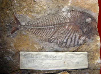
“Coelodus” gridellii d’Erasmo, 1952 (Fig. 1). Originally described from three specimens of small size, about 12 cm of total length, from an outcrop near Polazzo, Carso Isontino, northeastern Italy. The outcrop was destroyed during World War I, so the precise provenance is unknown. Similar out-crops in the nearby area are considered early San-tonian (Late Cretaceous). Amidst the three original type specimens, one of the paratypes (“Esemplare N. 3” in d’Erasmo 1952) seems to be currently lost. The other paratype (MCSNT 12361, “Esemplare N. 2” in d’Erasmo 1952) was reassessed to Polazzodus coronatus by Poyato-Ariza (2010), together with an additional specimen in the same collection (MCSNT 12447). The holotype, MCSNT-12366, was explicitly designated as “tipo” by d’Erasmo (1952: 84, “Esem-plare N. 1”). It is, therefore, the only currently known specimen of this species (Fig. 1). For further details, see Poyato-Ariza (2010: 650-651, 662).
Haqelpycnodus Taverne & Capasso 2018a. Erected for two complete, apparently well preserved specimens from late Cenomanian (Late Cretaceous) deposits of Haqel, Lebanon. Considered to present some, but not all, characters of the Pycnodontinae,
hence “immediate plesiomorphic (sic) sister-taxon of Pycnodontinae” and their “direct plesiomorphic sister-lineage (sic)” by Taverne & Capasso (2018a: 117, 131; no cladistic analysis). Both specimens in a private collection, therefore coded from the infor-mation in the original publication.
Libanopycnodus Taverne & Capasso, 2018b. Based on a single nearly complete, imperfect spec-imen from the late Cenomanian (Late Cretaceous) of En Namoura, Lebanon. Considered as “the most basal member of the Pycnodontinae” by Taverne & Capasso (2018b: 27; no cladistic analysis). Specimen in a private collection, therefore coded from the in-formation in the original publication.
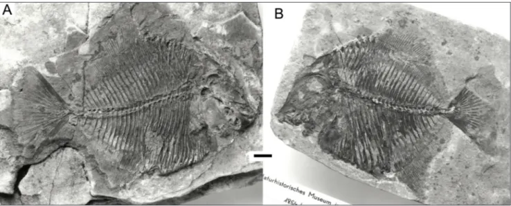
Oropycnodus Poyato-Ariza & Wenz, 2002 (Fig. 2). Known from several specimens of small size, about 14 cm of total length, from the Montian (Paleocene) of Mont Aimé in Chalons-sur-Marne, northeastern France. The type and only species is Oropycnodus ponsorti (Heckel, 1854). Although the original spelling of the specific name was ponsortii, it was changed to ponsorti following ICZN Article 33.3.1 for prevailing usage. For further details see Poyato-Ariza & Wenz (2002: 150).
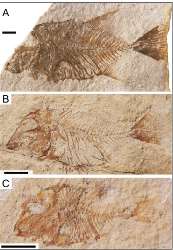
Polazzodus Poyato-Ariza, 2010 (Fig. 3). Initial-ly described from abundant, well preserved, mostInitial-ly complete specimens from the early Santonian (Late Cretaceous) near Polazzo, Carso Isontino, north-western Italy. An isolated vomerine dentition was subsequently cited as cf. Polazzodus sp. by Križnar
Fig. 1 - Polazzodus gridellii n. comb. (formerly “Coelodus” gridellii).
Holo-type and only specimen, MCSNT-12366. Late Cretaceous, probably early Santonian, near Polazzo, Italy (see text for de-tails). Photo A. Colla, archives MCSNT. The total length of the preserved part of the specimen is around 11 cm.
(2014) from the Albian-Cenomanian of Mrzlek near Solkan, Slovenia.
Pseudopycnodus Taverne, 2003. Replacement genus created for its type and only species, Pycnodus nardoensis Taverne, 1997, after revision with some new material (Taverne 2003) from the Campanian-Maastricthian (Late Cretaceous) of Nardò, Apulia,
southeastern Italy. Initially known from scarce, frag-mentary, partially disarticulated, very incomplete specimens of medium size. The longest one was part of a body without head, the preserved portion some 22 cm long. Later on, it was re-described by Taverne & Capasso (2012) on the basis of addition-al specimens in the private collection of the latter.
Pycnodus Agassiz, 1833. Type genus (Fig. 4). Abundant specimens, including ontogenetic series; adults of medium size, up to some 26 cm. Comes from the early-middle Eocene of Bolca, Verona, and Vicenza Provinces, northern Italy, where it represents the last reliable fossil record of the Pycnodontiformes. It is part of a diversified fauna that also includes Nursallia veronae and Palaeobalistum in strict sense (Ariza & Wenz 2002; Poyato-Ariza 2005a). The type species, Pycnodus apodus (Vol-ta, 1809) is currently regarded as the sole species of this genus known from articulated remains (Poyato-Ariza 2013). For further details on the nomencla-tural history of this taxon, see Blot (1987: 11-15) and Poyato-Ariza & Wenz (2002: 152).
Scalacurvichthys Cawley & Kriwet, 2018. A sin-gle species, S. naishi, based on an incomplete, imper-fectly preserved, partially disarticulated specimen from the early-mid Cenomanian, Late Cretaceous, of ‘Ein Yabrud in Israel. A member of the subfam-ily Pycnodontinae according to Cawley & Kriwet (2018).
Sigmapycnodus Taverne & Capasso, 2018b. Based on a single incomplete, imperfect specimen, artificially restored, from the late Cenomanian (Late
Fig. 2 - Oropycnodus ponsorti. A) Lectotype, NMW 1854/XXXIX/38; B) paralectotype NMW 1854/XXXIX/40. Photos Schumacher, courtesy
O. Schultz (modified from Poyato-Ariza & Wenz 2002). Scale bar equals 1 cm for both specimens.
Fig. 3 - Polazzodus coronatus. A) Holotype, MPCM-13464, standard
length 7,6 cm; B) paratype MPCM 12214, standard length 6,6 cm. Photos M. Tentor, MPCM (modified from Poyato-Ariza 2010; see p.650-651 in this reference for comments on the apparent differences of body shape among specimens of this taxon due to preservational artifacts).
Cretaceous) of Haqel, Lebanon. Specimen in a pri-vate collection, therefore coded from the informa-tion in the original publicainforma-tion.
Sylvienodus Poyato-Ariza, 2013 (Fig. 5). Abun-dant specimens of very small size, up to about 6 cm, from the upper Cenomanian of Laveiras, near Lisbon, west central Portugal. It only contains the species Sylvienodus laveirensis (Veiga Ferreira, 1961), initially assessed to Pycnodus. It was noted to show part of the diagnostic characters of the Pycnodon-tinae but not all, very much like Polazzodus and Ter-gestinia, thus triggering the present revision of the subfamily (Poyato-Ariza 2013: 98-99).
Tergestinia Capasso, 2000 (Fig. 6). Abundant specimens of very small size, up to 4-5 cm, from the early Paleocene of Trebiciano, near Trieste, northeastern Italy. Originally assessed to a “family Tergestiniidae” created ad hoc by Capasso (2000). It was noted to show significant similitudes with Pyc-nodus and Polazzodus by Poyato-Ariza (2010), and in-cluded in the Pycnodontinae by Taverne & Capasso (2012) and Taverne et al. (2019) without the benefit of a phylogenetic analysis.
Fig. 4 - Pycnodus apodus, syntype of the type species,
MNHN-BOL-0095 (the only syntype currently available; counterpart is labelled 0094 in the same collection). Photo D. Serrette, courtesy S. Wenz. The standard length of the specimen is 21,5 cm, total length 24 cm.
Fig. 5 - Sylvienodus laveirensis. A) Lectotype, LNEG-MG 6659.
Mod-ified from Poyato-Ariza (2103); B) subadult specimen IST-MDT 592-6; C) juvenile specimen IST-IST-MDT 592-1. B, C, photos M. F. Costa-Pereira (IST-MDT). Line bars equal 5 mm.
Fig. 6 - Tergestinia sorbinii, holotype. A) Part, MCSNT-T.203; B)
counterpart, MCSNT T-204. The total length of the indi-vidual is 43,3 mm. Photos A. Colla, archives MCSNT.
MaterIalandMethods
The revision attempted in this paper is based on articulated, complete, well preserved specimens. Isolated dentitions are purpose-fully excluded from this revision in order to avoid the numerous pa-rataxonomical problems involved, and also because dental characters alone are not useful to clarify the interrelationships of the Pycnodon-tidae (Poyato-Ariza 2003).
All specimens were studied in their corresponding institu-tions and/or from the literature, with one exception. It was not possi-ble to study the type specimen of “Pycnodus” nardoensis (Taverne 1997)
or any subsequently assessed material (Taverne 2003) directly. Photo-graphs were not made available for study either because of problems with the allocation of the fishes from Nardò (Zorzin pers. comm.: 2015). In turn, the specimens described by Taverne & Capasso (2013) belong to the private collection of the latter. Therefore, this fish was studied only from the descriptions and illustrations by Taverne (1997, 2003) and Taverne & Capasso (2012). Unfortunately, all illustrations showing anatomic detail are idealised restorations. Photographs only show general views without detail, and camera lucida drawings are al-together absent. In addition, detailed comparisons of the illustrations reveal relevant discordant features among the different specimens. For instance, the last arcocentra before the caudal endoskeleton ar-ticulate with each other and show crenulated medial borders in the restoration by Taverne (2003: fig. 8), but are largely separated from each other and show smooth borders in the restorations by Taverne & Capasso (2012: figs. 10 and 11, quite different in the morphology of the epichordal elements of the caudal endoskeleton). Vomerine teeth are largely separated from each other and those on the main row are elongated in lateral sense in Taverne (2003: fig. 2), but they are tightly packed and elongated in meso-distal sense in Taverne & Capasso (2013: fig.7). All specimens are considered to be from the same locality of Nardò, but the type specimen comes from Porto-selvaggio, whereas most of the other material comes from the ap-parently different sites of Canale and Alessano del Capo (Taverne 2003: 15; Taverne & Capasso 2012: 31-33; no further explanations provided). In this confusing situation, there is no point on trying to code such heterogeneous material into the data matrix for the pre-sent analysis. This problematic taxon will remain in need of revision until the taxonomic status of all particular specimens is solved (see Discussion below).
Institutional abbreviations
IST-MDT, Instituto Superior Técnico, Museu Décio Tha-deu, Lisbon, Portugal; LNEG-MG, Laboratorio Nacional de Energia e Geologia, Museu de Geologia, Lisbon, Portugal; MCSNT, Museo Civico di Storia Naturale di Trieste, Italy; MCSNV, Museo Civico di Storia Naturale di Verona, Italy; MNHN, Muséum national d’Histoire naturelle de Paris, France; MPCM, Museo Paleontologico Cittadino di Monfalcone, Italy; NHMUK, Natural History Museum, London, UK; NMW, Naturhistorisches Museum in Wien, Vienna, Austria.
Material
“Coelodus” gridellii: MCSNT-12366, holotype and only
speci-men. Transferred to the genus Polazzodus in the present paper. Oropycnodus ponsorti: NMW 1854/XXXIX/38 (lectotype),
1854/XXXIX/39 and 1854/XXXIX/40 (paralectotypes); MNHN MTA 3-7, 9-13, 15-17 and 38-49 ; NHMUK 30035-30040, 30042-30047, P1638.
Polazzodus coronatus: MPCM-13464 (holotype), 2536, 9724,
10856, 10879, 10880, 11333, 11360, 11849, 11897, 12035, 12045, 12046, 12050, 12174, 12214, 12215, 12264, 12268 (paratypes), 2531, 2532, 9655a/b, 9687, 9729, 11796a, 11949a/b, 12042, 13539;
MCSNT-12361 (paratype, specimen “number 2” in D’Erasmo, 1952) and 12447 (paratype).
Pycnodus apodus: MNHN-BOL-0094 and 0095 (syntype,
part and counterpart), 0124-0127, 0130-31, and 0134-35; MCSNV B1, II.D.167-68, 170-71, 180, T.998-999, I.G.135608-09, 135664; NHMUK P.1634, P.44520.
Sylvienodus laveirensis: LNEG-MG 6659 (lectotype), 4716
(pa-ralectotype), 6658 (paralectotype); IST-MDT 527 (pa(pa-ralectotype), 580, 583.1-6, 589, 591, 592 (paralectotype), 617.
Tergestinia sorbinii: MCSNT T.203-204 (holotype, part and
counterpart); T.11, T.17, T.19, T.66, T.201-202, T.206-207, T. 211-212 (all paratypes).
Nomenclature
In this paper “pycnodont” is used to refer to any taxon of the order Pycnodontiformes and “pycnodonts” is used to refer to all taxa or the order Pycnodontiformes.
The superordinal rank of the pycnodonts as proposed by Nursall (2010) is not kept in the pre-sent paper following Poyato-Ariza (2015).
The length of the teeth is referred to the re-lative orientation of the teeth in the jaw, not as the major measure regardless of orientation. That is, “length” is always the meso-distal measure, not ne-cessarily the longest measure in absolute value.
List of characters
The characters used in the present analy-sis are based on the characters by Poyato-Ariza & Wenz (2002) as revised and re-polarised for the Pyc-nodontoidei by Poyato-Ariza & Wenz (2004, 2005). Some of those characters have been deleted because they do not show variation within the ingroup gen-era of the present analysis (i.e., they are present only in Coccodontoidea, which are not included herein). In turn, other characters are added for the present analysis in order to account for the variation ob-served in the revised genera. The new character list below includes the former number of each charac-ter in parenthesis (as in Poyato-Ariza & Wenz 2005), so that they can be traced back to previous analyses. Other than this, and in order to avoid confusion, all references to character numbers in the present paper refer exclusively to their current number in the list below.
Small modifications on a few characters have been made by eliminating the character states that are not present in the ingroup genera included in the present analysis (in characters 5, 38, 51, 54, 56, 59, 62-64, 66, and 70-84). The variation observed among the genera revised for this analysis has
re-sulted on the addition of new derived states in char-acters 13 (also reworded), 15, 19 (with rewording of previous states), 22, 23, 26, 51, and 61. Finally, there are changes for a better explanation of ana-tomical variation in characters 3, 6, 8, 15, 46, 71, 72, 78, and 79. A typing mistake from previous analyses has been corrected in character 45. New states in previously existing characters are commented cor-respondingly in each particular character. Charac-ters 14, 27, and 67 are new, in order to account for new features observed in the material revised herein (mostly Polazzodus and Sylvienodus).
The taxa included in the present analysis are all based on articulated specimens; they include those in Poyato-Ariza & Wenz (2005). Anomoeodus has been added for the present analysis by revising its characters from Poyato-Ariza & Wenz (2002). Flagellipinna is coded with the information from Cawley & Kriwet (2019); Haqelpycnodus, Libanopyc-nodus, and Sigmapycnodus from Taverne & Capasso (2018a,b); Potiguara as in Machado & Brito (2006); Scalacurvichthys according to Cawley & Kriwet (2018); Thiollierepycnodus as in Ebert (2019); and Turboscinetes as in Ebert (2016) Unclear or disagreeing interpre-tations are discussed in the corresponding charac-ters below. “Coelodus” gridellii, Polazzodus, Sylvienodus, and Tergestinia are coded into the pycnodontid data matrix for the first time. Therefore, particular com-ments in the list below refer mostly to these taxa. For character coding of the other genera included in the analysis, see Poyato-Ariza & Wenz (2004, 2005). The complete data matrix used in the pres-ent analysis is prespres-ented in Table 1.
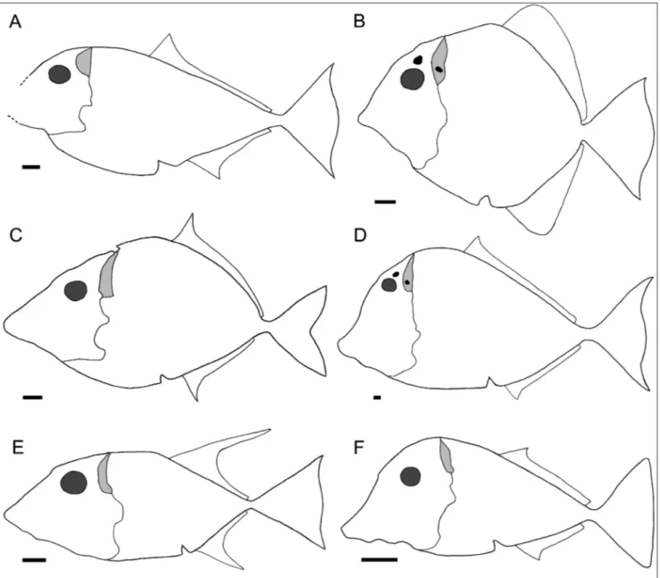
1. Body shape (as measured by ratio maxi-mum body height/standard length): discoid, 70– 100% (0); intermediate, 40–70% (1); fusiform, less than 40% (2); deep, more than 100% (3). See Fig. 7A-F for comparative outlines of the body in the genera revised herein. The state in Flagellipinna is coded as in the adult specimen (Cawley & Kriwet 2019: Table 1; state 0).
2. Relative position of dorsal apex: between the skull and the point of insertion of dorsal fin (0); apex absent (1); in point of insertion of dorsal fin (2); in skull (3). Many characters involving the body contour of Sigmapycnodus are coded as ? because it is not preserved in the only specimen known. The lectotype of Sylvienodus laveirensis, among other spe-cimens, shows that, although the contour scales are often preserved slightly disarticulated, there really is
no distinct dorsal apex because the dorsal border of the body is sub-horizontal between the skull and the dorsal fin (state 1; Figs. 5, 7E). Correct orientation of the holotype of Thiollierepycnodus (Ebert 2019: fig. 2) and Turboscinetes (Ebert 2016: Fig. 3, specimens should be shifted clockwise to make caudal fin com-pletely vertical) indicates that the dorsal apex is pla-ced in the point of insertion of the dorsal fin in the former (coded as 2) and between the skull and the dorsal fin in the latter (coded as 0). Character state 3, previously unknown in pycnodonts, is added in the present analysis because it was found to be pre-sent in Tergestinia, resulting on a specially truncated overall shape (Figs. 6, 7F).
3. Dorsal prominence: absent (0); present, curved, dorsally oriented (1).
4. Relative position of ventral apex: apex absent (0); before point of insertion of anal fin (1); in point of insertion of anal fin (2).
5. Mouth gape: horizontal or subhorizontal (0); inclined (1). The mouth gape in Turboscinetes as shown by Ebert (2016: Fig. 5) is subhorizontal ra-ther than inclined (coded as 0).
6. Prognathism: absent (i.e., anterior part of mouth gape not distinctively projected, although the whole ethmoid region is hypertrophied) (0); present by elongation of mesethmoid, vomer and prearticular in the horizontal plane, marking a con-cavity in the anterior border of the head (1); present by expansion of premaxilla and dentary (2). Flagel-lipinna is not considered to be prognathous (unlike Crawley & Kriwet 2019) because neither the vomer or the prearticular are elongated, the mesethmoid is hypertrophied as in other pycnodonts, and there is no concavity in the anterior border of the head (coded as 0).
7. Caudal pedicle: not differentiated (0); dif-ferentiated (1).
8. Morphology of frontal bones: narrow abo-ve the orbit (0); broad, expanded all throughout their length, nearly hemispheric in overall shape (1).
9. Prefrontal bones: absent (0); present (1). A paired prefrontal bone is present in Flagellipinna according to Cawley & Kriwet (2019: text, figs. 3, 4) and in Haqelpycnodus and Libanopycnodus according to Taverne & Capasso (2018a: 123, Fig. 9; 2018b: 6, Fig. 4). However, whenever a prefrontal bone is present, it is clearly distinct from, and observed in addition to, the superficial, ornamented portion of the mesethmoid, which is T-shaped in section
(e.g., Poyato-Ariza & Wenz 2005: Figs. 3, 5; see Nursall 1996: 129, figs. 5, 6 and 1999: 193, fig. 3, for the structure of the pycnodont mesethmoid). Such portion of the mesethmoid is restored absent in these three genera, which strongly indicates that their supposed prefrontal corresponds in fact to the shallow portion of the mesethmoid present in all pycnodonts (coded as 0). Most characters of the incomplete, very poorly preserved skull of Sigmapyc-nodus are coded as ? because the restoration and description by Taverne & Capasso (2008b: 17-23, fig. 17) appear unreliable when compared with the photos (op. cit.: figs. 15-16).
10(11). Dermocranial fenestra: absent (0); present (1). Personal observations confirm that the dermocranial fenestra is absent in Tergestinia (coded as 0 for this genus).
11(13). Parietal process: absent (0); present (1). This character is not observable in all taxa in-cluded in the analysis; when it is, the pectinated pa-rietal process is always present. The character has been kept because it is a very particular, traditionally remarked feature of many pycnodonts.
12(16). Extrascapular(s) fused to parietal: no (0); yes (1).
13(17). Endocranium (supraoccipital bone): completely covered by dermal skull in lateral view (0); exposed posteriorly (1); largely exposed poste-riorly (2). The type specimen of “C.” gridellii (Figs. 1, 7A) shows that the endocranium is very largely ex-posed posteriorly, comparatively more than in other pycnodonts (state 2). This is due partly to a poste-rior expansion of the endocranium and partly to the presence of a remarkable concavity in the posterior border of the dermal skull. The result is that the ex-posed portion of the endocranium is about as wide as it is deep (Fig. 7A). In other pycnodonts such as Polazzodus and Pycnodus the exposed portion is at least twice as high as it is wide (Fig. 7C, D; Poyato-Ariza 2010: fig. 3). The small ossifications identified as basioccipital and exoccipital, even intercalar, in Haqelpycnodus and Libanopycnodus (Taverne & Capas-so 2018a: Fig. 9; 2018b: Fig.4) do not correspond to the supraoccipital as exposed all throughout the posterior border of the parietal in Pycnodus or Po-lazzodus (e.g, Poyato-Ariza 2010: Fig. 3; coded as 0 for Haqelpycnodus and Libanopycnodus). Scalacurvichthys is reported to have a posteriorly exposed endocra-nium (Cawley & Kriwet 2018). However, the as-sessment of this character is not clear. In the only
specimen, this region is not well preserved and par-tially covered by matrix; the zone pointed as endo-cranium (Cawley & Kriwet op. cit.: figs. 2A, 3) looks hemispheric and massive, like an arcocentrum or an exoccipital rather than the thin, laminar supraocci-pital of other genera such as Pycnodus or Polazzodus. Scalacurvichthys was therefore conservatively coded as (?) for the present analysis; the homologies of the bones exposed in this region need confirmation in further, better preserved material.
14 (new). Postcephalic lacuna in endocranium (supraoccipital): supraoccipital not visible in late-ral view (0); lacuna absent (1); lacuna present (2). This character is considered independent from the previous one because, whenever the supraoccipital is exposed in lateral view, the lacuna can be either absent or present. The presence of a lacuna of os-sification in the posteriorly exposed portion of the endocranium was firstly reported by Blot (1984) in Pycnodus. Ulterior observations have confirmed the presence of such a lacuna in Oropycnodus (Poyato-Ariza & Wenz 2002: fig. 11B) and its absence in “C.” gridellii, Polazzodus, Sylvienodus, and Tergestinia (Fig. 7A-F). The poor state of preservation of this region as commented in the previous character does not allow assessment in Scalacurvichthys (coded as ?).
15(19). Infraorbitals: reduced to tubular os-sifications around infraorbital sensory canal (0); anterior infraorbital enlarged (1); small plates (2); absent (3). This character is coded as absent (3) in some taxa revised for the present analysis becau-se, even though these bones are tubular and easily lost during fossilization, complete absence of any indication of their presence in the numerous, well preserved specimens of Polazzodus, Sylvienodus, and Tergestinia strongly indicates that they have been al-together lost in these genera (coded as 3, state ad-ded for the present analysis). They also seem absent in the holotype of “C.” gridellii, but, since this is the only known specimen, it has been conservatively coded as unknown (?) in this form.
16(18). Anterior portion of infraorbital sen-sory canal: closely surrounding orbit (0); descen-ding towards ethmoid region (1). Not applicable in Polazzodus, Sylvienodus, and Tergestinia (see previous character).
17(20). Infraorbital ornamentation: present in all infraorbitals (0); present only in posteriormost one (1); absent in all infraorbitals (2). See comments on previous character.
18(21). Suborbitals: mosaic of small plates (0); absent as independent ossifications (1).
19(22). Preopercular and hyomandibular: preopercular single, hypertrophied; hyomandibular deep, unornamented (0); one high preopercular in close contact with small ornamented portion of hyomandibular, at same superficial level (1); preo-percular reduced, about as high as the ornamented portion of hyomandibular (2); preopercular additio-nally reduced, clearly lower than ornamented por-tion of hyomandibular (3). State 3 has been added for the present analysis because the preopercular bone is especially reduced in height in some genera, to the point that its main body (i.e., not including the anterior ascending process) is unmistakably lo-wer than the ornamented portion of the hyoman-dibular. This is clearly visible in all well preserved specimens of Polazzodus (Poyato-Ariza 2010: figs. 2, 4) and Pycnodus (e.g., Poyato-Ariza & Wenz 2002: fig. 10). Unfortunately, this region is not very well pre-served in the holotype and only specimen of “Coe-lodus” gridellii, but observable portions of the dorsal border of the preopercular bone indicate that it is not especially low, about as high as the ornamented portion of the hyomandibular (state 2). Although the anterior part of the preopercular bone is uni-quely partially fused to the hyomandibular in Syl-vienodus, its unfused surface clearly corresponds to state 2 (Poyato-Ariza 2013: fig. 3). The region is never well preserved in the numerous, but delica-tely ossified, specimens of Tergestinia, but the visible portions of the dorsal border of the preopercular bone indicate that it is very reduced, lower than the ornamented portion of the hyomandibular (state 3; e.g., holotype, MCSNT T.204).
20(23). Condyle in articular head of hyoman-dibular: absent (0); present (1).
21(24). Opercular bone: reduced (0); extre-mely reduced (1). An extreextre-mely reduced, narrow, opercular bone is observable in “C.” gridellii, Polaz-zodus Ariza 2010: 655), Sylvienodus (Poyato-Ariza 2013: fig. 3) and specimen MCSNT T.204 of Tergestinia (counterpart of the holotype; coded as 1 for this genus).
22(25). Branchiostegal rays: two, thin and se-parated (0); two, relatively large and in contact (1); absent (2); three (3). Poyato-Ariza & Wenz (2002: 167) pointed that when branchiostegal rays are not observed, such small, delicate bones may be absent due to taphonomic reasons. In some cases, at least
fragments of them can be observed in Oropycnodus (Poyato-Ariza & Wenz: fig. 17) and Pycnodus (Blot 1987: 35; pers. obs.). The large number of well-ossified and well-preserved specimens of Polazzo-dus observed with no traces of branchiostegal rays (Poyato-Ariza 2010: 655) is a reasonable indication that they are lost in at least this genus and, for the same reason, in Sylvienodus and Tergestinia (coded as 2, state added for the present analysis). The im-perfect preservation of the only specimen of “C.” gridellii prevents for such criterion to be applied to this genus (coded as ?). State 3 (three branchiostegal rays) is added for Turboscinetes according to Ebert (2016).
23(26). Morphology of premaxillary and den-tary teeth: robust, barely incisiform (0); very flatte-ned, fully incisiform (1); molariform (2); incisiform & bifurcated (3). The anterior region of the skull is unfortunately missing in the type and only spe-cimen of “C.” gridellii, so all characters concerning dentition and other features of the oral region are coded as unknown [?]. Haqelpycnodus presents small but robust, not fully incisiform teeth (Taverne & Capasso 2018a: Figs. 10, 11; coded as 0).
24(90). Morphology of premaxilla: small, with long, slender process continuous with anterior border of main body of bone (0); large, rounded, horizontally expanded, with short, robust process emitted by posterior region of the bone (1).
25(27). Number of premaxillary teeth: 2 (0); 3 (1); at least 8, arranged in at least two rows (2); 1 (3). State 3, only one premaxillary tooth, is added for the present analysis. This state is present in Po-lazzodus, Sylvienodus, and Tergestinia (see Poyato-Ariza 2010: 656 for a justification that this is not a preser-vational artifact).
26(28). Morphology of maxilla (outline): ovoid (0); reniform (1); straight oral border (2); elongated oval (3); oval with dorsal notch (4); axe-blade like (5). See Poyato-Ariza (2010: 656, fig. 5B; 2013: 94-95) for a full description of the very par-ticular morphology of the maxilla in Polazzodus and Sylvienodus (state 5, added for this analysis; see Poya-to-Ariza 2013: fig. 2 for a comparison of the maxil-lary outline in these two genera and Pycnodus). This confirms the remarkable variability of this bone in pycnodonts, in which it is always edentulous and very loosely articulated to the premaxilla; it is re-latively rare for it to be preserved. The maxilla in Thiollierepycnodus as illustrated by Ebert (2019: fig. 4)
is ovoid rather than reniform (i.e., there is no con-striction in the middle; coded as 0).
27(new). Crenulations on anteroventral bor-der of maxilla: absent (0); present (1). All pycno-donts whose maxilla is known exhibit a smooth border, whatever the contour morphology. Only Sylvienodus presents crenulations on the anteroven-tral border of this bone (see Poyato-Ariza 2013: 94-95, fig. 2 for additional details).
28(29). Morphology of vomerine teeth: cir-cular to subcircir-cular contour (0); oval contour (1);
re-niform contour (2); triangular contour on the main row; (3) oval, very elongated (4). Among the pyc-nodonts included in this analysis, state 3 is present in Polazzodus only (Poyato-Ariza 2010: 656, fig. 5D).
29(30). Arrangement of vomerine teeth in regular rows: present (0); absent anteriorly, present posteriorly (1).
30(31). Number of vomerine tooth rows: 5 (0); 3 (1); 6 (2).
31(32). Number of teeth in principal vomeri-ne tooth row: 10 or more (0); 8 or 9 (1); 7 or less (2).
Fig. 7 - Simplified body outline in: A) Polazzodus gridellii n. comb., formerly “Coelodus” gridellii (type and only known specimen anteriorly
incom-plete); B) Oropycnodus ponsorti; C) Polazzodus coronatus; D) Pycnodus apodus; E) Sylvienodus laveirensis; F) Tergestinia sorbinii. The eye is shown
in dark grey and the posteriorly exposed portion of the endocranium in light grey. The parietal process, present in all genera, is omitted for clarity; the dermocranial fenestra and the postcephalic lacuna are depicted in black when present. The pectoral and pelvic fins, very similar in all genera, are omitted for clarity. Idealized restorations based mostly on the corresponding type material, completed with observations on specimens MNHN MTA-42 and NHML P.30037 (Oropycnodus), NHMUK P.1634 (Pycnodus), IST-MDT 592-6 & 580
(Sylvienodus), and MCSNT T.201-202 (Tergestinia). See text and Poyato-Ariza & Wenz 2002: 189 for comments on the shape of the anal
Unlike Cawley & Kriwet (2017), this character is co-ded as unknown (?) for Scalacurvichthys because the vomer of the single known specimen is only partial-ly exposed in lateral view and is covered to a degree by the lower jaw posteriorly (Cawley & Kriwet 2017, figs. 2-3), so that the principal tooth row is not fully visible and there may be more than the seven teeth reported by those authors. Vomerine dentition cha-racters are also coded as ? for Libanopycnodus becau-se only the most lateral row is expobecau-sed, and only in lateral view (Taverne & Capasso 2018b, figs. 3, 4).
32(33). Alternation of teeth on main vomeri-ne tooth row: absent (0); present (1).
33(34). Number of dentary teeth: 4 (0); 3 (1); 2 (2). There are currently no known pycnodonts with only one dentary tooth. Polazzodus and Terge-stinia have a single premaxillary tooth (see character 27 above), but they clearly show two teeth on the dentary (state 2).
34(35). Morphology of prearticulary teeth: oval contour (0); circular contour (1); sigmoid to drop-shaped contour (2); extremely elongated in contour, long axis perpendicular to row axis (3); oval, elongated, long axis of teeth coincident with row axis (4). Characters 35-38 are unknown in Syl-vienodus because the prearticulary teeth are not suffi-ciently exposed in any observed specimen.
35(36). Arrangement of prearticular teeth in regular rows: present (0); absent anteriorly, present posteriorly (1); absent (2). Only the posterior part of the prearticulars is exposed in Libanopycnodus (Ta-verne & Capasso 2018b, figs. 5, 6; coded as ?).
36(37). Number of prearticular tooth rows: 3 (0); 2 (1); 5 or 6 (2). The prearticular tooth rows are different in the holotype and the paratype of Haqelpycnodus (Taverne & Capasso 2018a: 124, Figs. 10, 11, 13; coded as ?). “There are three rows of teeth present in the prearticular bone” in Scalacur-vichthys according to Cawley & Kriwet (2018: 665, figs. 1-3). However, the left prearticular of the ho-lotype and only specimen, partially visible in medial view, is largely covered by the right prearticular and the main tooth row is not exposed. Therefore, it is not possible to be certain of the number of prearti-cular tooth rows and it is coded as unknown (?) for this genus.
37(38). Number of teeth on main prearticular tooth row: 8 or 9 (0); 7 or fewer (1); 10 or more (2). See character 35 for Libanopycnodus. Incompletely prearticular dentitions in articulated specimens of
Polazzodus have 7 teeth at the most, but they are ne-ver observable in their entirety; in turn, complete specimen MPCM 10879 clearly shows 8 (Poyato-Ariza 2010: 657, fig. 5F; state 0). Although the pre-articular tooth plate is never observed in Tergestinia in its entirety, there is no room before the mandibu-lar articulation for more than 7 teeth on the main row. Coded as ? for Scalacurvichthys (see previous character).
38(39). Coronoid process: high, straight dor-sal border (0); high, club shaped (1). The morpho-logy that corresponds to state 0 is suggested by the observable mandibular portion of the lectotype and fully observable in specimen IST-MDT-580 of Syl-vienodus. As shown by Cawley & Kriwet (2018: figs. 2-3), the coronoid process of Scalacurvichthys is cle-arly not club-shaped, as it lacks the bulky head and the narrowing that define such morphology (com-pare with Poyato-Ariza & Wenz 2002: fig. 23B and 2004: figs. 5, 9B), so it is coded as 0 for this genus.
39(40). Crenulations in vomerine and prear-ticular teeth: occasionally present, weak (0); absent (1); present in most teeth, strong (2). Vomerine and especially prearticular teeth are seldom exposed in Sylvienodus. Nonethess, paralectotype LNEG-MG 6658 shows three teeth, two on the vomer and one on the prearticular, all three with strong crenula-tions. See Poyato-Ariza (2013: 95) for additional details on the strong crenulations observed in this taxon (state 2).
40(41). Groove on vomerine and prearticular teeth: absent (0); present (1).
41(42). Number of vertebrae (epichordal ele-ments excluding those of the caudal endoskeleton): 30–34 (0); 35 or more (1); 25–29 (2); 24 or fewer (3). The number of vertebrae in Sylvienodus, 22-23, is quite reduced for a pycnodont (Poyato-Ariza 2013: Table 1; state 3). A maximum number of 29 is consistently observed in Tergestinia (13 abdominal, 15-generally 16 caudal excluding those of the cau-dal endoskeleton).
42(43). Neural and haemal corresponding ar-cocentra: not surrounding notochord (0); surroun-ding notochord partially (1); surrounsurroun-ding notochord completely (2). Adult specimens of Flagellipinna are reported to have the arcocentra surrounding the no-tochord completely but restored with all arcocentra surrounding the notochord partially only (Cawley & Kriwet 2019: figs. 1, 2; coded as 1). Same applies to Sigmapycnodus (Taverne & Capasso 2008b: 23, figs.
12-14; coded as 1). The lectotype of Sylvienodus la-veirensis clearly shows that they surround the noto-chord completely (state 2).
43(44). Neural and haemal adjacent arcocen-tra: simple contact (0); complex contact (1); hyper complex contact (2); expanded and imbricate (3). The last caudal vertebrae show a hyper complex contact in Flagellipinna, but it is puzzling restored with overlapping arcocentra (Cawley & Kriwet 2019: figs. 7 & 2 respectively; coded as 2 & 3). This cha-racter is unclear in Sigmapycnodus too; it is described as “hypercomplex” by Taverne & Capasso (2008b: fig. 23), but their photos and restoration (op. cit.: 12-14, 20), show only 2-3 interdigitations (complex contact only), and some vertebrae appear separated, without contact (coded as 1 for the vertebrae actual-ly in contact).
44(45). Sagittal flanges on neural and haemal spines: anterior, small and short (0); anterior, large and long (1); anterior and posterior (2). Modified
in Potiguara from 0&1 to 1 because these flanges
cannot be simultaneously short and long, and they are long as described by Machado & Brito (2006: 2) in relation with the character as originally used by Poyato-Ariza & Wenz (2002, 2004).
45(46). Number of autogenous anterior neu-ral spines: 10 or more (0); 7–9 (1); 6 or fewer (2). At most six autogenous neural spines can be counted in the holotype of “C.” gridellii (state 2). This cha-racter is difficult to determine in Sylvienodus; howe-ver, paralectotype LNEG-MG 6658 shows that the seventh neural spine is autogenous and the eighth is fused (state 1).
46(47). Relative length of last neural spine not supporting precurrent caudal fin rays: slightly shor-ter than previous ones (0); less than half as long as preceding spines (1); vestigial (2).
47(48). Number of epichordal elements of caudal endoskeleton: primitive (?); 6 to 8 (1); 4 or 5 (2); 3 or fewer (3). The state of preservation in Libanopycnodus does not allow verification (Taverne & Capasso 2018b: fig.8). Most caudal skeleton cha-racters are coded as ? in Sigmapycnodus because of the incompleteness and very poor state of preservation (Taverne & Capasso 2008b: figs. 21, 22). It is diffi-cult to discern between 6 and 7 epichordal elements in Tergestinia; specimen MCSNT T.17 seems to show a maximum of 6 (state 1 in any case). There seem to be 6 epichordal elements in the caudal skeleton of the holotype of “C.” gridellii (state 1). According to
Ebert (2016: Fig. 11), there are 4, maybe 5, epichor-dal elements supporting the fin exoskeleton, so it is coded as 2 for this genus.
48(49). Relative development of hypochor-dal elements of cauhypochor-dal endoskeleton: only slightly enlarged (0); enlarged, plate-like (1); one hypertro-phied element (2); two hypertrohypertro-phied elements (3). For the present analysis, we consider that “hyper-trophied elements” correspond to those that are clearly larger than the normal size for their position in the series and clearly show at least one longitudi-nal ridge (suggesting a possible compound origin by fusion of two plates). In this sense, only Oropycnodus and Pycnodus among the genera re-evaluated for this analysis present state 3. Scalacurvichthys is coded as 0 after Cawley & Kriwet 2018 (fig. 4A, B; the camera lucida drawing on C does not seem to correspond clearly to the photos on A and B and is confusing and hard to interpret). Turboscinetes is coded as 1 be-cause hypochordal element 6 and very likely 7 (party under the fin rays) of the caudal endoskeleton are enlarged and plate-like (Ebert 2016: Fig. 11).
49(50). Number of hypochordal elements of caudal endoskeleton: 9–11 (0); 12–13 (1); 6–8 (2). See character 47 for Libanopycnodus.
50(51). Diastema: absent (0); present (1). 51(52). Cleithrum: curved, anteroventral limb subvertical, expanded (0); two small posterior ex-pansions framing a shallow notch for the insertion of the fin (1); two large posterior expansions fra-ming a deep notch for the insertion of the fin plus anterior elongation of the anteroventral limb (2). The morphology of the cleithrum in the genera re-evaluated for the present analysis has shown in-teresting variations (Fig. 8). The simple, subvertical cleithrum broadly present in other Pycnodontidae is not observed among the revised genera. State 1 corresponds to Pycnodus, whose cleithrum presents two short posterior processes, upper and lower, that frame a high but shallow notch for the inser-tion of the fin (e.g., Poyato-Ariza & Wenz 2002: fig. 10; present paper: Fig. 8A); this also seems to be the morphology in Oropycnodus (although the pre-servation of this bone in this taxon is not good), Sylvienodus, and Tergestinia (Fig. 7). The morphology of the cleithrum in Polazzodus presents remarkable additional modifications; the two posterior proces-ses are more robust and expanded, so that the notch for the insertion of the fin is quite deeper. As a mat-ter of fact, the lower process is formed as an
ex-pansion of the whole posteroventral angle between the limbs of the bone. In addition, the anteroven-tral part of the bone is elongated anteriorly, so that the bone is crescent-shaped rather than subvertical (Poyato-Ariza 2010: fig. 6; present paper: Fig. 8B). The holotype of “C.” gridellii shows a cleithrum whose morphology is similar to that of Polazzodus (state 2). The cleithrum of Scalacurvichthys, as seen in Cawley & Kriwet (2018: figs. 2A-3) corresponds to the morphology of the primitive state (coded as 0).
52(54). Position of pelvic fins (ratio prepel-vic distance/standard length): 45–55% (0); more than 55% (1); less than 45% (2). Although the type and only specimen of “C.” gridellii is incomplete, an estimation (for comparative purposes) of 55mm of prepelvic length and of 104 mm in standard length results in 52,3% (state 0).
53(55). Position of dorsal fin (predorsal length/standard length): 60%–69% (0); less than 49% (1); 50%–59% (2); 70%–79% (3). Althou-gh the type and only specimen of “C.” gridellii is incomplete, an estimation of 53mm of predorsal length and of 104 mm in standard length results in
51% (state 2). Correct orientation of the holotype
of Thiollierepycnodus (Ebert 2009: fig. 2) places the
predorsal length at about 67-68% (state 0).
54(56). Number of dorsal axonosts: 30–39 (0); 40–49 (1); 50–59 (2); 60 or more (3). The number of dorsal axonosts in Tergestinia is variable, but ne-ver more than 52 or less than 50 (state 2). The state of preservation prevents from an accurate account in “C.” gridellii, but an estimation of some 52-54 ele-ments places it safely within the interval of state 2. In Scalacurvichthys “(…) disarticulation seems likely” in the dorsal fin (Cawley & Kriwet 2018: 666). The posteriormost dorsal fin rays and axonosts, faintly preserved as impressions, shown by the holotype and only specimen (Cawley & Kriwet op. cit.: fig. 1) are progressively more separated from their anato-mic position; the whole posterodorsal border of the body appears completely eroded out and blurred in the specimen. The partial preservation and disarti-culation are indications that the posteriormost por-tion of the fin, maybe even quite long, has been lost during fossilization. As a consequence, it is not likely that there are only 11 axonosts in its dorsal fin (coded as ? for this genus).
55(57). Dorsal axonost not supporting lepi-dotrichium (free axonost): absent (0); present (1).
56(58). Morphology of dorsal and anal fins: primitive (?); strip-like (1); falcate to acuminate (2); sigmoid outline (3); rounded in center (4); rounded anteriorly (5); extremely acuminate, higher anteriorly than it is long (6). There is a remarkable variability in shape among the different genera, but the dor-sal and anal fin of each particular taxon normally present the same contour shape regardless of their different length. Although this may not seem the case in the type material of Oropycnodus (Fig. 2), this is a preservational artifact; whenever well observa-ble in its entirety, the anal fin of this genus has the same shape than that of the dorsal fin (e.g., NHML P.30037; Poyato-Ariza & Wenz 2002: 189), that is, rounded anteriorly; so it has been restored on Fig. 7B. Most lepidotrichia of these fins in Libanopycno-dus are lost, none preserved in its entirety; the resto-ration and description of the fins shape cannot be corroborated (Taverne & Capasso 2018b: figs. 1,2; coded as ?). See Poyato-Ariza (2013: 96-97; fig. 4) for a detailed description of the peculiar dorsal and anal fins of Sylvienodus. See also Fig. 7 in the present paper for a comparison of different genera. Due to inadequate preservation of this region,
Scalacur-Fig. 8 - Restored comparative outline of the cleithrum in: A) Pycno-dus apoPycno-dus, based on a camera lucida drawing of transferred
specimen NHMUK P.1634; B) Polazzodus coronatus, based on
a camera lucida drawing of the holotype, MPCM-13464. Ae) anteroventral elongation of the anteroventral limb of the bone; Lpp) lower posterior process; N) large notch for the articulation of the endochondral girdle and insertion of the pectoral fin; Upp) upper posterior process. Notice the small notch, practically closed, in the anterodorsal border of the lower posterior process in B. Line bars represent 2 mm.
vichthys is coded as ? (see comments in character 54). 57(59). Position of anal fin (preanal length/ standard length): 70%–79% (0); 50%–59% (1); 60%–69% (2); 80%–89% (3). The deformations presented to some extent by most specimens of Po-lazzodus makes this percentage quite variable. The lowest percentage is 61,74% in a clearly deformed specimen and 64,90-68,33% in apparently non-de-formed specimens (Poyato-Ariza 2010: table 1). The highest percentages are 71,33% in the holotype and 74,75% in an apparently non-deformed specimen (idem). Such variation makes advisable to code this character as 0 & 2 simultaneously in this genus (this character is processed as unordered). For analogous reasons, it has been coded also 0&2 in Sylvienodus (see Poyato-Ariza 2013: Table 1 for further details). Correct orientation of the holotype of Thiollierepyc-nodus (Ebert 2009: fig. 2) places the preanal length at about 61-62% (state 2).
58(60). Number of anal axonosts: 20–29 (0); 10–19 (1); 30–39 (2); 40–49 (3); 50 or more (4); 9 or fewer (5). The holotype of Tergestinia shows 34-35 anal axonosts (state 2). The state of preservation prevents from an exact account in “C.” gridellii, but an estimation of some 35-37 gets it safely within the interval of state 2. The holotype of Polazzodus coro-natus has 41 anal axonosts, although all other speci-mens have less than 39 (Poyato-Ariza 2010: table 1; coded as 2&3).
59(61). Urodermals: one (0); two (1); absent (2). When present, urodermals are paired elements placed lateral to the proximal portions of the fin rays and/or the hypochordal elements (e.g., Poyato-Ariza & Wenz 2002: Figs. 24-27, 29, 30). However, the element described and illustrated as a urodermal by Taverne & Capasso (2018a,b) in Haqelpycnodus is an unpaired osseous element not lateral to fin rays or hypochordal elements; could be a fragment of the broken last epichordal element. For these reasons, it has been coded as unknown (?) in this genus. See Poyato-Ariza (2013: 97; fig. 5) for the description of the single urodermal of Sylvienodus.
60(62). Number of caudal principal fin rays: primitive (?); 9 or fewer (0); 10–19 (1); 20–25 (2); 26–35 (3); 36 or more (4). Whenever accurately observable, specimens of Tergestinia show 8 caudal principal rays in the upper lobe and 10-11 in the lower lobe, accounting for a total of 18-19 (mostly 19; state 1). The holotype of “C.” gridellii seems to show 21-22 principal caudal fin rays (9 in the upper
lobe and 12-13 in the lower lobe; state 2).
61(63). Morphology of caudal fin: distal bor-der convex (0); distal borbor-der concave (1); double emarginated (2); vertical (3); forked (4); distal bor-der vertical, straight (5). See Fig. 7 for comparison among the genera revised for this analysis. Polaz-zodus has a forked caudal fin that is unique among Pycnodontidae (state 4, new in the present analysis; Fig. 7C). In turn, Tergestinia has a caudal fin with a distal border that is vertical and straight (Fig. 7F), so state 5 was added for the present analysis as well. The distal border of the caudal fin is not perfectly preserved in the holotype of “C.” gridellii (Fig. 1), but the observable portion strongly suggests it is double emarginated (Fig. 7A). This is not the case of Libanopycnodus, where only the basal portion of the fin rays is preserved (Taverne & Capasso 2018b: fig. 8; coded as ?).
62(64). Ossification of scales: complete in all scales (0); complete in abdominal scales, incomplete in caudal scales (1); complete in ventral scales and in some dorsal scales (2); complete in ventral sca-les, incomplete in dorsal scales (3); incomplete in all scales (4).
63(65). Distribution of scales: only abdomi-nal region (0); whole body except caudal pedicle (1); abdominal region plus part of caudal region (2); whole body (3).
64(66). Arrangement of scales: rows in same direction (0); rows in different directions (1).
65(67). Ornamentation: tubercles (0); ridges (1); reticulation (2). Some taxa show different or-namentations simultaneously, such as the tubercles and reticulation of Sylvienodus (IST-MDT 592). Be-cause this character refers to ornamentation on the dermal skull bones (not only on scales, which are rarely ornamented in pycnodontids), it is coded as 0 for Haqelpycnodus (Taverne & Capasso 2018a: 123) and 1 for Turboscinetes (Ebert 2016: Fig. 6).
66(69). First dorsal ridge scale: about same size as subsequent ridge scales (0); larger than subse-quent ridge scales (1). In Scalacurvichthys (Cawley & Kriwet 2018: figs. 1-3), the gap between the dermal supraoccipital and the first visible dorsal ridge scale strongly suggest that the latter is actually the second dorsal ridge scale, and the first dorsal ridge scale, normally occupying the area of that gap, has been lost during fossilization in this partially disarticula-ted specimen. This interpretation is suppordisarticula-ted by the fact that the whole dorsal ridge scale series is
partially disarticulated, broken, not entirely preser-ved (coded as ? for this genus). The argument that this scale contacts the dermal supraoccipital ante-roventrally and that it is incorporated into the skull roof (Cawley & Kriwet 2018: 667) does not support its interpretation as the real first dorsal ridge scale, because such contact also occurs in the second dor-sal ridge scale of other pycnodontids, such as Polaz-zodus (Poyato-Ariza 2010: fig. 3), whereas the real incorporation of the actual first dorsal ridge scale into the skull roof involves a tight structural conti-nuity with the dermal supraoccipital bone, with no gap between them (e.g., Poyato-Ariza & Wenz 2002: figs. 6-9; 2004: figs. 4-5; Poyato-Ariza 2010: fig. 3).
67(new). Morphology of second dorsal rid-ge scale: similar to subsequent dorsal ridrid-ge scales (0); very long and large, with a central longitudinal ridge and a conspicuous anterior hook (1). State 1 accounts for the particular morphology of the se-cond dorsal ridge scale in Polazzodus (for additional information see Poyato-Ariza 2010: 660-661; fig. 8). The second ridge scale (“first dorsal ridge scale” in Cawley & Kriwet 2018: figs. 1-3) of Scalacurvichthys is broken, only partially preserved; the restoration in Cawley & Kriwet (op. cit.: figs. 2B, D) is not straightforward when compared to the photos (op. cit.: figs. 2A, 3). For this reason, it has been coded as ? for the present analysis.
68(70). Scutellum-like contour scales: absent (0); present, dorsal only (1); present, ventral only (2); present, dorsal and ventral (3).
69(71). Number of differentiated dorsal ridge scales: 18 or more (0); 15 to 17 (1); 10 to 14 (2); 7 to 9 (3). The number of dorsal ridge scales is about 11 in Tergestinia. Although 9 dorsal ridge scales are shown by the holotype and only specimen of Sca-lacurvichthys (Cawley & Kriwet 2018: fig.1), the loss of the first dorsal ridge scale (as discussed in cha-racter 66 above) and the imperfect preservation of the whole series, with a complete loss of the poste-riormost elements, are strong arguments for a total number of dorsal ridge scales well over 9. In other words, there can be no certainty that there were only 9 (coded as ?).
70(72). Arrangement of dorsal ridge scales: dorsal contour scales in close contact with each other (0); point contact (1); separated from each other (2). This character is rarely observable in Syl-vienodus laveirensis; paralectotype LNEG-MG 6658 shows dorsal ridge scales that are in point contact
with each other. It is coded as 2 for Turboscinetes be-cause this character refers to the standard scales of the series, not including the anteriormost, enlarged ones (accounted for in characters 66 and 67) and mostly in contact because they are enlarged.
71(73). Number of spines on midline of dorsal ridge scales: 3 or more (0); midline serrated (1); 1 or 2 (2); no spines on dorsal contour scales (3). Consistently coded according to the maximum number observed in any scale of any specimen (the number of spines increases with age of specimen). As for the previous character, it is seldom visible in Sylvienodus laveirensis, but paralectotype LNEG-MG 6658 shows dorsal ridge scales that present a ser-rated midline (see also Poyato-Ariza 2013: fig. 6A). See Taverne & Capasso (2018b: figs. 1, 10) for the serrated midlines of the posteriormost scales of the series in Libanopycnodus (coded as 1).
72(74). Distribution of spines on midline of each dorsal ridge scale: all along midline, or cente-red if only one spine present (0); no spines on dor-sal contour scales (1); posterior region (at most two thirds) of midline (2); anterior region (at most two thirds) of midline (3).
73(75). Contact of spines on each dorsal rid-ge scale: separated from each other (0); no spines on dorsal contour scales (1); in contact with each other (2).
74(76). Relative size of anterior and posterior spines on each dorsal ridge scale: similar in size (0); no spines on dorsal contour scales (1); spines of increasing size in cephalocaudal sense (2).
75(91). First ventral keel scale: smaller than subsequent scales (0); larger than subsequent scales (1).
76(77). Number of ventral keel scales: 22 or more (0); 18 to 21 (1); 15 to 17 (2); 10 to 14 (3). The holotype of “C.” gridellii shows at most 14 ven-tral keel scales: 11-12 before the cloaca, remarkably longer in caudal sense, the anteriormost ones less perfectly preserved, plus 2 after the cloaca (coded as 3). Because of imperfect preservation and un-certainty of description of the ventral keel scales in Flagellipinna, Haqelpycnodus and Libanopycnodus, their corresponding characters are all coded as unknown. A complete series of ventral keel scales is never cle-arly observed in any specimen of Tergestinia, but a maximum estimation of 12 of them can be made on the basis of the number of corresponding flank scale rows. With the 2 post-cloacal scales, this
ac-counts for an estimated maximum number of 14 ventral keel scales (coded as 3 for this genus; simi-lar estimation for “C.” gridellii). The disarticulation and loss of the anteriormost ventral keel scales in Scalacurvichthys (Cawley & Kriwet 2018: fig. 1) ren-ders the number impossible to be estimated becau-se flank scales rows are also disarticulated and in-complete (coded as ?).
77(78). Arrangement of ventral keel scales: close contact with each other (0); point contact (1). The ventral keel scales are remarkably elongated in “C.” gridellii, resulting on a point contact with each other. A close contact can be observed in the ven-tral keel scales shown by specimen MCSNT T.11 of Tergestinia (coded 0 for this genus).
78(79). Number of spines on the midline of ventral keel scales: 4 or more (0); 1 to 3 (1); no spi-nes on ventral keel scales (2). Consistently coded as the maximum number observed in any scale of any specimen, usually in the postcloacal ventral keel scales of larger individuals (the number of spines increases with age). Characters 78-80 in “C.” gri-dellii: at least one postcloacal scale of the holotype shows two spines on the posterior region of the midline, separated from each other and increasing in size cephalocaudally.
79(80). Distribution of spines on midline of ventral keel scales: all along midline, or centered if only one spine present (0); no spines on ventral keel scales (1); posterior region (at most two thirds) of midline (2). Although this character is coded as 1 (no spines) by Ebert (2016) for Turboscinetes this must be a typing mistake, because the author states that “Spines on the ventral keel scales are only pre-sent on the posteriormost two thirds…” (op. cit.: p. 33). It has therefore been coded as 2 in this genus for the present analysis.
80(81). Contact of spines on each ventral keel scale: separated from each other (0); no spines on ventral keel scales (1); in contact with each other (2); imbricate (3).
81(82). Relative size of anterior and posterior spines on each ventral keel scale: spines of increa-sing size cephalocaudally (0); no spines on ventral keel scales (1).
82(83). Several scales attached to contour scales: no (0); yes (1).
83(84). Number of post-cloacal ventral keel scales: more than 6 (0); 5 or 6 (1); 3 or 4 (2); two (3). The holotype of “C.” gridellii and quite a
num-ber of specimens of Tergestinia, including paratypes MCSNT T.66 and T.201/202, exhibit 2 ventral keel scales posterior to the cloaca (state 3 for both taxa). Coded as state 2 (three scales) for Turbosci-netes, being the number shown by adult specimen in Ebert (2016: fig. 16). Scalacurvichthys is reported to have only one (Cawley & Kriwet 2018), but the ventral region between the cloaca and the anal fin is disarticulated, blurry, only partially preserved, so that it is impossible to be sure that there was only one post-cloacal keel scale. This is supported by the fact that there is an ample space before the anal fin; even a fragment of matrix in this area is completely lost (Cawley & Kriwet op. cit.: figs. 1, 5A), so that the actual number cannot be precisely established (coded as ?).
84(85). Number of anterior cloacal modified scales: mosaic of little scales (0); two (1); one (2); anterior cloacal scales not modified (3); three (4). Characters 85-89 in Sylvienodus: see Poyato-Ariza (2013: 98; fig. 6B) for a detailed description and restoration of the cloaca. The holotype of Tergesti-nia (MCSNT T.203) shows a single modified ante-rior cloacal scale (state 2). Only the roofing cloacal scales of Flagellipinna are described and restored by Cawley & Kriwet (2019), so that the complete structure of the cloaca is actually unknown. The inadequate preservation of the broken, partial-ly lost cloaca in Scalacurvichthys (Cawley & Kriwet 2018: fig. 5A; their restoration on fig. 5B is difficult to make correspond to the photograph) does not allow an accurate estimation (conservatively coded as ? for the present analysis).
85(86). Number of posterior cloacal modi-fied scales: mosaic of little scales (0); three (1); two (2); one (3); no scales, posterior part of anal notch formed by a rib (4). See comments on previous character for Scalacurvichthys (coded as ?).
86(87). Bifid scale in cloaca: absent (0); pre-sent (1); prepre-sent plus several comma-shaped scales (2). A short, thin scale, ventrally bifid, is forming the roof of the cloacal notch in Nursallia, Oropyc-nodus (Poyato-Ariza & Wenz 2002: fig. 42), Polazzo-dus (Poyato-Ariza 2010: fig. 9), PycnoPolazzo-dus (Blot 1987: pl.22, fig.2), and Sylvienodus (Poyato-Ariza 2013: fig. 6). The bifid scale in the cloaca of Tergestinia is cle-arly shown by the holotype and also, in different degrees of preservation, in a number of paratypes. The cloaca of the type and only specimen of “C.” gridellii is badly preserved. The presence of a
bi-fid scale is strongly indicated by the morphology and arrangement of the preserved fragments, yet it is conservatively coded as unknown (?) becau-se there is no direct evidence. This ventrally bifid scale is not to be mistaken with the large, dorsally bifid long structures reported by Cawley & Kriwet (2018) flanking the cloaca in the single known spe-cimen of Scalacurvichthys (op. cit.: fig. 5; coded as 0). Not only their relative size and structure is comple-tely different, also the fact that they are poorly pre-served and dubiously restored in their illustration. They also cite such structures in Stemmatodus and Proscinetes (op. cit.: figs 7, 8), in a single specimen each, also poorly preserved and illustrated. Such structure may simply represent standard flank sca-les ventrally fused in particular individual variations. The future of these structures as an useful cha-racter in pycnodont phylogeny requires previous confirmation and individual variation screening in further, better preserved specimens. The restora-tion of the cloacal remains, very poorly preserved,
in Libanopycnodus (Taverne & Capasso 2018b: fig.
11) shows a sort of broken, partially preserved sca-le described as “bifid”, but the poor preservation of the region suggest strong caution (coded as ?). In addition, the morphology of this scale, as tenta-tively restored, does not correspond to that of the actual bifid scale of the other genera.
87(88). Post-cloacal notch: absent (0); pre-sent (1).
88(89). Supracloacal scale: absent (0); pre-sent, contacting only cloacal scales (1); prepre-sent, contacting also non-differentiated scales adjacent to cloacal scales (2). This character is coded as 0
in Turboscinetes because there is no supracloacal
scale as defined by Poyato-Ariza & Wenz (2004: 364). This is so because the scale contacting se-veral scales, as seen in Ebert (2016: Fig. 16), does not contact cloacal scales; the scales that the sup-posed supracloacal scale is contacting ventrally are not cloacal scales, because they are not part of the actual border of the cloaca. This feature simply se-ems to correspond to a slight alteration of the sca-le rows between the pelvic fin and the cloaca; the cloacal scales of Turboscinetes do not contact a single scale dorsally.
Ebert (2016: 53) added three new characters. These have not been taken into account for dif-ferent reasons: his number 92, about fringing and
basal fulcra, because it does not take into account the very relevant individual variation in these struc-tures; his number 93, about ethmoid commissure, because the very delicate bones in this region are easily lost, so that this can mislead to an absence that is just preservational; and his number 94, com-plete scale rows posterior to the cloaca, because it is wrongly polarized (i.e., basal pycnodonts cer-tainly have more than one complete row posterior to the cloaca).
Analysis
The data matrix (Table 1) was processed by PAUP program 3.1.1 in an iMac 8 computer at the Unidad de Paleontología, Universidad Autónoma de Madrid. General heuristic search was used to run the analysis.
Multiple states are interpreted as polymorphi-sms (default settings), according to the observa-tions in the list above. Because the Pycnodontidae is a monophyletic clade (Poyato-Ariza & Wenz 2002), the ingroup is made a monophyletic sister group to the outgroup.
Additivity
Characters 13, 17, 19, 31, 33, 35, 42-46, 48, 51, 54, 62, 69, 70, 76, 78, 83, 85, 86, and 88 were processed as ordered. All other multistate characters were processed as unordered. For a justification on ordering particular characters in pycnodonts, see Poyato-Ariza & Wenz (2002: 142); for a discussion on the methodological sense of ordering characters in a cladistic analysis, see Poyato-Ariza (2005b: 544, with abundant references). Further arguments to fa-vour the additivity of multistate characters can be provided at present. The relevance of ontogenet-ic restrontogenet-ictions in evolutionary processes has been widely established in the last decades, stressing the relevance of heterochrony in explicit phylogenetic contexts as a mechanism for constraining direction-ality and patterns in morphological evolution (e.g., Alberch 1980; Maynard Smith et al. 1985; Alberch & Blanco 1996; Baguñà & García-Fernández 2003; Smith 2003; Laubichler & Maienschein 2007). Ad-mittedly, the relationships between ontogeny and phylogenetic systematics have long been regarded as enormously complicated (e.g., De Queiroz 1985; Kluge 1985; Kluge & Strauss 1985), way far of the scope of the present paper. It is worth noticing, though, that ontogeny provides empirical evidence