Summary X Chapter 1 - Fundamentals of tissue engineering
1.1 Introduction 2
1.2 Importance of cell-matrix interaction 3
1.3 Biomaterials in tissue engineering application 7
1.3.1 Design criteria of biomaterials 10
1.3.2 Surface modification 12
1.4 Semipermeable polymeric membrane as a biomaterial 14
1.4.1 Membrane properties in a bio-hybrid system 16
Experimental design and aim of the work 21
References 23
Chapter 2 - Membrane approaches for liver tissue engineering
2.1 Introduction 28
2.2 Hepatic structure and function: bal issues design 29
2.2.1 Cell source 30
2.3 Culture system 31
2.4 Bioreactor 33
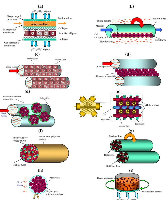
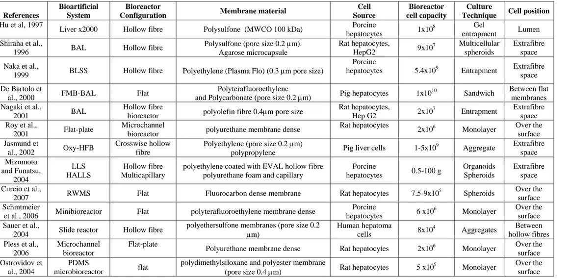
2.4.1 Membrane bio-hybrid artificial liver (BAL) systems in clinical evaluation 37 2.4.2 Membrane BAL system in preclinical and in vitro evaluation 40
2.5 Membranes for liver reconstruction 46
2.6 Concluding remarks 48
References 49
Chapter 3 - Membrane approaches for neuronal tissue engineering: state of the art
3.1 Introduction 56
3.2 Nervous system: injury and repair 57
3.2.1 Neurotrophic factors to promote regeneration 60
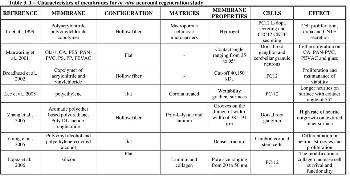
3.5 Membranes used in in vivo neuronal regeneration 73
3.6 Concluding remarks 78
References 79
Chapter 4 - Rat embryonic liver cell expansion and differentiation on NH3
plasma-grafted PEEK-WC-PU membranes – Paper 1 85
Chapter 5 - Biodegradable and synthetic membranes for the expansion and functional differentiation of rat embryonic liver cells – Paper 2 95
Chapter 6 - 2-D and 3-D membrane systems for the reconstruction of hyppocampal neuronal network - Manuscript Submitted
Abstract 108
6.1 Introduction 109
6.2 Materials and methods 111
6.2.1 Preparation of flat and hollow fiber (HF) membranes 111
6.2.2 Membrane characterization 112
6.2.3 Cell isolation and culture 113
6.2.4 Immunostaining of neuronal cells and quantitative analysis 114
6.2.5 Sample preparation for SEM 115
6.2.6 Metabolic assays 115
6.2.7 Extraction of total RNA and preparation of cDNA 116
6.2.8 Quantitative real time PCR (QPCR) 116
6.3 Results 117
6.3.1 Membrane properties 117
6.3.2 Neuronal morphological and morphometric evaluation 119
6.3.3 Neuronal metabolism 120
6.3.4 MAP2 and GLUR2 mRNA expression 121
Chapter 7 - Influence of micropatterned PLLA membranes on outgrowth and
orientation of hippocampal neurites – Paper 3 151
Conclusions 164
Other publications i
Scientific activity iii
Sommario
I notevoli progressi nel campo dei biomateriali hanno reso possibile l’impiego di polimeri sintetici e naturali in svariate applicazioni dell’ingegneria tissutale e della medicina rigenerativa.
Il microambiente tridimensionale che circonda le cellule in vivo esercita un ruolo fondamentale nei processi di rigenerazione, mantenimento e riparazione tissutale, pertanto una dettagliata conoscenza a livello strutturale e funzionale del tessuto di interesse è fondamentale per un efficente contributo dell’ingegneria tissutale.
Un approccio interessante nell’ambito della medicina rigenerativa è rappresentato dall’uso di diversi biomateriali, costituiti da componenti biologiche, sintetiche, o da entrambre, capaci di agire come “materiali instruttivi” simulando e ricreando il naturale microambiente cellulare. L’impiego delle membrane polimeriche nell’ambito dell’ingegneria tissutale, grazie alle loro caratteristiche di stabilità, biocompatibilità nonchè di permeabilità selettiva, suscita notevole interesse e si rivela particolarmente promettente.
Il vantaggio fornito dalle membrane polimeriche consiste infatti, nella capacità di simulare la matrice extracellulare, consentendo e promuovendo il ripristino di una citoarchitettura tridimensionale ed il trasporto selettivo di metaboliti e nutrienti verso il compartimento cellulare e l’allontanamento dei cataboliti dallo stesso.
È importante sottolineare come, nell’ambito delle applicazioni dell’ingegneria tissutale, il tipo di membrana polimerica da adoperare dipende strettamente dalle proprietà e caratteristiche istologiche, fisiologiche e biomeccaniche possedute dal tessuto da ingegnerizzare. Le membrane polimeriche semipermeabili forniscono un adeguato supporto meccanico e chimico, capace di modulare i processi fondamentali alla base della rigenerazione tissutale quali l’adesione, la proliferazione e la differenziazione cellulare; esse infatti, agendo come barriere selettive, garantiscono un trasferimento altamente controllato di materia, da e verso il compartimento cellulare, ricreando un microambiente in vitro le cui caratteristiche riproducono la peculiarità del microambiente presente in vivo.
All’interno del nostro corpo le cellule sono naturalmente circondate dalla matrice extracellulare, la quale fornisce al tessuto, mediante segnali chimici e topografici,
l’idoneo supporto fisico e meccanico per il mantenimento di un’architettura tridimensionale; pertanto, riproducendo il ruolo svolto dalla matrice extracellulare, le membrane polimeriche possono fornire alle cellule gli stessi segnali chimici, fisici e topografici forniti in vivo dalla complessa matrice extracellulare. Ciò che rende ancora più vantaggioso l’impiego delle membrane polimeriche è la possibilità di modificare ed ingegnerizzare le loro superfici mediante legame di sequenze peptidiche, proteine e specifici fattori di riconoscimento, al fine di migliorare l’interazione cellula-substrato, elicitando così le specifiche risposte cellulari, con conseguente mantenimento delle integre funzionalità del distretto tissutale. Il punto focale delle applicazioni a lungo termine di ingegneria tissutale e medicina rigenerativa è, quindi, lo sviluppo di nuovi biomateriali capaci di consentire il differenziamento cellulare così come di evocare specifiche risposte cellulari.
Le acquisite e sempre crescenti conoscenze nell’ambito delle tecniche di preparazione delle membrane polimeriche e la capacità di monitorare e variare le loro caratteristiche in funzione del tipo di applicazione hanno reso possibile la realizzazione di nuove membrane da impiegare per l’allestimento di colture cellulari in sistemi bioibridi a membrana; tali dispositivi, definiti biobridi perchè costituiti da una componente biologica quali le cellule, ed una sintetica, cioè la membrana, possono essere utilizzati per fini terapeutici, o come ottime piattaforme in vitro per studiare l’effetto di diversi farmaci e sostanze xenobiotiche sul metabolismo cellulare.
Sulla base delle precedenti considerazioni il principale obiettivo di questa tesi è stato lo sviluppo di diversi sistemi bioibridi a membrana per applicazioni nel campo dell’ingegneria tissutale epatica e nel campo delle neurobiotecnologie. Come componente cellulare sono stati usati rispettivamente progenitori epatici e cellule neuronali, allo scopo di sottolineare l’ampio spettro di applicazione e il grande contributo dato dai sistemi a membrana nel campo della medicina rigenerativa.
Nella prima parte del lavoro sperimentale (capitoli 4 e 5), sono stati impiegati diversi tipi di membrane per l’espansione e la differenziazione di progenitori epatici, mentre nella seconda parte (capitolo 6) è stato realizzato un sistema bioibrido a membrana, in due diverse configurazioni, per la crescita ed il mantenimento in coltura di neuroni piramidali isolati dall’ippocampo. Per la realizzazione del sistema bioibrido riguardante l’impiego di progenitori epatici, sono state utilizzate cellule embrionali di ratto
(RLC-18) come modello di studio alternativo all’impiego di cellule progenitrici umane il cui utilizzo avrebbe sollevato problematiche dal punto di vista etico. Il primo stadio della strategia sperimentale è stato focalizzato all’espansione e differenziazione di cellule epatiche embrionali di ratto su membrane polimeriche di nuova sintesi. In particolare, è stata adoperata una membrana bioattiva sviluppata a partire da un blend di soluzione polimerica costituita dal polietereterchetone modificato (PEEK-WC) e Poliuretano (PU). L’impiego di questa membrana è doppiamente vantaggioso grazie alla combinazione delle ottime proprietà dei polimeri utilizzati (biocompatibilità, resistenza termica e meccanica, elasticità) con quelle intrinseche delle stesse membrane (permeabilità, selettività e geometria ben definita).
Poichè i progenitori epatici sono cellule ancoraggio-dipendenti e, pertanto, altamente sensibili al milieu fornito dalla matrice extracellulare, è stata messa a punto una modifica della superficie della suddetta membrana al fine di migliorare l’adesione cellulare e, conseguentemente la vitalità. In letteratura sono riportate diverse tecniche per la modifica delle superfici delle membrane; esse spaziano dal semplice coating con proteine della matrice extracellulare, alla coniugazione di molecole peptidiche o galattosio, fino al grafting superficiale con specifici gruppi funzionali.
In questo studio, la superfice della membrana di PEEK-WC-PU è stata funzionalizzata con un coating stabile di gruppi NH3 mediante tecnica di deposizione al plasma, al fine
di investigare se la biofunzionalizzazione della mebrana potesse elicitare l’espansione, il differenziamento e, pertanto l’acquisizione delle funzionalità specifiche tipiche delle cellule epatiche adulte. Tra le varie modifiche di membrana la funzionalizzazione mediante gruppi NH3, consente di aumentare la polarità della superfice e di avere su di
essa e quindi a diretto contatto con le cellule in coltura, gruppi chimici, tipici delle proteine, capaci di favorire l’adesione e le funzioni cellulari. La membrana di PEEK-WC-PU modificata e non modicata, è stata utilizzata su scala di laboratorio in un bioreattore piano permeabile all’ossigeno al fine di valutare la morfologia e le specifiche attività funzionali delle cellule epatiche una volta a contatto con le diverse membrane. Per testare la validità delle due membrane nel mantenere cellule adese e vitali.sono stati effettuati la quantizzazione della lattato deidrogenasi (LDH), e l’analisi al microscopio a scansione elettronica (SEM).
L’acquisita capacità delle cellule progenitrici di svolgere specifiche funzioni tipiche del tessuto epatico è stata investigata in termini di produzione di albumina e sintesi di urea. Al fine di valutare se i progenitori epatici abbiano effettivamente raggiunto un fenotipo prossimo a quello delle cellule epatiche mature è stata investigata l’espressione genica dell’alfafetoproteina (AFP) e dell’albumina. L’effetto del nuovo microambiente, rappresentato dalla membrana, è stato ulteriormente approfondito mediante la valutazione della senescenza e dell’ invecchiamento cellulare attraverso monitoraggio dell’attività telomerasica, mentre l’influenza esercitata sulla proliferazione è stata valutata mediante analisi del ciclo cellulare (FACS). Tutte le operazioni sperimentali sono state parallelamente condotte su substrati di riferimento, quali il collagene e le tradizionali piastre di polistirene. I dati ottenuti in questa prima parte, hanno evidenziato che i bioreattori adoperanti sia la membrana nativa che quella funzionalizzata sono capaci di supportare l’espansione dei progenitori epatici e soprattutto di indurre e sostenere la loro integrità funzionale. Le cellule hanno mostrato un incremento dell’attività telomerasica su entrambe le membrane, acquisendo peculiari caratteristiche delle cellule epatiche come evidenziato dai livelli di produzione di albumina e sintesi di urea. Inoltre, l’analisi dell’espressione genica ha evidenziato un incremento dell’espressione del gene dell’albumina parallelamente ad un decremento osservato per l’AFP, indicando chiaramente l’acquisizione di un fenotipo diffrenziato durante il periodo di coltura.
Nelle moderne applicazioni dell’ingegneria tissutale, i polimeri biocompatibili e, soprattutto, biodegradabili sono stati proposti come materiali innovativi capaci di promuovere e supportare la crescita e la differenziazione cellulare, contribuendo, auspicabilmente, alla realizzazione di un costrutto epatico ingegnerizzato da impiegare in applicazioni cliniche e farmaceutiche. A tale scopo durante questo lavoro di tesi, è stata realizzata una membrana biodegradabile di chitosano. In particolare è stata investigata e messa a confronto la capacità delle membrane di chitosano e PEEK-WC di agire come materiale “instruttivo”, che fornendo alle cellulle embrionali di ratto gli idonei stimoli chimici e fisici, ne sostiene l’espansione e ne guida la diffrenziazione funzionale. Il collagene e le tradizionali piastre di polistirene sono stati utilizzati come substrati di riferimento. Il chitosano è composto da residui di N-acetilglucosammina uniti tra di loro da legami β-1,4; si tratta di un polisaccaride naturale sintetizzato da
diversi organismi viventi (lo si ritrova per esempio nell’ esoscheletro dei crostacei) ed è naturalmente degradato dagli enzimi del nostro corpo. Questo biopolimero è stato ampiamente utilizzato nel campo delle applicazioni biomedicali per le sue caratteristiche di biocompatibilità, rivelandosi particolarmente vantaggioso per la coltura di cellule epatiche grazie alla sua similarità strutturale con i glicosamminoglicani, componenti fondamentali della matrice extracellulare epatica. Le membrane di chitosano e di PEEK-WC hanno subito una caratterizzazione chimico fisica; lo swelling e il grado di dissoluzione sono state parallelamente analizzati poichè queste due proprietà sono di fondamentale importanza per una buona performance a lungo termine di un costrutto ingegnerizzato.
Per il mantenimento di cellule in coltura vitali e funzionalmente attive è necessario che esse mantengano una morfologia simile a quella posseduta in vivo ed, a tal fine, i cambiamenti morfologici delle cellule in coltura sono stati monitorati mediante microscopia a scansione elettronica (SEM) e, più approfonditamente mediante microscopia confocale (LCSM) in seguito ad immunofissazione di proteine del citoscheletro e della matrice extracellulare (ECM). Gli effetti dei diversi substrati sono stati anche analizzati in termini di incidenza sulla proliferazione cellulare, tramite analisi quantitativa della stessa. Attraverso la determinazione della produzione di albumina e sintesi di urea è stata valuata l’acquisizione della differenziazione funzionale da parte delle cellule progenitrici epatiche. Poichè il fegato èl’organo deputato alla detosifficazione delle sostanze xenobiotiche, come ulteriore funzione specializzata è stata indagata la capacità delle cellule di metabolizzare il Diazepam; a tal fine le cellule sono state incubate con Diazepam (10 μg/ml) in diverse fasi della coltura, e successivamente ne è stata valutata l’eliminzione e la formazione dei suoi metaboliti. La differenziazione funzionale è stata ulteriormente investigata attraverso tecnica di Western Blot che ha consentito di identificare il profilo di espressione proteico di alcuni markers epatici, come l’albumina, espressa dagli epatociti maturi, l’alfafetoproteina (AFP) che è tipicamente espressa dagli epatoblasti, ed infine la citocheratina 18 (CK18) una proteina espressa in diverse cellule del tessuto epatico. Dalla conduzione di questi esperimenti sono stati ottenuti risultati promettenti come si evince dall’osservazione della ricreazione di una citoarchitettura ben organizzata e dal mantenimento di una morfologia cellulare simile a quella del parenchima epatico; le cellule hanno esibito
infatti, una morfologia poligonale tipicamente posseduta dagli epatoci in vivo. Tali evidenze sottolineano che le membrane impiegate hanno fornito ai progenitori un ottimo microambiente nel quale riorganizzarsi secondo una morfologia consona al mantenimento della funzionalità cellulare. L’elevata attività metabolica in termini di produzione di albumina e sintesi di urea, osservabile specialmente sulla membrana di chitosano, ha indicato inoltre come questa membrana abbia effettivamente indirizzato le cellule verso la differenziazione. Tale evidenza è stata avvalorata dall’abilità delle cellule di metabolizzare il diazepam; é bene evidenziare come solo le cellule piastrate su chitosano e su collagene, il substrato naturale delle cellule epatiche, siano state capaci di formare l’intero pool di metaboliti del diazepam sin dalla prima somministrazione. L’ulteriore conferma dell’acquisite funzionalità specifiche del tessuto epatico, è stata dimostrat dalla forte espressione, evidenziata mediante western blot, della proteina albumina. Negli ultimi anni la ricerca nel campo della bio-ingegneria si è concentrata sulla realizzazione di sistemi artificiali costituiti da scaffold e cellule, da impiegare in fenomeni altamenti complessi come la riparazione e rigenerazione neuronale.
L’ultima fase del lavoro sperimentale è consistito nella realizzazione di un sistema bioibrido a membrana per applicazioni nel campo delle neurobiotecnologie. Le membrane polimeriche semipermeabili giocano un ruolo fondamentale in questo settore poichè che costituiscono un valido supporto per le cellule all’interno del sistema bioibrido. Esse contribuiscono, quindi, in maniera sostanziale alla realizzazione di dispositivi per la ricostruzione in vitro di un network neuronale che esibirà le tipiche caratteristiche morfologiche e funzionali delle cellule neuronali e potrà essere utilizzato come modello per lo studio della fisiologia neuronale. È in questo contesto che si inserisce la fase finale del lavoro sperimentale, che mira, pertanto, alla realizzazione di un sistema bioibrido a membrana per la coltura di neuroni piramidali isolati dall’ippocampo, regione cerebrale coinvolta in importanti funzioni neurofisiologiche, quali l’apprendimento e la memoria. L’utilizzo di membrane in configuarazione differente ha permesso di confrontare in vitro la crescita assonale e la ricostruzione di un network neuronale in sistemi bidimensionali (membrane piane) e tridimensionali (membrane in configurazione a fibra cava, HF ) e di confrontare lo sviluppo e la direzionalità dei processi neuritici nei due sistemi. Le membrane sia in configurazione piana che a fibra cava sono state realizzate attraverso la tecnica dell’inversione di fase a
partire da due differenti polimeri: il poliacrilonitrile (PAN) e il polietereterchetone modificato (PEEK-WC). Le membrane così realizzate sono state caratterizzate allo scopo di valutare le loro proprietà morfologiche, chimico-fisiche e di trasporto, e previo piastramento cellulare, sono state modificate con un coating di poli-L-lisina (PLL) allo scopo di favorire l’adesione cellulare e ricreare superfici aventi gli stessi gruppi funzionali a contatto con le cellule. Per valutare la validità del sistema bioibrido a membrana e paragonare gli effetti del sistema bidimensionale e tridimensionale, sono stati investigati lo sviluppo, il differenziamento e l’attività metabolica dei neuroni ippocampali isolati. Lo sviluppo ed il differenziamento delle cellule in coltura sono stati valutati attraverso l’osservazione dei possibili cambiamenti morfologici. L’analisi al microscopio a scansione elettronica ed al microscopio confocale all’ottavo e dodicesimo giorno di coltura, ha fornito importanti informazioni sui meccanismi di adesione al substrato, così come sull’organizzazione spaziale delle cellule in coltura. L’adeguato sviluppo del network neuronale è stato monitorato mediante localizzazione, distribuzione e quantizzazione di marcatori strutturali come la β-tubulina III, una proteina associata al citoscheletro e presente nel soma e in tutti i peolungamenti neuronali, e la GAP43, una proteina specifica dei prolungamenti assonali. La valutazione della morfologia è stata completata mediante analisi morfometrica per quantizzare la lunghezza assonale e dendritica e valutare la l’area della membrana ricoperta dalle cellule. La vitalià ed il metabolismo cellulare è stato indagato in termini di consumo di glucosio e produzione di lattato nel mezzo di coltura; la specifica attività funzionale è stata studiata mediante quantizzazione della secrezione di un fattore neurotrofico specifico, quale il Brain Derived Neurotrophic Factor (BDNF). Infine mediante real time PCR è stato studiato il profilo di espressione genica della MAP2 (microtubule-associate protein) e del GluR2 (glutammate receptor subtype 2), la cui regolazione è altamente critica per lo sviluppo dei neuroni ippocampali, intervenendo nel corretto sviluppo citoscheletrico e nel processo di formazione sinaptica rispettivamente.
I risultati conclusivi di questo lavoro evidenziano come sia le membrane in configurazione che a fibra cava sono in grado di supportare l’adesione e la crescita dei neuroni ippocampali, inducendone la loro differenziazione e polarizzazione. In particolare i migliori risultati sono stati ottenuti utilizzando la membrana di PAN a
configurazione tubulare; le prestazioni superiori del PAN HF, rispetto alle altre membrana in configurazione piana sono probabilmente da addurre alla sua geometria tridimensionale. A parità di conformazione, il PAN HF è risultato superiore al PEEK-WC HF, per la sua maggiore permeabilità, caratteristica, questa, che favorisce lo scambio e la diffusione di metaboliti e cataboliti, promuovendo l’adesione e la crescita delle cellule piramidali.
Un innovativo approccio nel campo delle neurobiotecnologie è basato sull’introduzione di specifiche modifiche strutturali al fine di ricreare un microambiente che possa fornire gli adeguati stimoli topografici, promuovendo l’adesione, la crescita e la polarizzazione dei neuroni. Poiché i neuroni in coltura sono capaci di modulare la loro risposta al variare dello stimolo topografico fornito, nell’ultima parte del lavoro sperimentale (capitolo 7) e’ stato indagato il comportamento di neuroni piramidali ippocampali coltivati su membrane di acido polilattico le quali presentano sulla superficie specifici e ripetuti micropattern strutturali. La citoarchitettura e l’attività metabolica dei neuroni e’ stata esaminata e messa a confronto sia coltivando le cellule su membrane strutturalmente modificate sia su membrane non modificate, utilizzando il polistirene come substrato di riferimento. L’acido polilattico e’ un polimero biodegradabile, ampiamente utilizzato per le applicazioni biomedicali grazie alla sua comprovata biocompatibilità. Le membrane strutturalmente non modificate esibiscono una superfice piane, quelle invece modificate possiedono una superficie tridimensionale in seguito alla presenza di unità strutturali ripetute sulla loro superficie, quali canali e righe canali interconnessi. La morfologia e l’adesione cellulare e’ risultata essere particolarmente suscettibile ed influenzabile dalla forma della ripetizione strutturale presente sulle diverse superfici di membrana. Infatti, sui substrati strutturalmente omogenei e, quindi piani, non si osserva una specifica direzionalità dei neuriti, che invece risultano omogeneamente distribuiti su tutta la superficie, al contrario, sui substrati strutturalmente modificati i neuriti si distribuiscono linearmente lungo le scalanature presenti sulla superfice. Questa differenza è essenzialmente dovuta alla presenza dei ripetuti microelementi sotto forma di canali, solchi e righe e canali interconnessi, che in seguito alla loro presenza riduco l’area di adesione cellulare e inducono il preciso orientamento dei microtubuli e dei filamenti di actina, direzionando lo sviluppo dei prolungamenti neuronali. L’organizzazione spaziale e l’area ricoperta
dalle cellule sui diversi substrati e’ stata investigata mediante microscopia confocale e microscopio a scansione elettronico, rivelando una significativa crescita con il progredire dei giorni di coltura. La localizzazione e quantizzazione della GAP 43, uno specifico marker assonale, ha consentito di valutare la crescita assonale. La vitalità e la funzionalità cellulare è stata investigata in termini di consumo di glucosio, produzione di lattato e secrezione della neurotrofina BDNF. I risultati ottenuti, stabiliscono il prominente ruolo svolto dalle unità microstrutturali ripetute presenti sulle membrane nell’indurre un preciso orientamento neuronale lungo le suddette, indicando che queste membrane sono capaci di ricreare in vitro una matrice neuronale altamente ordinata. Alla luce delle considerazioni riportate, si evince il vantaggio dell’impiego dei sistemi a membrana ed il potenziale contributo che questi sistemi possono offrire nella risoluzione di diversi problemi di interesse nel campo della tissue engineering e della medicina rigenerativa.
Summary
In recent years, rapid progress has been made in the field of the biomaterials, which utilize both natural and synthetic polymers in a variety of application in tissue engineering and regenerative medicine.
It became clear over the years that functional tissue engineering requires detailed knowledge on the structure and function of the tissue of interest at various length scales varying from the subcellular (nm-μm) to the tissue (mm-cm) level. In particular, the importance of the three-dimensional (3D) microenvironment of the cells is emphasized nowadays as key-regulator of tissue generation, maintenance and repair.
A promising approach for the in vitro re-creation of the cell-niche can be achieved by using different biomaterials, composed of either biological compounds, synthetic polymer, or a combination thereof, that acts as “INSTRUCTIVE MATERIAL”, inspired by the nature. Among polymeric materials, membranes are the most attractive in the use of bioartificial systems for their characteristics of stability, biocompatibility and selective permeability. Polymeric membranes could mimic the extracellular matrix with which cells interact allowing the organization of the cells into a three-dimensional architecture, and the selective transport of metabolites and nutrients to cells and the removal of catabolites and specific products from cells.
The suitability of polymeric membranes for tissue engineering purposes is highly dependent on the histological, physiological and biomechanical properties of the tissue that needs to be engineered. The membranes would be able to modulate the adhesion, proliferation and differentiation of cells, which are fundamental processes for tissue regeneration by governing the mass transfer of molecules that generate a precisely controlled microenvironment that mimic the specific features of in vivo environments.
In vivo cells are surrounded by the extracellular matrix (ECM) that provides physical architecture and mechanical strength to the tissue through patterns of chemistry and topography from macroscale to nanoscale. Bioactive membranes should provide to the cells chemical, physical and topographical features similar to those of the complex in vivo ECM. In addition, the membrane surface can be tailored with proteins, peptides and cell-specific
recognition factors by modification processes in order to stimulate specific cell responses and maintain differentiated functions.
The development of new biomaterials able to activate specific response of the cells and to maintain cell differentiation for long time is one of the most pertinent issues in the field of tissue engineering and regenerative medicine. Progress in polymeric membrane preparation and in the understanding and control of their properties make possible the design of novel membranes to be used for cell culture in biohybrid systems such as therapeutic device or as in vitro model systems for studying the effects of various drugs and chemicals on cell metabolism.
On the basis of these considerations, the main objective of this thesis is the development of membrane bio-hybrid systems for the realization of tissue engineered constructs by using progenitor liver cells and neuronal cells, demonstrating the wide range of applications of membrane systems, as well as their contribution, in the field of tissue engineering and regenerative medicine. In particular, in the first part, two different kinds of membrane approaches were used for the expansion and differentiation of progenitor liver cells, while in the second part, a membrane bio-hybrid system in two different configurations was used for the maintenance and growth of neuronal cells isolated from hippocampus.
As a first step of the experimental work, the expansion and differentiation of rat embryonic liver cells on novel bioactive membranes was carried out. Rat embryonic liver cells (17 day embryos) (RLC-18) were used in this study as an alternative model of human liver progenitor cells, since using cells from the foetal human liver is limited by major ethical issues.
A novel bioactive membrane has been developed from a polymeric blend of modified polyetheretherketone (PEEK-WC) and polyurethane (PU) as support for liver cell culture. This membrane combines advantageous properties of both polymers (biocompatibility, thermal and mechanical resistance, elasticity) with those of membranes (permeability, selectivity and well defined geometry).
Since the progenitor liver cells are anchorage-dependent cells and are highly sensitive to the Extra Cellular Matrix (ECM) milieu, a modification of the membrane surface with specific functional groups have been used to improve cell adhesion and viability. Several surface modification techniques have been proposed to optimize specific interactions between cells and substrates including coating substrates with ECM proteins (e.g., collagen, laminin,
fibronectin), conjugation with peptide or galactose moieties, or grafting with functional groups.
For this study PEEK-WC–PU membranes were modified with an NH3 glow discharge plasma, aiming to graft nitrogenated functionalities at their surface, in order to investigate whether the biofunctionalized membrane could elicit the expansion as well as to induce and maintain the differentiated specific function of the progenitor liver cells. Among several surface modification strategies, grafting of N-containing functional groups allows to increase the polarity of the surface and to have chemical groups typical also of proteins that could support cell adhesion and functions.
Both native and plasma-grafted membranes were used in a small-scale gas-permeable flat membrane bioreactor in order to compare the morphological behavior and specific functions of the liver cells on the modified and unmodified membranes. LDH assay and SEM analyses were used to verify if the membranes were useful for the maintenance of viable cells attached to the membranes. In particular, the ability of the progenitor cells of gaining the ability to perform their specific liver functions was investigated in terms of albumin production and urea synthesis. To further investigate if the cells were able to reach a differentiated state, losing their progenitor phenotype, alpha-fetoprotein (AFP) and albumin gene expressions of cells, expanded in the different investigated systems, was also carried out. The effect of the new microenvironment provided by the membranes on cellular behaviour was further investigated. The cellular senescence and aging, that are critical for cell viability, were measured through the evaluation of the telomerase activity of embryonic liver cells, employing a PCR–ELISA technique; finally the ability of the substrate to influence cell proliferation was evaluated by means of cell cycle analysis. In this work all the evaluations have been done in parallel with conventional substrates such as collagen and polystyrene culture dishes (PSCD) representing the reference substrates.
The complete pool of the obtained data, in this first part, showed that the bioreactors with the native and plasma grafted PEEK-WC-PU membranes were able to support the expansion of the progenitor liver cells and above all to induce and sustain their differentiated functional integrity. In fact, the cells displayed an increased telomerase activity on both membranes as well as a the gain of different liver features, as showed by the levels of albumin synthesis and urea production. Furthermore, the increased gene expression of albumin and the decresead of
AFP ones, proved that on the membranes, the progenitor liver cells were able to acquire a differentiated phenotype during the investigated culture time.
Nowadays biocompatible and biodegradable materials have been proposed to support cells and promote their differentiation, and proliferation towards the formation of an engineered liver tissue is desired in clinical and in pharmaceutical applications. To match this demand in this thesis a new biodegradable membrane made up of chitosan was realized. The ability of chitosan and PEEK-WC membranes to promote the expansion and functional differentiation of rat embryonic liver cells was investigated; these two membranes, providing the right physical and chemical signals can act as instructive material. Also in this case traditional systems such as polystyrene culture dishes (PSCD) and natural substrate were used as references.
Chitosan is the N- deacetylated derivative of chitin, a cationic polysaccharide composed of glucosamine and N-acetylglucosamine residues with 1,4-β-linkage, that is a component of the shell of crustacean, such as crab, shrimp, cuttlefish and is naturally degraded by the body enzymes.
Due to its excellent biocompatibility and bioadsorbility, chitosan has been used in biomedical applications (e.g., artificial skin, drug delivery vehicles, hemodialysis, nerve), and therefore membranes prepared from chitosan can be advantageous for liver tissue engineering because of their similar structure of glycosaminoglycans, which are components of the liver extracellular matrix. Furthermore, chitosan’s positive surface charge and biocompatibility enable it to effectively support the cell growth.
For the pivotal role played by the cell-membrane interaction, both membranes were fully characterized in terms of chemical and physical properties; the swelling behaviour and the degree of dissolution of the chitosan membrane were also evaluated since these parameters are crucial to achieve an efficient long-term performance of tissue-engineered construct. It is well established that the maintenance of a cell morphology similar to that in vivo is of primary importance for the development, expression and maintenance of hepatic differentiated functions, therefore the structural and morphological changes of the cells were monitored by SEM images and more deeply by LCSM after cytoskeleton and ECM protein immunostaining. The effect of the different substrates was also determined by quantitative analysis of cell proliferation. In order to determine if both chitosan and PEEK-WC membranes could commit the functional differentiation of the rat embryonic liver cells,
liver-specific functions were investigated in terms of albumin production and urea synthesis. The ability of the cells to perform drug biotransformation was evaluated after providing them diazepam (10 μg/ml), and evaluating its elimination and the formation of its metabolites over time. The functional differentiation of the progenitor liver cells was further assessed by Western Blot analysis, through the study of the expression pattern profile of some different specific proteins such as albumin, that is a typical marker of mature hepatocytes, the AFP, a marker of hepatoblast that are bipotent cells, and the CK18 a marker expressed by several liver cell type.
From these experiments promising results were obtained; indeed, the tissue architecture distribution and the maintenance of a parenchimal cell morphology with a polygonal shape typical of the hepatocytes in vivo, underlining that the membranes provided an optimal microenvironment to the progenitor liver cells. The high rate of metabolic activity of the specific liver functions performed by the cells, showed that the intrinsec characteristics of the chitosan membrane favoured and guided the cells towards the differentiations. This evidence is strongly supported by the ability of the cells to biotrasform the diazepam; moreover, this latter is worth to note that only the cells cultured on the chitosan membrane and on collagen, which is the natural hepatocytes substrate, were able to produce the complete pool of the diazepam metabolites from the first administration. The urea synthesis and albumin production was maintained at high levels, especially on the chitosan membrane. The functional differentiation is further confirmed by the strong immunodetection of albumin, that is a key features of the hepatic differentiation.
In the second part of the thesis the use of membrane system as a tool for neurobiotechnology application is proposed and developed. At present a great deal of attention is given towards the possibility of replacing or restoring the structural organization of damaged neural regions by developing artificial systems based on biomaterials scaffold and cells. In this context, once again the polymeric semi-permeable membrane could be the right support for the maintenance of the cell growth in a bio-hybrid system, representing also a valuable tool to be used as a physiological model in neuronal study. With this aim, taking into account, the highly structural anatomical organization in the nervous system, in this thesis the attention has been focused on the potential value of this biotechnological approach to a functionally key region of the brain such as the hippocampus, that is involved in several important neurophysiologic functions, such as learning and memory. Therefore, a membrane bio-hybrid system,
constituted of isolated pyramidal neurons and membranes, has been developed. New polymeric membranes in different configurations have been used in two- and three-dimensional culture systems in order to compare their performance in favouring and enhancing the reconstruction of a highly branched neuronal network as well as in inducing their orientation and the neuronal outgrowth. The membranes were prepared through the phase inversion technique by using two different polymer solutions made of the modified polyetheretherketone (PEEK-WC) and polyacrilonitrile (PAN) membranes, in both flat and hollow fiber configurations. These membranes were characterized evaluating the morphological, physico-chemical and transport properties, and were modified by coating with poly-L-lysine (PLL), in order to have the same functional groups interacting with the cells. In order to evaluate the suitability of the membrane bio-hybrid system for the reconstruction of the neuronal network and to compare the performance of the bidimensional and threedimensional system, the growth and the differentiation as well as the specific metabolic functions of the hippocampal neurons were investigated. The determination of structural and functional cell features have been carried out through the observation of possible morphological changes. Indeed, the electronical microscopy analysis gave interesting information on both the adhesion mechanisms to the substratum and space organization of neuronal cells. The morphological behaviour of hippocampal neurons was evaluated by SEM and confocal microscopy after 8 and 12 days of culture. The correct and stable hippocampal neuronal formation was supported by the evaluation, distribution and quantification, through LCSM analysis, of specific structural markers, such as GAP-43 and β-III tubulin. To further explore cell morphological features was performed a morphometric analysis in order to establish the axonal length as well as evaluating the membrane surface covered by the cells. The viability and metabolic functions were evaluated in quantitative terms via the determination of glucose consumption and lactate production. As specific differentiated activity was traced the secretion of the brain derived neurotrophic factor (BDNF), a neurotrophin that is an important indicator of the survival and differentiation of specific neuronal populations. Finally, through qRT-PCR, the expression of microtubule-associated protein (MAP2) and glutamate receptor subtype 2 (GluR2) gene expression were evaluated, which are known to be critically regulated for hippocampal neurons during the development of cytoskeleton and synaptic formations, respectively.
As a result from this study, it is possible to conclude that both 2D and 3D membranes support the adhesion and growth of hippocampal neurons, enhancing as well the neuronal differentiation and neurite alignment. In particular the best results were obtained by using the 3D PAN HF; this is probably due both to its geometry, with respect to the 2D membrane, and its higher permeability, with respect to the PEEK-WC HF membrane, that favours the diffusive exchange of metabolites and catabolites, enhancing the adhesion and outgrowth of the pyramidal cells.
Recent developments in biomaterial modifications are aimed to stimulate the microenvironment in which cells are cultured, in order to promote neuron adhesion, growth and especially polarization. The neurons have the ability to respond to topographical stimuli, and a particular application is represented by nano- and microtopographical features that can be incorporated into tissue-engineering design strategies to provide contact guidance for nerve regeneration. In the last part of the thesis, the behavior of hippocampal neuronal cells in terms of polarization and orientation on nonpatterned and micro-patterned biodegradable poly (L
-lactic acid) (PLLA) membranes was investigated. The PLLA is a biocompatible and biodegradable polymer, widely used in biomedical application thanks to its adjustable degradation rate that can match that one of the tissue formation. The non patterned membranes exhibit a flat structure, whereas the patterned membranes have a tree-dimensional one with channels plus ridge and bricks. Changes in morphological and functional behaviors of cells were evaluated and compared on both patterned and non patterned surface as well as on the traditional polystyrene culture dishes (PSCD) used as reference systems. The shape of the membrane pattern strongly affected the morphology and the adhesion of the cells as showed by SEM and LCSM analyses. Neurites on all investigated patterned membranes linearly extended along the grooves of micropatterns whereas no guidance of neurite outgrowths was observed on PSCD and on non patterned membranes. These differences are determined by the presence of the channel an the ridge that limits the adhesion area available for the cells and establish a precise direction towards the microtubules and act in filaments can accumulate and orient the filopodia.
The spatial organization of the cells on the different substrates was further investigated by confocal microscopy analysis quantifying the fluorescence average intensity of stained cytoskeletal protein and nucleic acid. The use of SEM images after 8 and 12 days of culture allowed to measure the area covered by the cells that increased significantly with time. The
staining of a specific axonal marker, GAP 43, allowed to measure the axonal length over the entire culture period. The metabolic activity was evaluated in terms of glucose consumption, lactate production and BDNF secretion, in order to obtain information about the viable status of the hippocampal cells on the different substrates. The results obtained strongly showed that the neuronal orientation follows the patterned surface of the PLLA membrane, indicating that this membrane is able to recreate in vitro and highly ordered neuronal cell matrix.
Concluding it is clear how the overall strategy of the work, therefore, was performed in order to highlight the important advantages of each of these membrane systems that might help in solving different problems of interest in the field of tissue engineering and regenerative medicine.
CHAPTER 1
1.1 INTRODUCTION
Tissue loss or end-stage organ failure caused by injury or other types of damage is one of the most devastating and costly problems in human health care.
Surgical strategies that have been developed to deal with these problems include organ transplantation from one individual to another, tissue transfer from a healthy site to the diseased site in the same individual, and replacement by using mechanical devices such as joint prosthesis or dialysis machines.
Although significant advances have been achieved in terms of health care by these therapeutic options, many limitations and unsolved issues remain [1]
Organ transplantation is extremely limited by a critical donor shortage and the necessity of lifelong immunosuppression, in addition of requiring complex surgical intervention. The difficulties encountered in repairing or replacing severely damaged tissue may be resolved through a new promising field called tissue engineering [2]. Tissue engineering is “an interdisciplinary field that applies the principles of engineering and of life science towards the development of biological substitutes that restore, maintain or improve tissue or organ function” [3].
There are three general strategies for the application of tissue engineering: (i) use of an instructive environment (eg. bioactive material) to recruit and guide host cells to regenerate a tissue, (ii) delivery of repair cells and/or bioactive factors into the damaged area and (iii) cultivation of cells on or within biomaterial matrices in a culture system (bioreactor), under conditions designed to engineer a functional tissue [4].
All these strategies have the common and fundamental goal of reproducing an engineered environment that is able to recreate the structure and function of a specific tissue.
Currently, tissue engineering allows for several exciting possibilities, including the following three: (i) to create functional grafts for implantation and repair of failing tissues, (ii) to study the behavior and developmental processes of stem cells in the context of controllable three-dimensional (3D) models of engineered tissues, and (iii) to employ engineered tissues as models for physiology and disease studies [5–6].
The recreation of an experimental platform as a model system for studying biological mechanisms and testing the efficacy of potential therapies is a very promising
application that involves the in vitro seeding and attachment of cells. These cells then proliferate, migrate, and differentiate into the specific tissue while secreting the extracellular matrix (ECM) components required to create the tissue. It is evident, therefore, that the choice of the material is essential for the development and replication of an appropriate microenvironment for controlling and directing the cellular behaviour and promoting specific cell interactions.
1.2 IMPORTANCE OF CELL-MATRIX INTERACTION
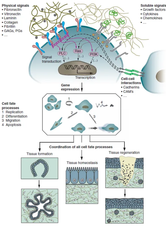
Cells are inherently sensitive to physical, biochemical and chemical stimuli from their surrounding. In vivo, the local cell environment or “niche” provides specific environmental cues that determine cell-specific recruitment, migration, proliferation, differentiation and the production of the numerous proteins needed for hierarchical tissue organisation.
As a consequence, tissue dynamics, that is, tissue formation, function and regeneration after damage, as well as its function in pathology, is the result of an intricate temporal and spatial coordination of numerous individual cell fate processes, each of which is induced by a myriad of signals originating from the extracellular microenvironment. The growth and differentiation of most cell types is regulated by the interplay of different major signaling, environmental stress and physical cues from the biological matrix which surrounds the cells in vivo. In particular the extracellular microenvironment is a highly hydrated network hosting three different main effectors: (i) insoluble hydrated macromolecules (fibrillar proteins like collagens, noncollagenous glycoproteins such as elastin, laminin or fibronectin, and hydrophilic proteoglycans with large glycosaminoglycan (GAG) side chains) called physical signals (Figure 1.1), (ii) soluble macromolecules (growth factors, chemokines and cytokines), and (iii) proteins on the surfaces of the neighboring cells that establish the cell-cell interaction [7].
The biological response is influenced by multiple cellular interactions with the individual and specific ECM molecules and often with multiple sites within the same molecule as well as by a highly dynamic and complex array of biophysical an biochemical properties of the ECM. The cells are able to receive the external signals through different cell surface receptor of the integrin family and integrate it by an
intracellular signaling pathway that affect the cellular response in terms of gene expression, ultimately establishing the cell phenotype.
Thus, the final decision of a cell to differentiate, proliferate, migrate, apoptose, or perform other specific functions is a coordinated response to the molecular interactions with these ECM effectors [8]. It is worth to note that the flow of information between cells and their ECM is highly bidirectional, as, for example, observed in processes involving ECM degradation and remodeling. It is evident how native ECM exhibits macroscale to nanoscale patterns of chemistry and topography [9], and it is therefore somewhat unsurprising that cells respond to these various scales of chemically and/or topographically patterned features.
Figure 1.1 The behavior of individual cells and the dynamic state of multicellular
tissues is regulated by intricate reciprocal molecular interactions between cells and their surroundings. This extracellular microenvironment is a hydrated protein and proteoglycan-based gel network comprising soluble and physically bound signals as well as signals arising from cell-cell interactions. Adapted from : M P Lutolf and J A Hubbell Nature Biotechnology 2005; 23 (19) , 47-55 [7].
However, when cells are cultured in vitro or when materials are implanted into the body, cells encounter very different, unfamiliar surfaces and environments.
Since the ECM is the optimized milieu that nature has been developed for maintaining homeostasis and directing tissue development, a great effort has been made to mimic the ECM to guide morphogenesis in regenerative medicine. In this context the biomaterials play a pivotal role in the field of tissue engineering as designable biophysical and biochemical enviromental conditions that direct cellular behaviour and functions [10-11].
The guidance provided by biomaterials may facilitate restoration of the structure and function of damaged or dysfunctional tissues. Such materials should provide a provisional three dimensional (3D) support for a biomolecular interaction with cells to control their function, guiding the spatially and temporally complex multicellular processes of tissue formation and regeneration.
Different biomaterials have been proposed to support cells and promote their differentiation and proliferation toward the formation of a new tissue. Clearly, the design and selection of a biomaterial is of critical importance in the development of a construct for tissue engineering application.
An appropriate biomaterial must exhibit good biocompatibility with extremely low inflammatory, immunogenic, and cytotoxic responses, promoting favourable cellular connection and tissue development. In addition, it should be biodegradable and bioresorbable with the ideal rate of the new tissue formation, having at the same time good mechanical and chemical properties in order to promote an adequate cell-substrate interactions. Figure 1.2 shows the various characteristics desired for an ideal scaffold for tissue regeneration [12]. All these requirements can be achieved by innovative nanotechnology approaches, which allow the design and the modification of suitable biomaterials controlling and directing the cellular behaviour. Therefore, the development of biomaterials currently does, and will continue to, impose significant challenges in the field and history of tissue engineering
IDEAL
SCAFFOLD
Radical scavenging ability Biodegradability
Cell Delivery Vehicle Growth factor delivery
Minimize secondary
progression of injury Eliminate chronic inflammation
Multiple cues (topographical, mechanical, biochemical
and electrical cue
Replace lost cells/tissue Simulate endogeneous
stem progenitor cells
• Mimics native extracellulare matrix •Promote cell adhesion, viability and proliferation
Figure 1.2 Ideal properties of scaffold. Adapted by Subramanian et al. [12].
1.3 BIOMATERIALS IN TISSUE ENGINEERING APPLICATION
The early or first-generation of biomedical materials, during the 1960s and 1970s, were developed for use inside the human body with a common feature of biological “inertness” for reducing the immune response to the foreign body to the minimum possible. The original goal was to obtain a suitable combination of physical properties that match those of the replaced tissue.
With the second-generation of biomaterials the field began to shift from achieving a bioinert tissue responses to instead producing bioactive components that could elicit a controlled action and reaction in the physiological environment. By the mid-1980s, bioactive materials had reached clinical use in a variety of different applications. A further improvements in this second generation was the development of resorbable biomaterials exhibiting clinically relevant, controlled chemical breakdown and resorption. In this manner, the interface problem is resolved, because the foreign material is ultimately replaced by regenerating tissue, and eventually there is no discernable difference between the implant site and the host tissue [13].
Improvements of first- and second–generation biomaterials are limited in part because all man-made biomaterials used for repair or restoration of the body represent a compromise. In fact, living tissue can respond to changing physiological loads or biochemical stimuli, a property not shared by synthetic materials. This limits the lifetime of artificial body parts, motivating the introduction of more biologically based method for the repair and regeneration of tissue: the third generation of biomaterials The third–generation of biomaterials are being designed to stimulate specific cellular responses at the molecular level, for instance activating genes that stimulate the regeneration of living tissue. The separate concepts of bioactive materials and resorbable materials have converged; resorbable materials are being made bioactive. Molecular modifications of resorbable polymer systems elicit specific interaction with cell integrins and thereby direct cell proliferation, differentiation, and extracellular matrix production and organization.
Polymeric materials have greatly contributed to the development of bioactive and biodegradable scaffold that can enhance tissue regeneration. Several classes of polymer have proved to be most useful in biomedical applications, but to select appropriate polymers for tissue engineering, it is necessary to understand the influence of the polymer in cell viability, growth and function.
Cell interaction with polymers are usually studied using cell culture techniques. Cells in culture are planted over a polymer surface and the extension of cell adhesion and spreading on the surface is evaluated. The maintenance of the cell culture for long periods is strongly influenced by the nature of the substrate that influence cell viability, function and motility. Most tissue-derived cells are anchorage-dependent and require attachment to a solid surface or viability and growth are compromised. For this reason, the initial events that occur when a cell approaches a surface are of fundamental interest. In tissue engineering cell-substrate adhesion is a multistep process that involves, in sequence: adsorption of an ECM onto the surface; recognition of the ECM components by cell receptors; cytoskeletal rearrangement. The proper accomplishment of these phases, leads the cells to gain the differentiated functions of the specific district with the subsequent tissue formation.
The biomaterials used for enhancement of tissue regeneration can be classified as natural, synthetic, and semi-synthetic materials.
Natural polymers can be considered as the first biodegradable biomaterials used clinically, possessing several properties that make them attractive for tissue engineering application. Natural materials such as collagen, gelatin, elastin, fibronectin, the linear glycosaminoglycan hyaluronic acid (HA), as well as carbohydrate polymers like chitosan, have been used for tissue engineering and as vehicles for cell delivery. Since ECM plays an instructive role in cell activities, such biomolecules would mantain the biological information and other physico-chemical features; exhibiting similar properties to the tissue replaced by them. Natural ECM polymers possess several inherent advantages such as bioactivity, the ability to present receptor-binding ligands to cells and the susceptibility to cell-triggered proteolytic degradation and natural remodeling.
Plant, animal, and insects components have also been explored to develop natural biomaterials such as silk, chitosan, alginate and matrigel. These polymers possess several properties that make them attractive for tissue engineering applications; for instance the chitosan is a natural biopolymer that consists of glucosamine and N-acetylglucosamine with a structure similar to GAGs which are components of the liver ECM consequently is a biomaterial with promising aplications in liver tissue engineering [14].
However, the rate of in vivo degradation of these natural and therefore enzymatically degradable polymers varies significantly with the site of implantation depending on the availability and concentration of the enzymes. Chemical modification of these polymers also can significantly affect their rate of degradation. The inherent bioactivity of these natural polymers has its own downsides. Natural materials, depending of the extraction methodology and tha batch characteristics, may induce immunological and inflammatory responses due to undefined factors and pathogens, which may still be present, even after purification [15].
Synthetic biomaterials on the other hand are generally biologically inert, they have more predictable properties, batch-to-batch uniformity, high reproducibility and the unique advantage of having property profiles tailored for specific applications, devoid of many of the disadvantages of natural polymers. Clearly, synthetic biodegradable polymers are preferable to nonbiodegradable polymers because of the advantage of avoiding a second surgery to remove the device.
At the present the most common biodegradable polymers in use or being studied include polylactic acid (PLA), poly-L-lactic acid (PLLA), polyglycolic acid (PGA), polycaprolactones (PCL) and polycarbonates [16-17]. Another group of synthetic polymers attracting attention in the field of regenerative medicine is polyurethanes. These are one of the most broadly used polymers in implantable biomedical devices such cardiac pacemakers and structural tissue replacements. The greatest disadvantage of synthetic material, however is the lack of cell recognition signals, resulting therefore in few cellular interaction. To overcome this limitation many researchers are focusing their efforts on the creation of semi-synthetic biomaterials by modifying the synthetic biomaterials with cell recognition sites such as incorporating cell adhesion petides.
1.3.1 Design Criteria of Biomaterials
Current interest has been focused on attempts to find new biomaterials suitable for realizing novel designs of engineered tissue. There are key parameters in selecting and engineering biomaterials such us the bulk and the surface properties. Bulk material selection is the first consideration of a matrix design, from biological effect to processability. Several properties of bulk materials for new biomaterials design have to be taken into consideration, including biocompatibility, wettability, transparency, biodegradability, and other mechanical properties. As mentioned before, a critical and important parameter that must be considered is the biocompatibility of the bulk material, because it determines the ability of materials to perform their desired functions with appropriate cellular or host responses. The degree of biocompatibility can vary from the lack of toxicity with respect to cell culture to the lack of immunological systemic response of human body. The strictest requirements are applied to implantable scaffold for avoiding undesired responses, such as a strong immune reaction or fibrous encapsulation. Ideally, the body should be able to metabolize the degraded substance; obviously, natural materials tend to show better biocompatibility than synthetic materials. A more relaxed definition of biocompatibility is applied to devices or substrates that will be used ex vivo, but the more complex the cellular system is, the more stringent the compatibility requirements are. Because natural ECM is a fully hydrated gel, wettability is a key consideration [18]. Indeed, materials with more hydrophilic chemistry are better at mimicking the aqueous in vivo environment.
Transparency of bulk material is another important parameter for in vitro modelling application in which cellular behaviours within the new matrix require microscopic detection. Thus, transparent materials are advantageous for a better monitoring and evaluation of the cell state during the culture period.
Controlled biodegradability is an essential requirement for implantable matrix and scaffold because the ideal tissue engineered construct is generally designed to disappear through degradation at the rate that in-growing tissue replaces it. Generally, synthetic materials degrade hydrolytically, and natural materials undergo an enzymatic degradation process. Hydrolytic degradation is more predictable and adjustable than enzymatic degradation. For example, the degradation profile of a poly (lactic-co-glycolic acid) (PLGA) matrix can be manipulated by adjusting the composition and the molecular weight of poly(lactic acid) (PLA) and poly(glycolic acid) (PGA) polymers. On the other hand, enzymatic degradation of natural materials is more dependent on the local enzyme concentration secreted from cells. As a consequence the degradation profile and mechanism under physiological conditions for the engineered construct materials, as well as the implantation site and desired function, need to be carefully considered during the design of a new implantable device [19]. Mechanical properties of bulk materials represent an important set of characteristics to consider in a biomaterial design or in choosing the right one. Bulk materials are fundamental contributors to the mechanical integrity of the matrix structure. In addition, the microscale mechanical properties are critical for determining the cell behaviour, including directing stem cell differentiation, cell migration and tissue growth, therefore, the new tissue construct should have mechanical properties resembling those of healthy tissue over the period of tissue regeneration. Cells not only adhere to surfaces, but also “pull” on the surface substrate and adjacent cells. The bulk mechanical properties directly shape the mechanical properties of the surface, such as stiffness or elasticity, which elicit clear cellular response. The relative substrate resistance encountered by the cells activates various mechanotransduction and cellular pathways, which in turn trigger gene expression. In vivo micromechanical stimuli are important environmental cues that enable cell attachment, migration and organogenesis. Matching the mechanical surface properties of the engineered tissue to the particular mechanical characteristics of the specific tissue site is therefore vital for controlling the cell behaviour. Cells on 2D
cultures initially recognize adhesive proteins on the substrate through the transmembrane integrin receptor receiving mechanical signals, which activate actin-filament polymerization and promote focal adhesion formation. Later, cells apply traction forces to pull the ligands from a substrate and sense the surface stiffness. For instance, it has been demonstrated that human mesenchymal stem cells (hMSCs) have a different phenotypic response on supports with different grades of elasticity. Human mesenchymal stem cells displayed a phenotype neurogenic lineage on the softest substrate, a myogenic phenotype on moderately stiff matrices, and an osteogenic phenotype on the stiffest substrates [20]. These results provide valuable informations for new biomaterial design introducing novel strategies suitable for many tissue regeneration applications.
Surface properties are also crucial in controlling the interaction between cells and a substrate. Although surface properties are often derived from the bulk properties of the materials, the bulk materials do not entirely define them, because the used substrates are coated with proteins almost immediately after implantation in the body or immersion in culture media. Surface chemistry and topography determine the identity, quantity, and conformational change of these adsorbed proteins. Surface properties include stiffness, charge, polarity, and chemistry, among a multitude of others. For example, the surface charge density determines the amount of protein adsorption and resultant cell adhesion. Greater surface charge brings a greater density of protein coating, which leads to better cell adhesion.
1.3.2 Surface Modification
When cells are cultured in vitro or when materials are implanted into the body, cells encounter very different and unfamiliar surfaces and environments. Several approaches have been introduced for modifying these unfamiliar substrates to promote desirable cell responses.
Surface modification of biomaterials, with the intent to improve not only biocompatibility but also the response of the target cell and/or tissue has been extensively studied to recreate the native tissue structure and to do it in the shortest time possible. Modifying material surface structures to mimic aspect of the ECM in order to provide the tissue-specific cues to direct cell behaviour and trigger tissue
regeneration, is an important aspect of many tissue-engineering strategies. Controlling the surface chemistry of materials enable, to some extent, to dictate the rate of protein absorption, the functionality of adsorbed proteins and subsequent cell adhesion.
The degree of biofunctional specificity exhibited on the material surface depends on the level and complexity of the surface-modification approach utilised [21].
One of the first approaches involves hybridizing natural and synthetic materials to improve the biological and physical properties of the substrate. For instance, limited bioactivity can be improved by covalently incorporating multifunctional ligands from natural materials, such as fibronectin, vitronectin, and laminin, onto synthetic polymers. Another technique involves chemical modification of the polymer, confering charged end-groups to its surface that may lead to protein adsorption and structural rearrangements via electrostatic interaction. Indeed, the surface of biomaterials can be modified via chemical reactions, to confer upon the material precise surface energy, charge or functional groups and tailor their levels of interaction with proteins and cells. For example, the radiation grafting of chemical group such as –OH, -COOH and NH2
onto the surface of relatively inert polymers has been used extensively to modify the surface of biomaterials. Energy sources like ionising radiation sources, ultraviolet radiation and high-energy electron beams are used to break the chemical bonds at the surface of the material to be grafted, allowing the formation of free radicals and other reactive species. The surface is then exposed to, and reacts with, molecules that will form the surface functional coating of the material.
Increasing biofunctionality can be achieved by attaching specific peptide motifs such as enzymes peptides, proteins which can bind to cell receptors inducing a “firm” cell anchorage. These biomolecules can be simply adsorbed onto the surface of the material, or covalently linked via chemical groups previously created on the surface. The biological response following the surface biomodification depends on structural parameter. The functionalisation of a material surface via absorption or chemical binding of other ECM elements is a common approach utilised to promote cell adhesion. For example a popular research strategy employed to improve the blood compatibility properties of vascular grafts comprises seeding endothelial cells onto their surface [22]. The complete coverage of the surface of the biomaterial by these cells (endothelialisation) inhibits thrombosis, prevents intimal hyperplasia, and thereby
increases the patency of vascular grafts. Furthermore, another example of mimickinng the natural ECM of surface modification is given by the incorporation of signal peptide such as RGD (Arg-Gly-Asp) which is found in many adhesion proteins and binds to many integrin receptors, modulating cell adhesion, and inducing cell migration. Finally, another approach is the incorporation of soluble signaling molecules within the scaffold, such as growth factor and deoxyribonicleic acid (DNA). For example, a larger biologically relevant protein can be produced using recombinant DNA technology incorporating the encoding sequence into the support.
On the basis of these important considerations it can be pointed out that the development of new biomaterials able to activate specific responses of the cells and to maintain cell differentiation for a long time is one of the most pertinent issues in the field of tissue engineering and regenerative medicine. Nowadays, it has been demonstrated that the suitability of polymers for tissue-engineering purposes is highly dependent on the tissue that needs to be engineered. The histological, physiological, and biomechanical properties of each tissue determine the success of the regenerative process, therefore restricting the choice of materials suitable for the specific application.
1.4 SEMIPERMEABLE POLYMERIC MEMBRANE AS A BIOMATERIAL
Among the biomaterials applied in the field of tissue engineering and regenerative medicine, polymeric semi-permeable membranes could provide the mechanical support required for the regulation of cell growth in bio-hybrid systems (Figure 1.3). The ultimate goal of this technique might be very well achieved by appropriate bio-interaction of desired cell responses which lead to the fabrication of the tissue structure. The realization of such bio-hybrid systems have a great impact in tissue engineering application since represent a key approach for studying both physiological and pathogenic states of the of the specific cells and tissue site as well as for the development of appropriate bio-molecules for therapeutic purposes. With the purpose of simulating in vitro biological phenomena, scientists have begun to construct artificial membranes that may be handled in both industrial and medical application. Artificial lungs (blood oxygenation) and kidneys (hemodialysis) are just the two oldest examples. The use of polymeric semi-permeable membranes with different physico-chemical and
![Figure 1.2 Ideal properties of scaffold. Adapted by Subramanian et al. [12].](https://thumb-eu.123doks.com/thumbv2/123dokorg/2884154.10639/29.892.159.778.144.580/figure-ideal-properties-scaffold-adapted-subramanian-et-al.webp)