INDEX
Summary……….…………..….. 1
Introduction………...……….. 5
1. The Testis….……….……….………..………. 6
1.1 Anatomy………...………...………… 6
1.2 Testicular function and its regulation ..…………..…....…….………… 1.3 Steroid production ….…..………….………..………… 6 9 2. Estrogen biosynthesis and action …..………....………...……… 11
2.1 The aromatase gene: structure and regulation ………….…………... 11
2.2 Estrogen receptors (ERs) …..……….….………...…... 13
2.3 Distribution of ERs in the male reproductive system ……….………… 2.4 Aromatase overexpression in rodents ……….……… 2.5 Exposure to excess of estrogens in animals ………...………….. 2.6 Exposure to excess of estrogens in humans …………...…...….……… 13 15 15 16 3. Testicular cancer ………...…… 17
3.1 Leydig cell hyperplasia and cancer …..…………..………... 18
Specific aim………...… 21
Materials and Methods………... 23
Cell cultures and animals …...………..…………...…… 24
Aromatase sctivity assay…...………...…………... 24
Radioimmuno assay ...………..…...……… 24
Chromatin immunoprecipitation (ChIP)………..………. 25
Real time RT-PCR ...………...……… 26
Western blot analysis ...………...……… 27
Data analysis and statistical methods ...………...……… 28
Results……….. 29 Estradiol induces Leydig cell tumor proliferation through an autocrine
mechanism ... 30 Aromatase overexpression is determined by constitutive activation of
transcription factors SF-1 and CREB ... 33 IGF-I is produced by R2C cells and induces aromatase expression through
PI3K- and PKCmediated activation of SF-1 ... 34 IGF-I induces aromatase expression and activity in R2C cells ... 37 Changes in IGF-I pathway activation status lead to changes in SF-1 binding to
the aromatase PII promoter ... 38
Discussion ... 40
References... 47
Scientific Publications
Differential expression of steroidogenic factor-1/adrenal 4 binding protein and liver receptor homolog-1 (LRH-1)/fetoprotein transcription factor in the rat testis: LRH-1 as a potential regulator of testicular aromatase expression.
Corticotropin-releasing hormone directly stimulates cortisol and the cortisol biosynthetic pathway in human fetal adrenal cells.
type 1 CRH receptors to stimulate dehydroepiandrosterone sulfate production in human fetal adrenal cells.
Antiestrogens upregulate estrogen receptor beta expression and inhibit adrenocortical H295R cell proliferation.
The AP-1 complex: a negative regulator of CYP17 transcription in adrenal cells.
IGF-I regulating aromatase expression through SF-1, supports estrogen dependent tumor Leydig cell proliferation.
Summary
Aim of this study was to investigate the role of estrogens in Leydig cell tumor proliferation. We used rat R2C Leydig tumor cells and testicular samples from Fischer rats with a developed Leydig tumor (FRTT). Both experimental models express high levels of aromatase and Estrogen Receptor alpha (ERα). Treatment with exogenous E2 induced proliferation of R2C cells and upregulation of cell cycle regulators cyclin D1 and E, that were blocked by addition of antiestrogens. These observations leaded us to suppose an E2/ERα dependent mechanism for Leydig cell tumor proliferation. Determining the molecular mechanism responsible for aromatase overexpression, we found that total and phosphorylated levels of transcription factors CREB and SF-1 were higher in tumor samples. Moreover, we found that R2C cells produce also high levels of IGF-I that increased aromatase mRNA, protein and activity as a consequence of increased total and phosphorylated SF-1 levels and that specific inhibitors for IGF-I receptor, Protein Kinase C and Phosphoinositol-3-kinase determined a reduction in SF1 and consequently in aromatase expression and activity. The same inhibitors were also able to inhibit the IGF-1 dependent-SF-1 recruitment to the aromatase PII promoter as shown with ChIP assays. We conclude that one of the molecular mechanims determining Leydig cell tumorogenesis is an excessive estrogen production stimulating a short autocrine loop determining cell proliferation. In addition, cell produced IGF-I amplifies estrogen signaling through a SF-1-dependent up-regulation of aromatase expression. The finding of this new molecular mechanism will be helpful in defining new therapeutic approaches of Leydig cell tumor.
Introduction
1. The testis
1.1 Anatomy
In mammalian species both testicular compartments consist of a variety of different cell types (1). The Sertoli cells comprise the main structural component of the seminiferous epithelium. They are responsible for the physical support of the germ cells, in addition to providing nutrients and growth factors. The germ cells are sequentially organised into several layers signifying the respective mitotic or meiotic processes and spermatid development. Each seminiferous tubule is surrounded by mesenchymal cells. Among these are the peritubular myoid cells whose contractile elements generate peristaltic waves along the tubules, but do not present a tight diffusion barrier. The interstitium is populated by androgen-producing Leydig cells which are heterogeneous in respect to their physiological and structural features. Vascular smooth muscle cells, macrophages and endothelial cell types are also located in the interstitial space of the testis. The physiological role of macrophages has long been underestimated. In the rat, the number of macrophages is one quarter of the number of Leydig cells and the presence of macrophages is crucial for (re)population of Leydig cells during development and after experimental depletion (2;3). Immune cells, known to secrete a number of growth factors and cytokines, are part of the intratesticular communication pathways (4).
1.2 Testicular function and its regulation
Testes are components of both the reproductive system (being gonads) and the endocrine system (being endocrine glands). The respective functions of the testicles are:
1. producing sperm (spermatozoa) 2. producing male sex hormones
These two functions occur in separate compartments within the testis: 1. the seminiferous tubules produce sperm and 2. the interstitial cells (i.e., Leydig cells) synthesize androgens (Fig. 1).
Introduction
Figure 1. Schematic representation of functions of the testis.
Both functions of the testis, sperm-forming and endocrine, are under control of gonadotropic hormones produced by the anterior pituitary: luteinizing hormone (LH) and follicle-stimulating hormone (FSH).
Synthesis and release of both FSH and LH is regulated by a single gonadotropin releasing hormone (GnRH) also referred to as LHRH. GnRH is a decapeptide secreted from hypothalamic neurons into the hypothalamic/hypophysial portal vessels. LH and FSH secretion is subject to negative feedback control by the testis. At least two products of the testis are involved. LH acts to stimulate Leydig cells to produce testosterone which, in turn, inhibits further secretion of LH by inhibition of GnRH release from the hypothalamus. Testosterone also decreases the responsiveness of the pituitary to GnRH (Fig. 2).
Introduction
Figure 2. Hypothalamic-Pituitary-Testicular axis.
LH, through specific receptors found on the surface of Leydig cells, controls the production and secretion of testosterone (5;6). The interaction of LH with its receptor, a seven transmembrane domain G protein coupled receptor, initiates signalling through the cyclic AMP pathway through GTP binding proteins (7;8). Signal transduction occurs through the protein kinase A pathway as its principal signal transduction mechanism. The testis is also able to produce growth factors that can act with an autocrine/paracrine manner (9). Some factors induce specific differentiation steps, while others act primarily as environmental or survival factors. Insulin-like growth factor (IGF) family which includes three structurally related peptides: insulin, IGF-I (also called somatomedin C) and IGF-II belongs to the second group of factors. The receptor for IGF-I has been found on most testicular cell types, including rat and human Sertoli cells, Leydig cells and pachytene spermatocytes (4;10-12), indicating a more general role such as the stimulation of
Introduction
steroidogenesis in Leydig cells (13-16). IGF-I is produced locally in the testis, in Sertoli, Leydig and peritubular cells derived from the immature rat testis and cultured in vitro (17;18). The crucial role of IGF-I in the development and function of Leydig cells was highlighted by studies on IGF-I gene knock-out mice (19;20). The failure of adult Leydig cells to mature and the reduced capacity for T production is caused by deregulated expression of testosterone (T) biosynthetic and metabolizing enzymes (21). Expression levels of all mRNA species associated with T biosynthesis were lower in the absence of IGF-I.
1.3 Steroid production
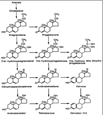
The pathway of testosterone synthesis from cholesterol and the conversion of testosterone to active androgen and estrogen metabolites is shown in Figure 3. The mobilization of cellular sources of cholesterol is achieved through the action of cholesterol ester hydrolase and subsequently, this is converted to pregnenolone by the enzyme cholesterol side-chain cleavage termed cytochrome P450SCC (22). The conversion of cholesterol to pregnenolone is a key step at which regulation of androgen production within the Leydig cells occurs. Availability of cholesterol substrate can be rate-limiting and the intracellular trafficking of cholesterol across mitochondrial membranes is dependent on the steroidogenic acute regulatory protein (STAR) (23-25). The role of this protein has been well demonstrated in patients with mutations in the gene encoding STAR in the disorder termed congenital lipoid adrenal hyperplasia wherein the mitochondria from the adrenals and gonads of these patients are unable to convert cholesterol to pregnenolone (26). Further, the results of studies involving targeted disruption of the mouse gene encoding STAR support the data derived from human studies (27).
Intracellular transport of steroid substrates involved in androgen production is the transport of cholesterol into the mitochondrion to form pregnenolone and the transport of pregnenolone to smooth endoplasmic reticulum for the remainder of the steps in the production of testosterone. Pregnenolone may progress to testosterone production through two pathways. It can be converted to progesterone through the enzyme 3β-hydroxysteroid
Introduction
dehydrogenase (the ∆4 pathway) or can be hydroxylated at the 17α position by the enzyme 17α-hydroxylase to form 17α-hydroxypregnenolone (the ∆5 pathway). The relative importance of these two pathways vary with the species and the physiological status of the male (28). The further conversion of 17α-hydroxypregnenolone through the ∆5 pathway involves the formation of the C19 steroid dehydroepiandrosterone catalyzed by the enzyme 17,20 lyase and both steps appear to be catalyzed by a single microsomal enzyme cytochrome P450c17 encoded by a single copy gene (29;30). The conversion of dehydroepiandrosterone to androstenediol is mediated by a microsomal enzyme 17β-hydroxysteroid dehydrogenase encoded by a single gene (31;32). The conversion of substrates from the ∆5 to the ∆4 pathway are catalyzed by the enzyme 3β-hydroxysteroid dehydrogenase (33). In the ∆4 pathway 17α-hydroxyprogesterone proceeds through the action of cytochrome P450c17 to androstenedione and testosterone. Testosterone can be converted to a dihydrotestosterone by the enzyme 5α-reductase (34) or can be metabolised to 17β-estradiol by the enzyme aromatase (35).
Introduction
2. Estrogen biosynthesis and action
2.1 The aromatase gene: structure and regulation
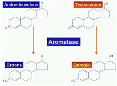
In males, estrogens derive from circulating androgens. Aromatization of the C19 androgens, testosterone and androstenedione, to form estradiol and estrone, respectively, is the key step in estrogen biosynthesis, which is under the control of the aromatase enzyme. The aromatase enzyme is a P450 mono-oxygenase enzyme complex present in the smooth endoplasmic reticulum which acts through three consecutive hydroxylation reactions, whose final effect is the aromatization of the A ring of androgens (Fig. 4).
Figure 4: Biochemical pathway of testosterone conversion into estrogens.
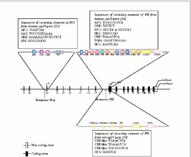
P450 aromatase is the product of the CYP19 gene which consists of at least 16 exons and is located on chromosome 15 in humans (36;37) (Fig. 5). Analysis of transcript and genomic sequences indicates that the tissue-specific expression of P450arom is regulated in part by alternatively spliced untranslated first exons (38;39). The proximal promoter PII
Introduction
regulates P450arom expression in mammalian gonads (40;41) as well as in Leydig cell tumors (42). Promoter PII activity (fig. 5) is regulated by cyclic AMP and requires the transcription factors cAMP responsive element binding protein (CREB), cAMP response element modulator (CREM) and steroidogenic factor-1 (SF-1). SF-1 belongs to the nuclear orphan receptor superfamily and regulates steroidogenic gene transcription.
Figure 5: Structure of the human Cyp19 gene showing the various untranslated first exons and their
corresponding promoters. The region around promoter PI.4 and PII from human and PII from rat are expanded to show the identified response elements. Sequences of these are shown in boxes.
Introduction
2.2 The Estrogen Receptors (ERs)
Estrogens actions are mediated through the specific binding to nuclear estrogen receptors (ERs), which are ligand-inducible transcription factors regulating the expression of target genes after hormone binding. Two subtypes of ERs have been described: estrogen receptor α (ERα) and the more recently discovered estrogen receptor β (ERβ). The two ER (α and β) proteins have a high degree of homology at the amino acid level (Fig. 6).
Figure 6: ERs genes and their products.
While it is clear that estrogens regulate transcription via a nuclear interaction after binding their receptors, a non-genomic action of estrogens has been recently demonstrated, suggesting that a different molecular mechanism accounts for some estrogen actions. In vitro studies showed a very short latency time between the administration of estrogens and the appearance of biological effects. These actions are thought to be mediated through cell-surface receptors, which are not believed to act via a transcriptional mechanism (43).
2.3 Distribution of ERs and aromatase in the male reproductive system
ERs and the aromatase enzyme are widely expressed in the male reproductive tract in both animals and humans, implying that estrogen biosynthesis occurs in the male reproductive
Introduction
tract and that both locally produced and circulating estrogens may interact with ERs in an intracrine/paracrine and/or endocrine fashion (43). The concept of a key estrogen action in the male reproductive tract is strongly supported by the fact that male reproductive structures are able to produce and respond to estrogens (44).
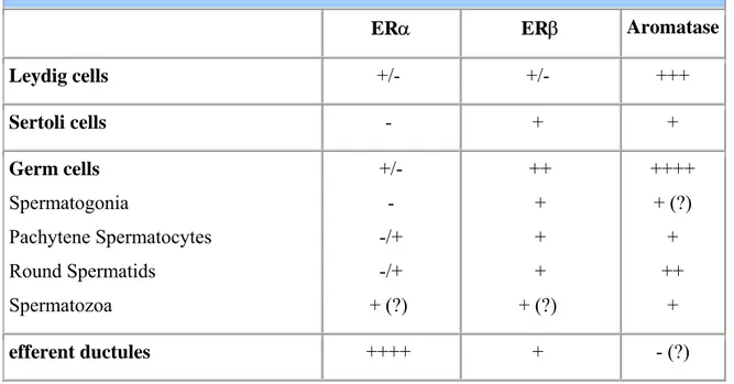
In particular in the adult rodent testisERα is expressed in the Leydig cells of both adult rats and mice (45) but not in Sertoli cells. Knowledge of the distribution of ERα is of great importance in understanding estrogen action on the male reproductive tract. ERα is highly expressed in the proximal reproductive ducts (rete testis, efferent ductules, proximal epididymis) and its expression progressively decreases distally (corpus and cauda of the epidydimis, vas deferens). The highest degree of ERα expression is seen in the efferent ductules of the rat (46) and accounts for one of the most well-documented estrogenic actions on male reproductive system, that of fluid reabsorption from the efferent ductules. It has to be remarked that the concentration of ERα in the male reproductive tract is opposite to that of ERβ, which is more concentrated in the distal tract (Table 1).
Table 1. ERs and Aromatase distribution in the adult rodent testis.
ERα ERβ Aromatase Leydig cells +/- +/- +++ Sertoli cells - + + Germ cells Spermatogonia Pachytene Spermatocytes Round Spermatids Spermatozoa +/- - -/+ -/+ + (?) ++ + + + + (?) ++++ + (?) + ++ + efferent ductules ++++ + - (?)
Introduction
ERβ is expressed in Leydig, Sertoli and germ cells in adult rodents (44;47-49) and has also been detected in primate germ cells (50). There is now considerable evidence that germ cells contain both ERβ and aromatase (44;50).
By adulthood, rodent Leydig cells show higher aromatase activity compared to every other age and in comparison to Sertoli cells (51). Aromatase is also expressed at high levels in germ cells throughout all stages of maturation, and its expression appears to increase as the germ cell becomes a mature spermatid.
2.4 Aromatase over-expression in rodents
Recently a transgenic line of mice overexpressing aromatase enzyme (AROM+) has been developed (52;53). These mice show highly elevated serum estradiol concentrations, with a reciprocal decrease in testosterone concentrations. About half of these male mice were infertile and/or had enlarged testis and showed Leydig cell hyperplasia and Leydig cell tumors (54) while the female of these mice revealed mammary glands hyperplasia associated with an altered expression of proteins involved in apoptosis, cell cycle, growth and tumor suppression (55).
2.5 Exposure to excess estrogens in animals
In order to evaluate the effect of estrogen excess on the reproductive tract, several studies have been performed in various animal species treated with diethylstilbestrol, a synthetic estrogenic compound. In male mice, the critical period for Müllerian duct formation is day 13 post-coitus. Prenatal exposure of fetal male mice to DES caused a delay in Müllerian duct formation by approximately two days as well as incomplete Müllerian duct regression with a female-like differentiation of the non-regressed caudal part (56). An increase in the expression of anti-Müllerian-Hormone (AMH) mRNA in male mice fetuses exposed to DES has also been demonstrated. This increase was not accompanied by a regression of the ducts. This data was interpreted to suggest that the asynchrony in the timing of Müllerian duct formation, with respect to the critical period of Müllerian duct regression, led to the persistence of Müllerian duct remnants at birth in male mice. Moreover DES
Introduction
exposure did not impair embryonal genetic development, but increased ERs number, and slightly prolonged the gestation time (cesarean sections were performed to rescue the litter and revealed no difference in size of fetuses from control and DES treated mothers). The timing of DES exposure is crucial to the induction of abnormalities of Müllerian duct development and regression (56).
Many studies in rodents suggest that inappropriate exposure to estrogen in utero and during the neonatal period impairs testicular descent, efferent ductule function, the hypothalamic-pituitary-gonadal axis, and testicular function (44). The latter effect can be a direct consequence of exposure to excess estrogen, as well as a secondary effect due to perturbations in circulating hormones or the ability of the efferent ductules to reabsorb fluid. Some studies show that low dose estrogenic substances given during puberty can actually stimulate the onset of spermatogenesis, likely due to stimulatory effects on FSH (57), highlighting the fact that the effects of excess estrogen on male fertility are often complex. The effects of excess estrogen in the neonatal period can impact upon the testis into adulthood, with permanent changes in testis function and spermatogenesis (44).
2.6 Exposure to excess estrogens in humans
The clinical use of diethylstilbestrol (DES) by pregnant women in order to prevent miscarriage resulted in an increased incidence of genital malformations in their sons (58). DES may have an effect on sex differentiation in men, as is the case in rodents (56). The risk of testicular cancer among men exposed to DES in utero has been a controversial issue and several meta-analyses showed no increased risk (59). However more direct evidence will be necessary in order to fully understand this issue.
While various studies suggest that environmental estrogens affect male fertility in animal models, the implications for human spermatogenesis are less clear (60). It has been demonstrated that male mice whose mothers have consumed a 29 ng/g dose of bisphenol A for seven days during pregnancy had a 20% lower sperm production as compared to control males (61). Various abnormalities in reproductive organs have also been described in males exposed to bisphenols (i.e. a significant decrease in the size of the epididymis and
Introduction
seminal vesicles and an increase in prostate gland volume), suggesting that bisphenols interfere with the normal development of the Wolffian ducts in a dose-related fashion. Exogenous estrogens could interfere with the development of the genital structures if administered during early organogenesis, by leading to both an impairment of gonadotropin secretion and by creating an imbalance in the androgen to estrogen ratio, which may account for impaired androgen receptor stimulation or inhibition according to the dose, the cell type and age (58;62-64).
An excess of environmental estrogens has been suggested as a possible cause of impaired fertility in humans (65-67). A progressive decline in sperm count has been reported in some Western countries during the past 50 years, suggesting a possible negative effect of environmental contaminants on male reproductive function (58;62;66;68). Data concerning the role of estrogens in male reproductive structure development remains conflicting. Animal studies suggest that exposure to estrogen excess may negatively affect the development of reproductive male organs. These effects, however, are considered to be the result of an impaired hypothalamic-pituitary function as a consequence of estrogen excess and of the concomitant androgen deficiency (63;64). Much of the knowledge on excess estrogen exposure and human fertility depends upon animal data and the validity of these concepts to humans has not been established.
3. Testicular cancer
Although cancer of the testes is rare, accounting for only about 1 percent of all cancers in men of all ages and about 5 percent of all male genitourinary system cancers, it is the most common cancer in men between the ages of 15 and 35, and the second most common malignancy in men ages 35 to 39 (69-72).
Because the incidence of testicular cancer has risen markedly in the past 20 years, numerous studies are being conducted to explore possible environmental causes, including the mother's diet during her pregnancy as well as her use of diethlstilbestrol (DES) to prevent miscarriage. Researchers are also looking at the increasing presence of
Introduction
mimicking pollutants in the environment. The most consistent occupational association has been the elevated rate among men in professional and white-collar occupation, which may be linked to an increased risk observed with lower levels of exercise. Other possible causes include hereditary factors, genetic anomalies, congenital defects involving the reproductive tract, testicular injury, and atrophy of the testes. Viral infections such as mumps, which cause inflammation of the testes, have not been proven to cause cancer.
Testicular cancer comprises a number of different diseases. Nearly all of the main cell types in the testis can undergo neoplastic transformation, but germ cell-derived tumors constitute the vast majority of cases of testicular neoplasms. Ninety-five percent of testicular cancers arise from sperm-forming, or germ cells and are called germinal tumors. The remaining 5 percent are nongerminal tumors. About 40 percent of germinal tumors are categorized as seminomas. Several other types of germinal tumors are referred to collectively as non-seminomas. Somatic cell tumors, known as sex cord-stromal neoplasms and Leydig cell tumors are relatively rare. However, being derived from endocrine active cells, they have endocrine manifestations.
3.1 Leydig cell hyperplasia and tumors
Although Leydig cells in adult men are considered to be a terminally differentiated and mitotically quiescent cell type, in various disorders of testicular function, focal or diffuse Leydig cell hyperplasia is very common. Micronodules of Leydig cells are frequently seen in certain conditions associated with severe decrease of spermatogenesis or germinal aplasia, such as the so-called Sertoli-cell-only syndrome (Del Castillo syndrome), cryptorchidism, or Klinefelter’s syndrome (73). A term “Leydig cell adenoma” is used when the size of a nodule exceeds several fold the diameter of a seminiferous tubule. It is unknown whether Leydig cell adenomas can progress further to form overt Leydig cell tumors, but even if it was the case, it is exceedingly rare. Morphological heterogeneity of hyperplastic Leydig cells is noticeable in some cases.
The mechanism of Leydig cell hyperplasia in the human male is still poorly understood. The disruption of hypothalamo-pituitary-testicular axis leading to an excessive stimulation
Introduction
of Leydig cells by LH can play a central role (73). However, molecular pathways remain largely unknown in the vast majority of cases. In a small subset of cases structural changes of the LH receptor (74;75) and G proteins (76;77) were detected. Constitutively activating mutations of LH receptor cause early Leydig cell hyperplasia and precocious puberty (74;78). Similarly, constitutively activating mutations of Gs-protein in Leydig cells lead into hyperplasia and endocrine hyperactivity (77;79). However, Leydig cell hyperplasia is distinct from tumors that are usually solitary, and the role of the LH receptor and G protein mutations in the tumorigenesis may be limited to few cases (75;77). Leydig cell hyperplasia and adenomas can be easily induced in rodents by administration of estrogens, gonadotropins and a wide range of chemical compounds. Whether or not humans would be similarly susceptible to environmental effects remains to be elucidated.
Leydig cell tumors account for one to three percent of testicular neoplasms and occur in all age groups (79-81). Approximately 20 % are found before the age of 10, most often between five and ten years of age. Precocious puberty is the presenting symptom in these cases. Tumors produce androgens, mainly testosterone in a gonadotropin independent manner, and therefore LH and FSH remain low in spite of external signs of puberty. Approximately 10 % of the boys also have gynecomastia that is caused by estrogens produced in excess due to aromatase activity. In adults, gynecomastia is found in approximately 30 % of patients (81). The excessive androgen secretion rarely causes notable effects in adults.
Leydig cell tumors are always benign in children and can be treated with surgical enucleation when the tumor is encapsulated (71), whereas in adults malignant tumors have been found in 10-15 % of patients, and inguinal orchidectomy remains the treatment of choice (80). The presence of cytologic atypia, necrosis, angiolymphatic invasion, increased mitotic activity, atypical mitotic figures, infiltrative margins, extension beyond testicular parenchyma, and DNA aneuploidy are associated with metastatic behavior in Leydig cell tumors (81;82). Malignant tumors are hormonally active only in exceptional cases. Benign tumors can be treated by orchidectomy, whereas an additional retroperitoneal lymphadenectomy should be considered when the gross or histological features suggest
Introduction
malignancy (82). Malignant tumors have not responded favorably to conventional chemotherapy and irradiation (82). Survival time has ranged from 2 months to 17 years (median, 2 years), and metastases have been detected as late as nine years after the diagnosis (81;82). Therefore follow-up of patients with malignant Leydig cell tumors has to be life-long. The remaining testis may be irreversibly damaged by longstanding high estrogen levels, resulting in both permanent infertility and hypoandrogenism (81-83).
Specific aim
Whereas the effects of estrogen on mammary gland tumorogenesis in human and in rodents is well known, the role of aromatase overexpression and in situ estrogen production in testicular tumorogenesis is not clearly defined. In this study we have investigated the molecular mechanisms causing aromatase overexpression and the effect of estradiol (E2) overproduction on rat Leydig cell tumor proliferation. As experimental model we used rat R2C Leydig tumor cell line and to validate our in vitro data in an in vivo model we used Leydig cell tumors from old Fisher rat testes in which the incidence of the spontaneous neoplasm is exceptionally high in aged animals (84).
We investigated the role of IGF-I a peptide also demonstrated to have a role in testicular growth and development, control of Leydig cell number (9). A previous study showed that in IGF-I gene knock-out mice (19;20) adult Leydig cells fail to mature, as a consequence these animals have a reduced capacity for testosterone (T) production caused by deregulated expression of T biosynthetic and metabolizing enzymes (21). Expression levels of all mRNA species associated with T biosynthesis were lower in the absence of IGF-I. However, this study did not investigate the effect on aromatase expression, even though an effect could be supposed.
Starting from these findings, in this study we investigated if a testicular overproduction of IGF-I could be one of the mechanisms determining aromatase overexpression in rat tumor Leydig cells through the activation of specific transcriptional factors. The high related Leydig cells E2 production through an autocrine/paracrine mechanism mediated by their own receptors, could contribute to the hormone dependence of testicular tumorogenesis stimulating Leydig tumor cell proliferation.
Materials and Methods
Cell cultures and animals.
TM3 cells (mouse Leydig cell line)were cultured in D-MEM/F-12 medium supplemented with 5% HS, 2.5% FBS and antibiotics (Invitrogen, S.R.L., SanGiuliano Milanese, Italy); R2C cells (rat Leydig tumor cell line) were cultured in Ham/F-10 medium supplemented with 15% HS, 2.5% FBS and antibiotics (Invitrogen, S.R.L., SanGiuliano Milanese, Italy). Male Fischer 344 rats (a generous gift of Sigma-Tau Pomezia, Italy), 6 (FRN) and 24 (FRT) months of age were used for studies. 24 months old animals presented spontaneously developed Leydig cell tumors absent in younger animals. Testes of all animals were surgically removed by qualified, specialized animal care staff in accordance with the Guide for Care and Use of Laboratory Animals (National Institutes of Health) and used for experiments.
Aromatase activity assay.
The aromatase activity in subconfluent R2C cell culture medium was measured by tritiated water-release assay using 0.5 µM [1β-3H(N)]androst-4-ene-3,17-dione (DuPont NEN, Boston, MA, USA) as a substrate (85). Incubations were performed at 37 °C for 2 h under a 95%:5% air/CO2 atmosphere. Obtained results were expressed as pmol/h and normalized to milligram of protein (pmol/h per mg protein).
Radioimmunoassay.
Prior to experiments, TM3 cells were maintained overnight in DME/F12 medium and R2C cells in Ham/F-10 (medium only). The Estradiol content of medium recovered from each well was determined against standards prepared in low serum medium using a radioimmunoassay kit (DSL 43100; Diagnostic System Laboratories, Webster, TX, USA). Results assay were normalized to the cellular protein content per well and expressed as pmol per mg cell protein. IGF-I content in medium recovered from each well of R2C and TM3 cells was determined following extraction and assay protocols provided with the mouse/rat IGF-I radioimmunoassay kit (DSL 2900; Diagnostic System Laboratories, Webster, TX, USA).
Materials and Methods
Chromatin Immunoprecipitation (ChIP).
This assay was performed using the ChIP assay kit from Upstate (Lake Placid, NY) with minor modifications in the protocol. R2C cells were grown in 100 mm plates. Confluent cultures (90 %) were treated for 24 h with AG1024 (Sigma St Louis, MO, USA), PD98059 (Calbiochem, VWR International S.R.L. Milano), LY294002 (Calbiochem, VWR International S.R.L. Milano), GF109203X (Calbiochem, VWR International S.R.L. Milano) or for increasing times with 100 ng/ml IGF-I (Sigma St Louis, MO, USA) or left untreated. Following treatment DNA/protein complexes were crosslinked with 1 % formaldehyde at 37 °C for 10 min. Next, cells were collected and resuspended in 400 µl of SDS lysis buffer (Upstate Technology, Lake Placid, NJ) and left on ice for 10 min. Then, cells were sonicated four times for 10 sec at 30 % of maximal power and collected by centrifugation at 4 °C for 10 min at 14 000 rpm. Of the supernatants 10 µl were kept as input (starting material, to normalize results) while 100 µl were diluted 1:10 in 900 µl of ChIP dilution buffer (Upstate Technology, Lake Placid, NJ) and immunocleared with 80 µl of sonicated salmon sperm DNA protein A agarose (Upstate) for 6 h at 4 °C. Immunocomplex was formed using 1 µl of 1:5 dilution of specific antibody anti-SF-1 (provided by Prof. Ken-ichirou Morohashi, Division for Sex Differentiation, National Institute for Basic Biology, National Institutes of Natural Sciences, Myodaiji-cho, Okazaki, Japan) overnight at 4 °C. Immunoprecipitation with salmon sperm DNA protein A agarose was continued at 4 °C until the day after. DNA/protein complexes were reverse crosslinked overnight at 65 °C. Extracted DNA was resuspended in 20 µl of TE buffer. 3 µl volume of each sample and input were used for PCR using CYP19 promoter II specific primers. The PCR conditions were 1 min at 94 °C, 1 min at 50 °C and 2 min at 72 °C for 30 cycles using the following primers: forward, TCAAGGGTAGGAATTGGGAC-3’; reverse, 5’-GGTGCTGGAATGGACAGATG-3’. Amplification products were analyzed on a 1 % agarose gel and visualized by ethidium bromide staining. In control samples, non immune rabbit IgG was used instead of specific antibodies.
Materials and Methods
Real-time RTPCR.
Prior to experiments, cells were maintained overnight in low serum medium. Cells were then treated or the indicated times and RNA was extracted from cells using the TRiazol RNA isolation system (Invitrogen). TRiazol was also used to homogenize total tissue of normal (FRNT) and tumor (FRTT) Fisher rat testes for RNA extraction. Each RNA sample was treated with DNase I (Ambion, Austin, TX), and purity and integrity of the RNA was confirmed spectroscopically and by gel electrophoresis prior to use. One µg of total RNA was reverse transcribed in a final volume of 30 µl using the ImProm-II Reverse transcription system kit (Promega, Promega Italia S.R.L. Milano, Italy), cDNA was diluted 1:3 in nuclease free water, aliquoted and stored at –20°C. Primers for the amplification were based on published sequences for the rat CYP19, rat CREB and rat SF-1 genes. The nucleotide sequences of the primers for CYP19 were: forward GAGAAACTGGAAGACTGTATGGAT-3’ and reverse 5’-ACTGATTCACGTTCTCCTTTGTCA-3’. For CREB amplification were used the following primers: forward AATATGCACAGACCACTGATGGA-3’ and reverse 5’-TGCTGTGCGAATCTGGTATGTT-3’; for SF-1 amplification primers have been previously published (86). PCR reactions were performed in the iCycler iQ Detection System (Biorad Hercules, CA, USA), using 0.1 µM of each primer, in a total volume of 30 µL reaction mixture following the manufacturer’s recommendations. SYBR Green Universal PCR Master Mix (Biorad Hercules, CA, USA) with the dissociation protocol was used for gene amplification, negative controls contained water instead of first-strand cDNA. Each sample was normalized on the basis of its 18S ribosomal RNA content. The 18S quantification was performed using a TaqMan Ribosomal RNA Reagent kit (Applied Biosystems, Applera Italia, Monza, Milano, Italy) following the method provided in the TaqMan Ribosomal RNA Control Reagent kit (Applied Biosystems, Applera Italia, Monza, Milano, Italy). The relative gene expression levels were normalized to a calibrator that was chosen to be the basal, untreated sample. Final results were expressed as n-fold differences in gene expression relative to 18S rRNA and calibrator, calculated following the ∆∆Ct method, as follows:
Materials and Methods
n-fold = 2 – (∆Ctsample – ∆Ctcalibrator)
where ∆Ct values of the sample and calibrator were determined by subtracting the average Ct value of the 18S rRNA reference gene from the average Ct value of the different genes analyzed.
Western-blot analysis
R2C and TM3 cells or total tissue of FRNT and FRTT were lysed in ice-cold Ripa buffer containing protease inhibitors (20 mM Tris, 150 mM NaCl, 1% Igepal, 0.5% sodium deoxycholate, 1 mM EDTA, 0·1% SDS, 1 mM PMSF, 0.15 units/ml aprotinin and 10 µM leupeptin) for protein extraction. The protein content was determined by Bradford method (87). The proteins were separated on 11% SDS/polyacrylamide gel and then electroblotted onto a nitrocellulose membrane. Blots were incubated overnight at 4 °C with 1. anti-human P450 aromatase antibody (1:50) (Serotec, Oxford, UK, MCA 2077), 2. anti-ERα (F-10) antibody (1:500) (Santa Cruz Biotechnology, Santa Cruz, CA, USA, sc8002), 3. anti-ERβ (H-150) (1:1000) (Santa Cruz Biotechnology, Santa Cruz, CA, USA, sc8974), 4. anti-cyclin D1 (M-20) antibody (1:1000) (Santa Cruz Biotechnology, Santa Cruz, CA, USA, sc718), 5. anti-cyclin E (M-20) antibody (1:1000) (Santa Cruz Biotechnology, Santa Cruz, CA, USA, sc481), 6. anti-CREB antibodies (1:1000) (48H2, Cell Signaling Technology, Celbio, Milan, Italy) and (1:1000) (AHO0842Biosource Inc. Camarillo CA USA); 7. pCREB ser133 (87G3) (1:1000) (Cell Signaling Technology, Celbio, Milan, Italy) or anti-pCREB Ser129/133 (1:1000) (Biosource Inc. Camarillo CA USA, 44-297G), 8. anti SF-1 (1:1000) provided by Prof. Ken-ichirou Morohashi, Division for Sex Differentiation, National Institute for Basic Biology, National Institutes of Natural Sciences, Myodaiji-cho, Okazaki, Japan), 9. anti-pSF-1 (1:1000) provided by Dr Holly A. Ingraham Department of Physiology, University of California, San Francisco, San Francisco, California 94143-0444, USA), 10. anti-actin (C-2) antibody (1:1000) (Santa Cruz Biotechnology, Santa
Materials and Methods
Cruz, CA, USA, sc8432). Membranes were incubated with horseradish peroxidase (HRP)-conjugated secondary antibodies (Amersham Pharmacia Biotech, Piscataway, NJ) and immunoreactive bands were visualized with the ECL western blotting detection system (Amersham Biosciences, Cologno Monzese, Italy). To assure equal loading of proteins membranes were stripped and incubated overnight with β-actin antiserum.
Cell-proliferation assay.
For proliferative analysis a total of 1x10⁵ cells were seeded onto 12-well plates in complete medium and let grow for 2 days. Prior to experiments, cells were maintained overnight in Ham/F-10 medium and the day after treated with ICI 182780, a gift from Astra-Zeneca (Basiglio, Milano, Italy), 4-hydroxytamoxifen (OHT) (Sigma St Louis, MO, USA) and Letrozole, a gift from Novartis Pharma AG (Basel, Switzerland) and 17β- estradiol (E2) (Sigma St Louis, MO, USA). Control (basal) cells were treated with the same amount of vehicle alone (DMSO) that never exceeded the concentration of 0.01% (v/v). [3H]Thymidine incorporation was evaluated after a 24-h incubation period with 1
µCi [3H]thymidine (PerkinElmer Life Sciences, Boston, MA, USA) per well. Cells were
washed once with 10% trichloroacetic acid, twice with 5% trichloroacetic acid and lysed in 1 ml 0.1 M NaOH at 37°C for 30 min. The total suspension was added to 10 ml optifluor fluid and was counted in a scintillation counter.
Data Analysis and Statistical Methods.
Pooled results from triplicate experiments were analyzed using one-way ANOVA with Student-Newman-Keuls multiple comparison methods, using SigmaStat version 3.0 (SPSS, Chicago, IL).
Results
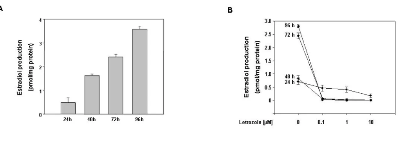
Estradiol induces Leydig cell tumor proliferation through an autocrine mechanism. We performed our study utilizing as model system R2C Leydig tumor cells. These cells have been demonstrated to have high aromatase expression and, consequently, activity (42), while we used another Leydig cell line, TM3 cells, as a normal control. We also analyzed testes from older and younger Fischer rats. Aged animals have a high incidence of spontaneous neoplasm of Leydig cells (84;88), a phenomenon not observed in younger animals, allowing us to use them as a good in vivo model to confirm results obtained in cell lines. Our first step was to measure estradiol content in culture medium of R2C and TM3 cells maintained in culture for increasing time. While E2 levels in TM3 medium were extremely low (data not shown) in R2C cells E2 levels after 24 h were 0.5 pmol/mg protein and increased by 7-fold at 96 h (Fig. 1A). This production was dependent on high constitutive active aromatase activity, since the presence of aromatase inhibitor Letrozole was able to decrease E2 production at all time points tested (Fig. 1B). E2 levels after 24 h treatment with Letrozole were still detectable, but were completely knock down when we removed the medium after 24 h and renewing the treatment for an additional 24 h. The same effect was maintained for the other two time points investigated (Fig. 1B).
Figure 1. E2 production in R2C cells. (A) Cells were cultured for the indicated times in serum free medium. (B) Cells were treated for the indicated times in HAM-F10 in the absence (-) or presence of aromatase inhibitor Letrozole (0.1, 1, 10 µM). Every 24h, before renewing treatment, cell culture medium was removed and analyzed for steroid content. E2 content was determined by RIA and normalized to the cell culture well protein content. Data represent the mean ± SEM of values from three separate cell culture wells expressed as pmol/mg protein.
Results
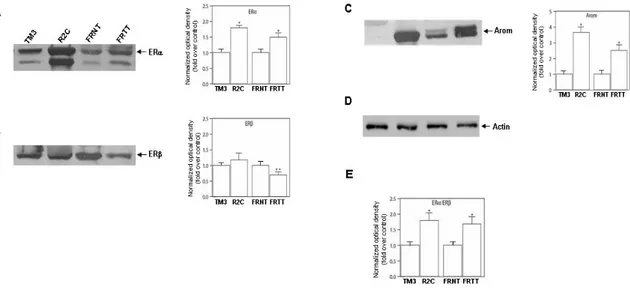
Once estradiol is produced it can exert its actions binding to specific receptors, the estrogen receptors α e β (ERα and ERβ). Analysis of the two receptor protein isoforms in our models demonstrated that tumor Leydig cells express both isoform of ERs (Fig. 2). Particularly the α isoform seems to be more expressed in R2C cells respect to TM3 and in FRTT respect to the its control FRNT (Fig. 2A) whereERβ is more expressed (Fig. 2B). In R2C as well as in FRTT was observed an increase in ERα/ERβ ratio (Fig. 2E). Moreover aromatase protein content is extremely high in tumoral samples (54) (Fig. 2C).
Figure 2. Expression of estrogen receptors and Aromatase in R2C cells. ERα (A) ERβ (B) and aromatase (C) western blot analysis
was performed on 50 µg of total proteins extracted from TM3 and R2C cells or from total tissue of normal (FRNT) and tumor (FRTT) Fisher rat testes. Results are representative of three independent experiments. β-actin (D) was used as loading control. Graphs depicted near western blots were obtained by averaging densitometric analyses of the three independent experiments. Protein expression in each lane was normalized to the β-actin content, and expressed as fold over control. (E) Graph was obtained calculating ERα/ERβ ratio of normalized optical density. (*,P < 0.001 and **,P <0.05 compared with basal).
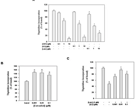
Our next experiments demonstrated that estrogen receptors are required for proliferation through a short autocrine loop maintained by endogenous E2 production in Leydig tumor cells. For instance, the use of both antiestrogens OHT and ICI and the use of aromatase inhibitor Letrozole determined a dose-dependent inhibition of cell proliferation (Fig. 3A). Among the different doses tested the highest dose of OHT (10 µM) was able to inhibit cell proliferation by 90%, ICI 10 µM by 86% and letrozole by 70%. In the same vein, starving
Results
cells for prolonged time and changing the medium every day in order to remove local E2 production, we found that addition of 1, 10 and 100 nM E2 stimulated Leydig tumor cell proliferation (Fig. 3B), and partially abrogated the inhibition induced by Letrozole (Fig. 3C).
Figure 3. Effects of antiestrogens, aromatase inhibitor Letrozole and estradiol on R2C cell proliferation. (A) Cells were treated for
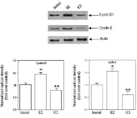
96h in HAM-F10 in the absence (-) or presence of antiestrogens hydroxytamoxifen (OHT) or ICI 182,760 (ICI) or aromatase inhibitor Letrozole at the indicated concentrations. (B) Cells were cultured for 48h in serum-free HAM-F10, every 24 h cell culture medium was removed and renewed. Cells were then treated for 24 h with estradiol at the indicated concentrations. (C) Cells were cultured for 24h in serum-free HAM-F10, cells were then treated for 48 h with letrozole (1µM) changing the culture medium and renewing treatment every 24h. For additional 24h cells were treated with letrozole (1µM) in combination with estradiol at the indicated concentrations. Proliferation was evaluated by [3H] Thymidine incorporation analysis. Values expressed as percent of untreated (basal) cells (100%) represent the mean ± SEM of three independent experiments each performed in triplicate. (*) P< 0.05 compared with basal cells. The stimulatory effect of E2 was concomitant with the increased levels of cell cycle regulator cyclin D1 and E, whose expression was inhibited by pure antiestrogen ICI (Fig. 4). All these results address how the classic E2/ERalpha signalling may control Leydig cell tumor growth and proliferation similarly to what observed in other estrogen-dependent tumors.
Results
Figure 4. R2C cells were cultured for 48h in serum-free HAM-F10, every 24 h cell culture medium was removed and renewed.
Cells were then treated for 24 h in the absence (basal) or in the presence with estradiol (1nM) and ICI (1µM) before extracting total proteins. Western blot analysis of Cyclin D1 and Cyclin E was performed on 50 µg of total proteins extracted from R2C cells. Blots are representative of three independent experiments with similar results. β-actin was used as loading control. Graphs depicted below western blots were obtained by averaging densitometric analyses of the three independent experiments. Protein expression in each lane was normalized to the β-actin content, and expressed as relative fold over basal. (*,P < 0.05 and **,P <0.01 compared with basal).
Aromatase overexpression is determined by constitutive activation of transcription factors SF-1 and CREB.
Aromatase gene transcription in rat Leydig cells is driven by the PII promoter, which is principally regulated through three CRE-like sites and one NRE site binding SF-1 and LRH-1 (42;86). Constitutive active levels of CREB have been previously demonstrated in R2C cells (89). Here we confirmed these data and demonstrated high phosphorylated status of CREB together with enhanced phosphorylation of SF-1 in FRTT (Fig. 5). Furthermore we demonstrated the presence of high expression levels of SF-1 with the protein present in a phosporylated status in R2C but not in TM3.
Results
Figure 4. Expression of total and phosphorylated forms of SF-1 and CREB. Western blot analysis was performed on 50 µg of total
proteins extracted from TM3 and R2C cells or from total tissue of normal (FRNT) and tumor (FRTT) Fisher rat testes. Blots are representative of three independent experiments with similar results. β-actin was used as loading control. Graphs depicted near western blots were obtained by averaging densitometric analyses of the three independent experiments. Protein expression in each lane was normalized to the β-actin content, and expressed as relative difference from controls. (*,P < 0.001 compared with basal).
IGF-I is produced by R2C cells and induces aromatase expression through PI3K- and PKC- mediated activation of SF-1.
Starting from previous findings demonstrating that SF-1 and CREB are activated by IGF-I and lead to an increase in StAR transcription and then steroidogenesis (90;91), we investigated the role of this factor locally produced in the testis in regulation of aromatase. Determination of IGF-I content in TM3 and R2C culture medium by RIA revealed a
Results
significant difference in the growth factor production with R2C cells producing about 4-fold higher IGF-I (Fig. 5).
Figure 5. IGF-I production in Leydig cells. IGF-I levels in culture medium of TM3 and R2C cells by RIA. TM3 and R2C cells were
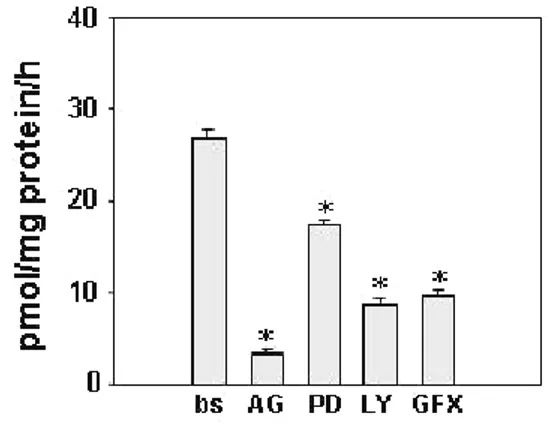
cultured for 24 h in serum free medium and IGF-I content was normalized to the cell culture well protein content. Data represent the mean ± SEM of values from three separate cell culture wells expressed as pmol/mg protein. (*) P< 0.01 compared with basal conditions. Upon binding to its receptor, IGF-IR, IGF-I activates three major transductional pathways: Ras/Raf/MAPK, PI3K/AKT, PLC/PKC, to demonstrate involvement of IGF-I transductional pathways in modulating aromatase expression in Leydig cell tumors, we used specific inhibitors: of IGF-I receptor (IGF-IR) [AG1024 (AG)], of ERK1/2 [PD98059 (PD)], of PI3K [LY294002 (LY)] and of PKC [GF109203X (GFX)]. IGF-I receptor inhibitor was able to inhibit aromatase activity in R2C cells by 85%, LY determined 65 % inhibition, PD 35 % and GFX 61 % (Fig. 6).
Figure 6. Aromatase activity in R2C cells in response to inhibitors of IGF-I pathways. Cells were treated with AG (20µM), LY (10
µM), PD (20 µM) and GFX (20 µM). Aromatase activity was assessed by using the modified tritiated water method. Results obtained are expressed as pmoles of [3H]H2O released per hour and are normalized to the well protein content (pmol/h/mg protein). Values represent the mean ± SEM of three independent experiments each performed with triplicate samples. * P < 0.01 compared to basal.
Results
The same inhibitory pattern was observed also on aromatase mRNA (fig. 7A) and protein content (Fig. 8). Parallely all of the different inhibitors but PD were able to reduce SF-1 mRNA, while CREB remained unchanged (Fig. 7). For SF-1 inhibition was 75% with AG, 90 % with LY and 80 % with GFX (Fig. 7). Analysis of protein levels by western blot confirmed the data on mRNA (Fig. 8). Treatments with increasing doses of AG, LY and GFX but not PD were able to induce a dose-dependent inhibition of total and phosphorylated levels of SF-1, on the other hand CREB was not affected by the presence of any of the inhibitors (Fig. 8).
Figure 7. Effects of inhibitors of IGF-I pathways on mRNA expression of CYP19, SF-1 and CREB in R2C cells. Total RNA was
extracted from R2C cells untreated (bs) or treated for 24h with AG (20µM), LY (10 µM), PD (20 µM) and GFX (20 µM). Real time RT-PCR was used to analyze mRNA levels of CYP19, SF-1, and CREB. Data represent the mean ± SEM of values from three separate RNA samples. Each sample was normalized to its 18S ribosomal RNA content. Final results are expressed as n-fold differences of gene expression relative to calibrator (basal) calculated with the ∆∆Ct method. * P < 0.001 compared to basal.
Results
Figure 8. Effects of inhibitors of IGF-I pathways on expression of Aromatase, total and phosphorylated forms of SF-1 and CREB in R2C cells. Western blot analyses were performed on 50 µg of total proteins extracted from R2C cells untreated (bs) or treated
for 24h with the indicated doses of AG (A), LY (B), PD (C) and GFX (D). Blots are representative of three independent experiments with similar results. β-actin was used as loading control. Graphs depicted near western blots were obtained by averaging densitometric analyses of the three independent experiments. Protein expression in each lane was normalized to the β-actin content, and expressed as relative difference from basal. (*,P < 0.01 compared with basal).
IGF-I induces aromatase expression and activity in R2C cells. To further demonstrate the prevalent role of SF-1 in IGF-I induced aromatase expression in Leydig cell tumor, we monitored the effect of IGF-I on CYP19 and SF-1 expression. Addition of exogenous amounts of IGF-I were able to induce aromatase activity by 1.8-fold (Fig. 9A). A significant effect of I treatment was seen also on CYP19 mRNA levels (Fig. 9B). IGF-I was able to induce a significant increase of 2- and 3.8-fold in aromatase mRNA at 12h and 24h, respectively (Fig. 9B). Aromatase protein levels under the same treatments reflected mRNA data (Fig. 9C). Analysis of expression levels of total and phosphorylated forms of transcription factors SF-1 and CREB showed an increase in SF-1 and pSF-1 in the
Results
presence of IGF-I after 4h while no difference was observed for CREB at any of the investigated times (Fig. 9C).
Figure 9. Aromatase activity and expression in R2C cells in response to IGF-I. (A) Cells were treated with IGF-I (100 ng/ml) for
24h. Aromatase activity was assessed by using the modified tritiated water method. Results obtained are expressed as pmoles of [3H]H2O released per hour and are normalized to the well protein content (pmol/h/mg protein). Values represent the mean ± SEM of three independent experiments each performed with triplicate samples. *P<0.05 compared to basal. (B) Total RNA was extracted from R2C cells untreated (bs) or treated for the indicated times with IGF-I (100 ng/ml). Real time RT-PCR was used to analyze mRNA levels of CYP19. Data represent the mean ± SEM of values from three separate RNA samples. Each sample was normalized to its 18S ribosomal RNA content. Final results are expressed as n-fold differences of gene expression relative to calibrator (basal) calculated with the ∆∆Ct method. *P<0.01 and **P<0.001compared to basal. (C) Western blot analyses were performed on 50 µg of total proteins extracted from R2C cells untreated (basal) or treated for the indicated times with IGF-I (100 ng/ml). Blots are representative of three independent experiments with similar results. β-actin was used as loading control. Graphs depicted below western blots were obtained by averaging densitometric analyses of the three independent experiments.. Protein expression in each lane was normalized to the β-actin content, and expressed as relative difference from controls. (*,P < 0.01 compared with basal).
Changes in IGF-I pathway activation status lead to changes in SF-1 binding to the aromatase PII promoter.
Finally we performed CHIP assay to investigate how IGF-I stimulation influence per se binding of transcription factors to the aromatase PII promoter. We evidenced how in basal condition all the different inhibitors but not PD reduced the amount of bound SF-1
Results
reflecting changes in SF-1 protein amount (Fig. 10 A). The increase in SF-1 protein content seen under IGF-I treatment (Fig. 9C) reflected an increase in SF-1 binding to the PII promoter (Fig. 10B).
Figure 10. IGF-I increases SF-1 recruitment to the aromatase PII promoter through PI3K and PKC. (A) R2C cells were
incubated for 24 h with AG (20µM), LY (10 µM), PD (20 µM) and GFX (20 µM). Untreated cells (basal, bs) were treated with the same amount of vehicle alone (DMSO) that never exceeded 0·01% (v/v). (B) R2C cells were incubated for the indicated times with IGF-I (100 ng/ml). In vivo binding of SF-1 to the aromatase PII promoter was examined using ChIP assay. Immunoprecipitated (SF-1) and total (10% input) DNA were subject to PCR using specific primers. Similar results were obtained in two additional experiments.
Discussion
Discussion
The current study was aimed to explain the molecular mechanism responsible for aromatase overexpression in tumor Leydig cells with a consequent excess of estradiol in
situ production sustaining tumor cell growth and proliferation.
Mammalian testis is capable of estrogen synthesis, whose production is regulated by different factors at different ages. In mature animals, aromatization of testosterone to estradiol is enhanced by LH/chorionic gonadotropin (CG) and not by FSH. The site of this synthesis appears to be age-dependent, at least in some species, such as the rat (92). Leydig cells are an elective target site of LH/CG which controls testosterone biosynthesis as well as its conversion to estradiol through aromatase activity. Leydig cell is also known to be the site of estrogen synthesis in several species, including mice (93), humans (94), suine (95), and sheep (96). Alterations in local estrogen synthesis may have significant consequences in malignancy of these cells. In the present study we observed that manteinance of R2C cells in the absence of serum induces a cospicous release of E2 from cellular storage in a time dependent manner. This synthesis was abrogated by treatment with Letrozole, an aromatase inhibitor, addressing how estrogen production is dependent on high constitutive aromatase activity. A strongly increased aromatase expression was observed in R2C cells respect to the normal cell line control TM3 as well as in FRTT respect to FRNT. These findings concord with a previous study on human tissues showing that the increase in estrogen synthesis, as a consequence of a more intense aromatase activity, is higher in Leydig cell tumor fraction than in normal tissue surrounding the tumor of the same patient (97).
Mediators of the physiological effects of estrogens are the estrogen receptors (ER) α and β. ERα appears to be confined to Leydig cells in testicular tissue (45), while ERβ has been detected immunohistochemically in several rat testicular cell types, including Sertoli cells, germ cells, and peritubular cells (98). An enhanced expression of ERα, resulting in an increased ERα/ERβ, ratio was observed in R2C compared to TM3 cell line as well as in FRTT respect to FRNT. This is in agreement with previous reports demonstrating that
Discussion
transgenic mice overexpressing aromatase have an enhanced occurrence of breast and Leydig cell tumors together with an enhanced expression of ERα in the tumoral tissue (54). The latter findings address reasonably how an estrogen short autocrine loop may be involved in breast and testicular tumorogenesis in the presence of an excess of locally produced estradiol. Indeed, an arrest of cell growth was observed following abrogation of local E2 production with Letrozole or after addition of ERα inhibitors ICI or OHT. Besides, only after remotion of medium every day along with prolonged R2C starvation abolishing local steroid production, we observed how exogenous E2 was able to display proliferative effects.
One mechanism through which estrogens induce cell proliferation is by increasing protein levels of G1 regulatory cyclins A, B1, D1, D3, and E in target cells (99). In our study we showed that the expression of two of the most important regulators of Leydig cell cycle, cyclin D1 and E can be increased by E2 and downregulated by treatment with antiestrogens. These data further confirm that aromatase overexpression and the consequent E2 production may be the cause of altered cell cycle regulation of Leydig tumor cells.
In the attempt to explain the molecular mechanism determining aromatase overexpression in our tumor cell line, we focused our attention on expression levels of transcription factors identified as crucial regulators of aromatase gene expression: CREB and SF-1. In the adult testis SF-1 is predominantly expressed in Leydig cells (100). The increase of total and/or phosphorylated protein can potentiate SF-1 transcriptional activity (101). In R2C cells and in FRTT compared to the normal controls we found higher SF-1 phosphorylated protein levels as a consequence of elevated protein content. Total CREB levels were similar in all samples but highly phosphorylated in tumor samples. Starting from these observations we investigated which pathways could be involved in the activation of these transcription factors.
The most important signal that initiates steroidogenesis in Leydig cells is the binding of LH to the LH receptor (102). It has been demonstrated that LH/LHreceptor signaling pathway is constitutively active in R2C cells and makes the phenotype of these cells
Discussion
constitutively steroidogenic (103). For instance in the presence of a specific PKA inhibitor, constitutive synthesis of Star mRNA and steroidswere significantly inhibited (104). These observations fit well with our findings evidencing how the presence of PKA inhibitor determined a strong decrease in aromatase activity together with a drop in CREB phosphorylation (data not shown). In the presence of a specific PKC inhibitor no effects were elicited on phosphorylation of CREB, while SF-1 dropped dramatically.
It has been shown that CREB in mouse Leydig cells can be phosphorylated also through the PKC pathway, activated by IGF-I (103). In this study we have revealed that R2C tumor Leydig cells release higher levels of IGF-I in the culture medium respect to TM3 cells. However, the exposure to I as well as the treatment with inhibitors of IGF-I signalling did not affect CREB phosphorylative status but decreased SF-1 phosphorylation, postulating a separate mechanism controlling CREB and SF-1 activation in modulating aromatase activity.
These findings led us to suppose that the IGF-I derived from tumor Leydig cells could act through an autocrine mechanism in activating aromatase expression. IGF-I receptors have previously been identified in Leydig cells of several species (10;105-107). It has been hypothesized that changes in IGF-RI expression can influence tumor cell progression. However in our cellular models, we did not reveal differences in IGF-RI expression between normal and tumor cells (data not shown), indicating that IGF-I level may be the determining factor in potentiating IGF-I signalling. A previous study investigating the effects of long term IGF-I treatment on Leydig cells did not reveal alterations in DNA synthesis, indicating that IGF-I may act as a differentiation factor rather than a mitogenic factor (12). In fact, expression levels of all mRNA species associated with T biosynthesis were shown to be lower in the absence of IGF-I, while treatment with IGF-I/insulin has been found to stimulate steroidogenesis and StAR expression in Leydig cells through a process that does not require cAMP signaling (9;107;108). In the same vein we may reasonable hypothesize that IGF-I could sustain, through an autocrine/paracrine mechanism, the elevated aromatase expression/activity in tumor Leydig cells. To verify this hypothesis we studied the various signalling pathways initiated by I through
Discussion
IR. Binding of IGF-I to its receptor causes receptor autophosphorylation and the activation of intrinsic tyrosine kinase that acts on various substrates including the insulin receptor substrate (IRS) and Shc adaptor proteins. These activated proteins recruit other factors, leading to activation of multiple signalling pathways including the phosphatidyl inositol 3-kinase (PI3K)/Akt and the mitogen-activated protein (MAP) 3-kinase cascade. In addition, it has been shown that IGF-I can activate also the phospholipase C (PLC)/protein kinase C (PKC) pathway (90;109). To demonstrate a role for IGF-I in mediating aromatase activation we used specific inhibitors for IGF-I signaling [AG1024 (AG)], ERK1/2 [PD98059 (PD)], PI3K [LY294002 (LY)] and PKC [GF109203X (GFX)] and showed a reduction of aromatase activity with all of them. Together these data confirm a role for IGF-I in mediating aromatase activation in tumor Leydig cells. All of the different inhibitors but PD were able to produce a similar inhibitory pattern on both aromatase and SF-1 mRNA and protein expression. Furthermore by ChIP assay we evidenced that SF-1 binding to the aromatase promoter II that was reduced by AG, LY, GFX but not by PD indicating a central role of this transcription factor in regulating aromatase gene transcription in tumor Leydig cells. This is the first report of a direct link between SF-1 transcription and IGF-I signalling pathway in regulating aromatase expression.
Furthermore, addition of IGF-I itself was able to increase aromatase activity and expression. These events were due to an increase in the amount of total and phosphorylated SF-1 levels whose binding to the aromatase promoter was shown to be rapidly augmented. So we postulate that an enhanced endogenous IGF-I local production may contribute to maintain an elevated aromatase activity sustained by a direct stimulatory effect of SF-1. For instance the inhibition of IGF-I signalling through inhibition of either PI3K/AKT and PLC/PKC pathways were able to block SF-1 expression and protein phoshorylation. Particularly treatment with AG blocked SF-1 phosphorylation more efficiently than the separate treatment of PI3K or PKC, addressing how both pathways may synergize in upregulating SF-1 activity. In the presence of PD, SF-1 expression remained unchanged together with unaffected aromatase mRNA and protein levels. Importantly aromatase activity appeared decreased in the presence of PD suggesting a potential stimulatory role of
Discussion
ERK1/2 on the enzyme at a post-transcriptional level. From our findings then emerges a double mechanism inducing enhanced expression of aromatase: 1. a constitutive activation of LH/cAMP/PKA pathway which determines CREB activation; 2. an enhanced IGF-I signaling potentiating SF-1 action. The enhanced expression of SF-1 may be maintained by the lack of DAX-1 (Dosage-Sensitive Sex Reversal, Adrenal Hypoplasia Congenita, Critical Region on the X Chromosome, Gene-1) in R2C cells (89). DAX-1 is a specific co-repressor of SF-1 (60-64) and inhibits StAR expression and steroidogenesis by 40-60% when overexpressed in R2C cells (89). The lack of DAX-1 expression in R2C cells may be due to the constitutive active PKA signalling, in fact since in a mouse Leydig cell line was shown a marked decrease of DAX-1 mRNA within 3 h after addition of LH or forskolin (110). Then, the activation of LH/LHr/PKA pathway at the same time decreases DAX-1 expession and promotes SF-1 activity.
Remains to explain which molecular mechanism(s) is responsible for the elevated IGF-I production in tumor Leydig cells. IGF-In vivo, administration of hCG increases IGF-IGF-IGF-I mRNA levels in rat Leydig cells (111). LH deprival determines a decrease in the BrdU incorporation as well as a decrease in mRNA levels of IGF-I and IGF-I receptor. These observations together with our data showing a decrease in IGF-I basal production after treatment with a PKA inhibitor (data not shown) suggest the possibility that LH can mediate its proliferative effects also by regulating IGF-I and its receptor in Leydig cells and that the altered LH/LHreceptor activated pathway in R2C cells could be the cause of oIGF-I overproduction (112).
Moreover the observation that in murine Leydig cells IGF-I is able to increase the LHr mRNA stability (113) together with data showing that the presence of an IGF-I antibody reduced the steroidogenic responsiviness to LH/hCG (114) suggest also the possibility of an IGF-I action in sustaining LH/LHr signalling. If the constitutive activation of LH/LHr/PKA signalling in R2C cells may be involved in upregulation of IGF-I expression remains also to be explored.
In conclusion, in this study we demonstrated that in tumor Leydig cells aromatase overexpression determines an excessive local estradiol production able to stimulate the
Discussion
expression of genes involved in cycle regulation and sustaining cell proliferation. Aromatase overexpression appears to be induced by the combined enhanced LH/LHr and IGF-I signalling (Fig. 11).
Particularly, LH/LHr signaling determines a constitutive active CREB phosphorylation on aromatase gene promoter while IGF-I overproduction stimulates through an autocrine mechanism SF-1 binding on the same promoter. The observation that antiestrogens and aromatase inhibitors as well as IGF-I signalling blockers are able to reduce R2C proliferation opens new perspectives on the adjuvant therapeutic approach of testicular cancer.
References
1. De Kretser DM, Kerr JB 1994 The cytology of the testis. ThePhysiology of Reproduction. E Knobil & JD Neill. New York: Raven Press ed.; 1177-1290
2. Gaytan F, Bellido C, Aguilar E, van Rooijen N 1994 Requirement for testicular macrophages in Leydig cell proliferation and differentiation during prepubertal development in rats. J Reprod Fertil 102:393-399
3. Gaytan F, Bellido C, Morales C, Reymundo C, Aguilar E, van Rooijen N 1994 Effects of macrophage depletion at different times after treatment with ethylene dimethane sulfonate (EDS) on the regeneration of Leydig cells in the adult rat. J Androl 15:558-564
4. Borland K, Mita M, Oppenheimer CL, Blinderman LA, Massague J, Hall PF, Czech MP 1984 The actions of insulin-like growth factors I and II on cultured Sertoli cells. Endocrinology 114:240-246
5. De Kretser DM, Catt KJ, Paulsen CA 1971 Studies on the in vitro testicular binding of iodinated luteinizing hormone in rats. Endocrinology 88:332-337
6. Loosfelt H, Misrahi M, Atger M, Salesse R, Vu Hai-Luu Thi M, Jolivet A, Guiochon-Mantel A, Sar S, Jallal B, Garnier J, al. e 1989 Cloning and sequencing of porcine LH-hCG receptor cDNA: variants lacking transmembrane domain. Science 245:525-528
7. Dufau ML 1988 Endocrine Regulation and Communicating Functions of the Leydig Cell. Annual Review of Physiology 50:483-508
8. Dufau ML, Dufau ML, Tsuruhara T, Horner KA, Podesta E, Catt KJ 1977 Intermediate role of adenosine 3':5'-cyclic monophosphate and protein kinase during gonadotropin-induced steroidogenesis in testicular interstitial cells. Proc Natl Acad Sci U S A 74:3419-3423
9. Saez JM 1994 Leydig cells: endocrine, paracrine, and autocrine regulation. Endocr Rev 15:574-626
10. Vannelli BG, Barni T, Orlando C, Natali A, Serio M, Balboni GC 1988 Insulin-like growth factor-I (IGF-I) and IGF-I receptor in human testis: an immunohistochemical study. Fertil Steril 49:666-669
11. Handelsman DJ, Spaliviero JA, Scott CD, Baxter RC 1985 Identification of insulin-like growth factor-I and its receptors in the rat testis. Acta Endocrinol 109:543-549