R E S E A R C H A R T I C L E
Open Access
Potential novel therapeutic strategies in cystic
fibrosis: antimicrobial and anti-biofilm activity of
natural and designed
α-helical peptides against
Staphylococcus aureus, Pseudomonas aeruginosa,
and Stenotrophomonas maltophilia
Arianna Pompilio
1,2, Valentina Crocetta
1,2, Marco Scocchi
3, Stefano Pomponio
1,2, Valentina Di Vincenzo
1,2,
Mario Mardirossian
3, Giovanni Gherardi
4, Ersilia Fiscarelli
5, Giordano Dicuonzo
4, Renato Gennaro
3and
Giovanni Di Bonaventura
1,2*Abstract
Background: Treatment of cystic fibrosis-associated lung infections is hampered by the presence of multi-drug resistant pathogens, many of which are also strong biofilm producers. Antimicrobial peptides, essential components of innate immunity in humans and animals, exhibit relevant in vitro antimicrobial activity although they tend not to select for resistant strains.
Results: Threeα-helical antimicrobial peptides, BMAP-27 and BMAP-28 of bovine origin, and the artificial P19(9/B) peptide were tested, comparatively to Tobramycin, for their in vitro antibacterial and anti-biofilm activity against 15 Staphylococcus aureus, 25 Pseudomonas aeruginosa, and 27 Stenotrophomonas maltophilia strains from cystic fibrosis patients. All assays were carried out in physical-chemical experimental conditions simulating a cystic fibrosis lung. All peptides showed a potent and rapid bactericidal activity against most P. aeruginosa, S. maltophilia and S. aureus strains tested, at levels generally higher than those exhibited by Tobramycin and significantly reduced biofilm formation of all the bacterial species tested, although less effectively than Tobramycin did. On the contrary, the viability-reducing activity of antimicrobial peptides against preformed P. aeruginosa biofilms was comparable to and, in some cases, higher than that showed by Tobramycin.
Conclusions: The activity shown byα-helical peptides against planktonic and biofilm cells makes them promising “lead compounds” for future development of novel drugs for therapeutic treatment of cystic fibrosis lung disease. Keywords: Cystic fibrosis, Antimicrobial peptides, Biofilm
Background
Physicians treating patients with cystic fibrosis (CF) are increasingly faced with infections caused by multidrug-resistant strains. Pseudomonas aeruginosa and Staphylo-coccus aureusare the most common bacterial pathogens isolated from the CF respiratory tract where they cause
persistent infections associated with a more rapid de-cline in lung function and survival [1,2]. In recent years, however, there has been an increasing number of reports on potentially emerging and challenging pathogens, probably due to improved laboratory detection strategies and to selective pressure exerted on bacterial popula-tions by the antipseudomonal antibiotic therapy [2]. In this respect, both the overall prevalence and incidence of intrinsically antibiotic-resistant Stenotrophomonas mal-tophilia isolations from CF respiratory tract secretions have been recently reported [3-5].
* Correspondence:[email protected]
1
Department of Biomedical Sciences,“G. d’Annunzio” University of Chieti, Via Vestini 31, 66100 Chieti, Italy
2
Center of Excellence on Aging,“G. d'Annunzio” University Foundation, Via Colle dell’Ara, 66100 Chieti, Italy
Full list of author information is available at the end of the article
© 2012 Pompilio et al.; licensee BioMed Central Ltd. This is an Open Access article distributed under the terms of the Creative Commons Attribution License (http://creativecommons.org/licenses/by/2.0), which permits unrestricted use, distribution, and reproduction in any medium, provided the original work is properly cited.
Efforts to treat CF infections are also hampered by the high microbial adaptation to the CF pulmonary environ-ment, resulting in an increased ability to form biofilms intrinsically resistant to therapeutically important anti-biotics such as aminoglycosides, fluoroquinolones, and tetracycline [6-10].
Novel antimicrobial agents that could replace or com-plement current therapies are consequently needed to fight chronic infections in CF patients.
Antimicrobial peptides (AMPs) are naturally occurring molecules of the innate immune system that play an im-portant role in the host defence of animals and plants [11-13]. Over the last years, natural AMPs have attracted considerable interest for the development of novel anti-biotics for several reasons [14,15]: i) the broad activity spectrum, comprised multiply antibiotic-resistant bac-teria; ii) the relative selectivity towards their targets (mi-crobial membranes); iii) the rapid mechanism of action; and, above all, iv) the low frequency in selecting resistant strains. Although the antimicrobial activity of AMPs has been extensively reported in literature [13-17], only few studies have been reported with respect to CF pathogens [18-21].
Hence, in an attempt to evaluate the therapeutic potential of AMPs in the management of CF lung infections, for the first time in the present study three cationic α-helical AMPs - two cathelicidins of bovine origin (BMAP-27, BMAP-28) and the artificial peptide P19(9/B) - were tested for their in vitro antibacterial effectiveness, as well as their in vitro anti-biofilm activity, against selected S. aureus, P. aeruginosa, and S. maltophilia strains collected from CF patients. The efficacy of the AMPs was compared to that of Tobramycin, selected as the antibiotic of choice used for chronic suppressive ther-apy in CF patients.
Since the conditions present in the CF patients’ airway surface liquid could counteract the potency of antibiotics such as Tobramycin [22,23], in the present study all in vitro antimicrobial assays were carried out under ex-perimental conditions simulating the physical-chemical properties observed in CF lung environment [24-26].
Results
Phenotypic features and clonal relatedness of CF strains
A total of 9 out of 25 P. aeruginosa strains tested showed mucoid phenotype on MHA, while 3 exhibited SCV phenotype. Among 15 S. aureus isolates tested, 7 were methicillin-resistant.
PFGE analysis showed 8, 21, and 12 different pulso-types among S. aureus, S. maltophilia, and P. aeruginosa isolates, respectively. Among S. aureus isolates, only the PFGE type 1 was shared by multiple strains, which com-prised 8 isolates and 7 PFGE subtypes. Among S.
malto-philia isolates, 2 multiple-strains PFGE types were
observed: PFGE type 23 (5 isolates, 2 PFGE subtypes), and PFGE type 73 (2 isolates with identical PFGE pro-file). Among P. aeruginosa isolates, 5 multiple-strains PFGE types were observed: PFGE type 5 (6 isolates, 2 PFGE subtypes), PFGE type 1 (4 isolates with indistin-guishable PFGE profile), PFGE types 9 and 11 (3 isolates each, with identical PFGE pattern), and PFGE type 8 (2 isolates, one PFGE subtype) (data not shown).
In vitro activity of AMPs and Tobramycin against planktonic cells: MIC, MBC
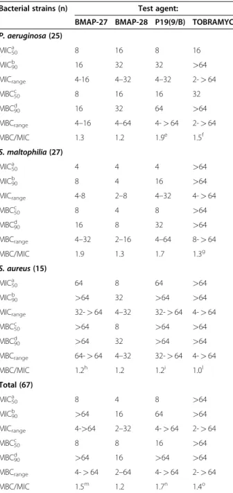
In order to determine the efficacy of AMPs, the anti-microbial activity was measured against 67 CF clinical isolates, and results are summarized in Table 1. Overall, BMAP-28 showed the widest activity spectrum among AMPs tested, as suggested by MIC90and MBC90values
(16 μg/ml, for both), although all of them exhibited a species-specific activity. In fact, although AMPs showed comparable activity against P. aeruginosa, BMAP-28 was found to be more active than P19(9/B) against S. maltophilia, and resulted the best active AMP against S. aureus (MIC90: 32 μg/ml; MBC90: 32 μg/ml).
Com-pared to AMPs, Tobramycin exhibited a lower activity (MIC90 and MBC90: >64 μg/ml) regardless of the
spe-cies considered. Killing quotient values, calculated as MBC/MIC ratio, were < 4 for all AMPs, as well as for Tobramycin, clearly suggesting a bactericidal activity. No differences in susceptibility levels to AMPs were found with regard to phenotype (mucoid, SCV, MRSA), pulsotype, or susceptibility to Tobramycin (data not shown).
MIC and MBC values obtained under
CLSI-recommended or“CF-like” experimental conditions (see Materials and Methods section) are shown in Table 2. Comparative evaluation of these values showed that
mean MICCF-like/MICCLSI and MBCCF-like/MBCCLSI
values obtained for Tobramycin (23.9 and 15.6, respect-ively) were significantly higher than those observed for BMAP-27 (1.5 and 1.2, respectively; p< 0.001), BMAP-28 (0.5 and 0.5, respectively; p< 0.001), and P19(9/B) (2.8 and 2.9, respectively; p< 0.001), regardless of spe-cies tested, indicating a reduced antibiotic activity of Tobramycin in CF-like conditions.
Bactericidal kinetics
Time-killing results have been summarized in Figure 1. BMAP-27, BMAP-28, and P19(9/B) exerted a rapid bac-tericidal activity against P. aeruginosa, reducing the num-ber of viable bacterial cells of at least 3 logs within 60 min of exposure. However, the bactericidal effect of BMAP-28 against P. aeruginosa was incomplete for two (Pa6 and Pa22) of the three strains tested, allowing bacterial regrowth after 24-h incubation, although at levels lower than those observed for untreated control. In parallel experiments, Tobramycin showed only a bacteriostatic
effect against P. aeruginosa, causing no more than 1-log reduction in viable count after 24 h.
BMAP-27, BMAP-28 and P19(9/B) exerted bactericidal activity also against S. maltophilia, although with streaking strain-specific differences. Particularly, BMAP-28 exhibited
only bacteriostatic effect against Sm192 strain, while P19(9/ B) showed a rapid bactericidal effect against Sm138 strain, causing more than a 4-log reduction in viable count after 10 min-exposure. Tobramycin exhibited a late (after 24-h exposure) bactericidal effect only against Sm138 strain.
AMPs activity against S. aureus was significantly strain-specific, ranging from the rapid bactericidal activity of BMAP-28 against Sa10 strain, to the bacteriostatic effect of P19(9/B) and BMAP-28 against Sa4 strain. Tobramycin
Table 1 In vitro activity of BMAP-27, BMAP-28, P19(9/B), and Tobramycin against P. aeruginosa, S. maltophilia and S. aureus CF strains
Bacterial strains (n) Test agent:
BMAP-27 BMAP-28 P19(9/B) TOBRAMYCIN P. aeruginosa (25) MIC50a 8 16 8 16 MIC90b 16 32 32 >64 MICrange 4-16 4–32 4–32 2-> 64 MBC50c 8 16 16 32 MBC90d 16 32 64 >64 MBCrange 4–16 4–64 4-> 64 2-> 64 MBC/MIC 1.3 1.2 1.9e 1.5f S. maltophilia (27) MIC50a 4 4 4 >64 MIC90b 8 4 16 >64 MICrange 4-8 2–8 4–32 4-> 64 MBC50c 8 4 8 >64 MBC90d 16 8 32 >64 MBCrange 4–32 2–16 4–64 8-> 64 MBC/MIC 1.9 1.3 1.7 1.3g S. aureus (15) MIC50a 64 8 64 >64 MIC90b >64 32 >64 >64 MICrange 32-> 64 4–32 32-> 64 4- > 64 MBC50c >64 8 >64 >64 MBC90d >64 32 >64 >64 MBCrange 64-> 64 4–32 32-> 64 4- > 64 MBC/MIC 1.2h 1.2 1.2i 1.0l Total (67) MIC50a 8 4 8 >64 MIC90b >64 16 64 >64 MICrange 4->64 2–32 4-> 64 2-> 64 MBC50c 8 8 16 >64 MBC90d >64 16 >64 >64 MBCrange 4-> 64 2–64 4-> 64 2-> 64 MBC/MIC 1.5m 1.2 1.7n 1.4o a, b
MIC50and MIC90: MIC (μg/ml) inhibiting 50 and 90% of the strains tested,
respectively.
c, d
MBC50and MBC90: MBC (μg/ml) eradicating 50 and 90% of the strains
tested, respectively.
Only isolates exhibiting in range MIC values were considered for killing quotient calculation (MBC/MIC):e
n = 24;f n = 12;g n = 3;h n = 6;i n = 2;m n = 58; n n = 57;o n = 17.
Table 2 Antimicrobial activity of BMAP-27, BMAP-28, P19 (9/B) and Tobramycin evaluated under different
experimental conditions:“CF-like” (5% CO2, pH 6.8,
SCFM) and“standard CLSI-recommended” (aerobiosis, pH 7.2, CAMHB)
Susceptibility (MICCF-like/MICCLSI) to:
Bacterial strains BMAP-27 BMAP-28 P19(9/B) TOBRAMYCIN P. aeruginosa Pa1 8/4 8/8 4/16 4/0.25 Pa5 8/4 16/16 8/8 16/2 Pa6 8/8 16/16 16/8 8/8 Pa9 8/4 16/16 16/8 64/1 S. maltophilia Sm109 4/8 4/16 4/8 128/64 Sm126 8/16 8/32 4/32 256/64 Sm143 8/8 4/8 4/4 8/2 S. aureus Sa1 128/64 8/16 128/16 256/64 Sa3 64/64 4/32 64/16 256/16 Sa4 64/64 4/16 32/8 32/2 Sa7 64/16 4/16 64/8 256/2
Mean MICCF-like/MICCLSI 1.5 0.5 2.8 23.9
P. aeruginosa Pa1 8/8 8/16 16/32 4/1 Pa5 16/8 16/32 16/16 16/4 Pa6 16/8 16/16 16/32 8/8 Pa9 8/8 16/32 64/16 128/2 S. maltophilia Sm109 8/16 8/16 8/8 256/128 Sm126 8/32 16/32 8/32 256/64 Sm143 16/8 8/8 4/4 8/8 S. aureus Sa1 128/64 8/16 128/16 256/64 Sa3 64/64 4/32 64/16 256/32 Sa4 64/64 8/32 32/8 32/2 Sa7 64/NDa 8/16 64/8 256/4
Mean MBCCF-like/MBCCLSI1.2 0.5 2.9 15.6
a
showed a bactericidal effect against all S. aureus strains tested, although allowing bacterial regrowth of Sa4 strain after 2-h exposure.
In vitro activity of Tobramycin-AMP combinations against planktonic cells
Results from checkerboard assays are summarized in Table 3. FICI values showed that all AMP + Tobramycin combinations tested showed an indifferent effect against P. aeruginosa and S. maltophilia strains. Conversely, BMAP-27 + Tobramycin (tested at 16 + 8, 16 + 4, and 16 + 2μg/ml, respectively) combination exhibited syner-gic effect against Sa4 strain (the only one tested, 100% synergy), while P19(9/B) + Tobramycin (tested at 4 + 2,
4 + 1, and 8 + 1 μg/ml, respectively) combination exhib-ited synergic effect against S. aureus Sa10 strain (1 out of 3 strains tested, 33.3% synergy).
In vitro activity of AMPs and Tobramycin against biofilm
All CF strains were screened for biofilm forming ability on polystyrene. A significantly higher proportion of bio-film producer strains was found in P. aeruginosa and S. aureus, compared to S. maltophilia (96 and 80% vs 55%, respectively; p< 0.01) (data not shown). However, efficiency in biofilm formation was significantly higher in P. aeruginosa than in S. aureus, as suggested by me-dian biofilm amounts produced (0.162 vs 0.109, respect-ively; p< 0.01) (data not shown).
Figure 1 Time-killing kinetic of AMPs against CF strains. BMAP-27 (■), BMAP-28 (▲), P19(9/B) (×), and Tobramycin (●) were tested at MIC value against representative P. aeruginosa (Pa6, Pa15, and Pa22), S. maltophilia (Sm138, Sm143, and Sm192), and S. aureus (Sa4, Sa10, and Sa13) CF strains. Controls (♦) were not exposed to drugs. Values are the mean of two independent experiments performed in triplicate. The dotted line indicates a 3-log reduction in viability.
Table 3 In vitro effect of AMP + Tobramycin (TOB) combinations against P. aeruginosa, S. maltophilia, and S. aureus CF strains
Drug combinations P. aeruginosa S. maltophilia S. aureus
Synergy Indifference Antagonism Synergy Indifference Antagonism Synergy Indifference Antagonism FICIa≤ 0.5 0.5 < FICI ≤ 4 FICI > 4 FICI≤ 0.5 0.5 < FICI ≤ 4 FICI > 4 FICI≤ 0.5 0.5 < FICI ≤ 4 FICI > 4
BMAP-27 + TOB 0 (0%) 12 (100%) 0 (0%) 0 (0%) 8 (100%) 0 (0%) 1 (100%)b 0 (0%)b 0 (0%)b
BMAP-28 + TOB 0 (0%) 12 (100%) 0 (0%) 0 (0%) 8 (100%) 0 (0%) 0 (0%)c 1 (100%)c 0 (0%)c
P19(9/B) + TOB 0 (0%) 12 (100%) 0 (0%) 0 (0%) 8 (100%) 0 (0%) 1 (33.3%)d 2 (66.7%)d 0 (0%)d
a
Fractional Inhibitory Concentration Index (FICI).
Only isolates exhibiting in-range MIC values were considered for checkerboard titration method: P. aeruginosa (n = 12), S. maltophilia (n = 8), and S. aureus (b
n = 1;
c
To determine if AMPs could be prophylactically used to prevent biofilm formation, we tested the effect of AMPs and Tobramycin at sub-inhibitory concentrations (1/2x, 1/4x, and 1/8xMIC) against biofilm formation (Figure 2). Tobramycin at 1/2x and 1/4xMIC caused a significantly higher reduction in biofilm-forming ability of S. maltophilia and S. aureus, in comparison with the three AMPs. This effect was more relevant with S. aureus, being observed also at 1/8xMIC. Tobramycin showed to be more effective than BMAP-27 against
P. aeruginosa at concentrations equal to 1/4x and
1/8xMIC. The activity of Tobramycin in reducing bio-film formation was not related to drug susceptibility (data not shown). Among AMPs, BMAP-28 and P19(9/B) at 1/2xMIC were significantly more active compared to BMAP-27, and BMAP-28 at 1/4xMIC was significantly more active than other AMPs against S. aureus.
We further evaluated AMPs as potential therapeutics for CF by testing their efficacy against preformed bio-films. To this, BMAP-27, BMAP-28, P19(9/B), and Tobramycin at 1xMIC and at bactericidal concentrations (5x, and 10xMIC) were assayed against preformed (24 h) biofilms by six representative P. aeruginosa strains selected for high biofilm formation ability (Figure 3).
The activity of AMPs and Tobramycin against pre-formed biofilms resulted to be similar in 5 out of 6 strains tested, causing a highly significant reduction of biofilm viability compared to the controls (biofilm not exposed; p< 0.0001), regardless of the concentrations tested (Figure 3). AMPs showed to be active at all concentrations, also against biofilms formed by P.
aeruginosa Pa32, against which Tobramycin was
ef-fective only at the highest concentration used
(10xMIC). The activity of Tobramycin against pre-formed biofilms was not related to drug susceptibility (data not shown).
Discussion
This study was aimed at verifying the potential of some α-helical AMPs as lead compounds for the development of novel antimicrobials to treat lung disease in CF patients. To this, we tested the in vitro susceptibility of P. aeruginosa, S. maltophilia and S. aureus CF isolates to the naturally occurring AMPs 27 and BMAP-28, as well as the rationally designed P19(B/9), and we compared their effectiveness with that of Tobramycin, the antibiotic of choice for the inhalation therapy of chronic airway infections in CF patients.
BMAP-27 and BMAP-28 are two cathelicidin-derived peptides of bovine origin that have a role in innate defence [27,28]. The hallmark of cathelicidins is the presence of a conserved N-terminal proregion associated with C-terminal antimicrobial sequences showing a remarkable diversity and considerable inter-species differences [13]. BMAP-27
and BMAP-28 are cationic (charge: +11 and +8, respect-ively) and both adopt anα-helical structure on interaction with the negatively charged bacterial surface [28]. Recent results have suggested that AMPs with these characteristics
Figure 2 Effect of AMPs at sub-inhibitory concentrations against biofilm formation by CF strains. BMAP-27 (white bars), BMAP-28 (light gray bars), P19(9/B) (dark gray bars), and Tobramycin (black bars) were tested at 1/2x, 1/4x, and 1/8xMIC against biofilm formation by P. aeruginosa (n = 24, 24, 25, and 17, for BMAP-27, BMAP-28, P19(9/B) and Tobramycin, respectively), S. maltophilia (n = 14, 14, 27, and 5, for BMAP-27, BMAP-28, P19(9/B) and Tobramycin, respectively), and S. aureus (n = 11, 11, 8, and 3, for BMAP-27, BMAP-28, P19(9/B) and Tobramycin, respectively) CF strains. Prevention of biofilm formation was plotted as percentage of strains whose ability in forming biofilm was significantly decreased (of at least 25%) compared to controls (not exposed), as analyzed by a crystal violet staining assay.* p< 0.05; ** p < 0.0001, Fisher’s exact test.
may be the most effective against strains producing exogen-ous polysaccharides that are known to inhibit the activity of other types of AMPs [19,29]. For this reason, we added to our study also a third peptide from this class which has been rationally designed, making use also of non-proteinogenic aminoacids, to optimize its propensity to as-sumeα-helical conformation [30].
Effort to treat CF are also hampered by the conditions present in patients’ airway surface liquid where the accu-mulation of large volumes of viscous sputum (mucus) providing bacteria with a nutritionally rich growth environment composed of host- and bacterial-derived factors which deeply change their phenotype and pos-sibly their susceptibility against AMPs [31]. Therefore, to accurately judge the feasibility of these peptides as potential anti-infectives in the context of CF, in this study we investigated the activity of AMPs under some CF-like experimental conditions, including acidic pH, reduced O2
tension, and a chemically defined medium mimicking the nutritional composition of CF sputum [24-26].
These conditions allow pathogens to assume a physiology similar to that shown in vivo in the CF lung [24] and constitute a more realistic model to assay their sensitivity to AMPs.
Evaluation of MIC and MBC values, as well as time-killing assays against planktonic forms of different CF isolates of P. aeruginosa, S. maltophilia, and S. aureus, have shown that all three AMPs are highly active in vitro against most tested strains, although BMAP-28 showed the widest spectrum of activity. It is noteworthy that all the three peptides exhibited an activity higher than Tobramycin. This observation is even more evident when considering the molar concentration (μM) of each
compound rather than that by weight (μg/ml), given that the peptides tested are at least six folds heavier than Tobramycin.
The poor activity showed by Tobramycin is probably due to the experimental conditions used in this study, as suggested by comparative evaluation of MIC values
observed in both“CF-like” and CLSI-recommended
con-ditions. On the contrary, the activity of AMPs tested resulted to be slightly enhanced (BMAP-28), unaffected (BMAP-27), or slightly reduced [P19(9/B)] in “CF-like” conditions, compared to CLSI-recommended ones, so they can be considered to be quite robust and medium insensitive.
MBC/MIC ratio clearly indicated that all AMPs exert a bactericidal effect against the CF isolates, in agreement with the known capability of BMAP-27, BMAP-28 and P19(B/9) to kill target cells by rapid permeabilization of their membranes [28]. Results of killing kinetic assays confirmed this mode of action, although bactericidal ac-tivity against S. aureus and S. maltophilia was strain-dependent. Again, the potency of AMPs was overall comparable or higher than that showed by Tobramycin.
Due to the different mechanism of action showed by AMPs and Tobramycin, we investigated the potential syn-ergy between them. Interestingly, Tobramycin exhibited synergy with both BMAP-27 and P19(9/B) against plank-tonic S. aureus Sa4 and Sa10 strains, both resistant to Tobramycin, thus suggesting that at least in these cases both AMPs may overcome resistance to Tobramycin by facilitating the internalization of the aminoglycoside into the bacterial cells. Further studies on a more representa-tive number of S. aureus strains will be mandatory to understand the mechanism of this synergy and the Figure 3 Activity of AMPs at bactericidal concentrations against preformed P. aeruginosa biofilms. BMAP-27, BMAP-28, P19(9/B), and Tobramycin were tested at 1x (white bars), 5x (gray bars), and 10xMIC (black bars) against preformed biofilm by 6 P. aeruginosa CF strains. Results are expressed as percentage of biofilm’ viability compared to control (not exposed, 100% viability). ** p < 0.0001, Fisher’s exact test.
feasibility to use these AMPs in association with trad-itional antibiotic treatments.
Within the CF lung, pathogens cells grow as biofilms, which are inherently recalcitrant to antimicrobial treat-ment and host response [32]. Even worse, it has recently been reported that some antibiotics may even stimulate biofilm formation at subinhibitory concentrations [7]. Biofilm resistance is mainly due to the slow growth rate and low metabolic activity of bacteria in such commu-nity. For these reasons, AMPs whose mechanism of ac-tion makes them active also on non-growing bacteria, should be able to efficiently inhibit or prevent biofilm formation.
Our results in fact indicate that the three α-helical peptides were all able to reduce biofilm formation, al-though generally at a less extent than Tobramycin. In particular, all peptides reduced the capacity of P. aerugi-nosa, S. maltophiliaand S. aureus to form biofilms when used at sub-inhibitory concentrations, with the strongest effects at about 1/2xMIC values, while Tobramycin was efficacious also at lower concentrations (1/4x, and 1/8x MIC). This effect was particular evident with the isolates of S. aureus. Interestingly, no planktonic growth inhib-ition was observed at concentrations able to reduce bio-film formation, and also AMPs with poor killing capacity against some planktonic cells showed anti-biofilm effects. These observations suggest that BMAP-27, BMAP-28 and P19(9/B) may interfere with biofilm formation by different mechanisms other than direct antimicrobial activity similarly to what observed with the human cathelicidin LL-37 [33], and recently reviewed by Batoni et al. [34].
Most CF patients are infected by P. aeruginosa whose persistence is due to the formation of antibiotic resistant biofilms in the lung [35]. Our results showed that BMAP-27, BMAP-28, and P19(9/B) were also as effective as Tobramycin in reducing cell viability of preformed bio-films formed by selected strains of P. aeruginosa. At MIC concentrations, and even more at 5xMIC values, the two cathelicidins caused highly significant reduction of bio-film viability of all six strains of P. aeruginosa whereas Tobramycin showed comparable results only for five iso-lates. It has previously been reported that extracellular DNA is an important biofilm component [36], and that in P. aeruginosa it is involved in cell-cell attachment and biofilm development [37]. Due to the high affinity of cat-ionic AMPs for DNA [38], it may be presumed that this binding might facilitate the detachment or disruption of otherwise-stable biofilm structures.
Conclusions
The overall results of this study shed new insights on the antibacterial properties of α-helical peptides, allow-ing the selection of those with the best properties to
cope with lung pathogens associated to CF. BMAP-27, BMAP-28 and also the rationally designed P19(9/B) may thus be considered useful not only as lead compounds for the development of novel antibiotics but also for compounds that may counteract bacterial biofilm forma-tion and eradicate preformed biofilms, reflecting the modern understanding of the role of biofilm formation in chronic CF infections. However, before applying these molecules in the future for early prophylactic and thera-peutic treatment of CF lung disease, further in vitro studies (against other CF pathogens, such as Burkhol-deria cepacia, and fungi), as well as in vivo studies are needed to evaluate their therapeutic potential.
Methods
Bacterial strains
Overall, 67 antibiotic-resistant bacterial strains were tested in the present study: 15 S. aureus, 25 P. aerugi-nosa, and 27 S. maltophilia. Strains were collected from respiratory specimens obtained from patients admitted
to the CF Operative Unit, “Bambino Gesù” Children’s
Hospital and Research Institute of Rome. Identification to species level was carried out by both manual (API System; bioMérieux, Marcy-L'Etoile, France) and auto-mated (BD Phoenix; Becton, Dickinson and Company, Buccinasco, Milan, Italy) biochemical test-based systems. Each isolate was collected from a single patient and re-sistant to at least three of the following groups of anti-biotics: β-lactams with or without β-lactamase inhibitor, aminoglycosides, fluoroquinolones, folate-pathway inhi-bitors (trimethoprim-sulphamethoxazole), tetracyclines, and macrolides. Strains were stored at−80°C in a Micro-bank system (Biolife Italiana S.r.l., Milan, Italy) and sub-cultured in Trypticase Soya broth (Oxoid S.p.A., Milan, Italy), then twice on Mueller-Hinton agar (MHA; Oxoid S.p.A) prior to the use in this study.
Phenotypic and genotypic characterization of CF strains
All strains grown on MHA were checked for mucoid phenotype and the emergence of small-colony variants (SCVs). Further, they were screened for their susceptibility to antibiotics by agar-based disk diffusion assay, according to the CLSI criteria [39], and by the Etest following the manufacturer’s instructions assays (Biolife Italiana S.r.l.; Milan, Italy).
All CF strains tested in this study were genotyped by Pulsed-Field Gel Electrophoresis (PFGE) analysis in order to gain clue on genetic relatedness of strains. DNA was prepared in agarose plugs for chromosomal macro-restriction analysis as previously described [40,41]. For
S. aureus isolates, agarose plugs were digested with
enzyme SmaI (40U). DNA from P. aeruginosa and S. maltophilia isolates was digested using XbaI (30U). PFGE profiles were visually interpreted following the
interpretative criteria previously described [27,40]: in particular, isolates with indistinguishable PFGE patterns were assigned to the same PFGE subtype; for S. aureus, isolates differing by 1 to 4 bands were assigned to dif-ferent PFGE subtypes within the same PFGE type; for S. maltophilia and P. aeruginosa, isolates were assigned to the same PFGE type with different PFGE subtypes when they differed by 1 to 3 bands.
Peptide Synthesis, purification and characterization
P19(9/B) (GZZOOZBOOBOOBZOOZGY; where Z = Norleucine; O = Ornithine; B = 2-Aminoisobutyric acid) was a kind gift of Prof. A. Tossi and was prepared as described previously [30]. BMAP-27 (GRFKRFRKKFK-KLFKKLSPVIPLLHL-am) and BMAP-28 (GGLRSLGRKI-LRAWKKYGPIIVPIIRI-am) were synthesised as C-terminal amides by solid-phase peptide Fmoc strategy on a Microwave-enhanced CEM Liberty Synthesizer on a Pal-PEG Rink Amide resin LL (substitution 0.18-0.22 mmol/ g). The peptides were purified by RP-HPLC on a Phe-nomenex preparative column (Jupiter™, C18, 10 μm,
90 Å, 250 × 21.20 mm) using a 20-50% CH3CN in
60-min gradient with an 8 ml/60-min flow. Their quality and purity were verified by ESI-MS (API 150 EX Applied Biosystems). Concentrations of their stock solutions, were confirmed by spectrophotometric determination of tryptophan (E280= 5500 M-1cm-1), by measuring the
dif-ferential absorbance at 215 nm and 225 nm [42] and by spectrophotometric determination of peptide bonds (E214
calculated as described by Kuipers and Gruppen [43]).
“CF-like” experimental conditions
In order to simulate the physical-chemical properties observed in CF lung environment [24-26], all in vitro antimicrobial assays against planktonic (MIC, MBC, time-kill kinetics, synergy testing) and sessile (biofilm formation, preformed biofilms) cells were performed in “CF-like” conditions: i) under reduced oxygen concen-tration (5% CO2); ii) at acidic pH (6.8); and iii) in a
chemically defined “synthetic CF sputum medium”
(SCFM), that mimics the nutritional composition of CF sputum [24]. SCFM was prepared by using Casamino Acids Vitamin Assay (BD Difco) mixture containing each amino acid at concentration not significantly differ-ent from that originally described by Palmer and co-workers [24], except for a reduced amount of glycine and ornithine, which were therefore added from ad hoc prepared stock solutions to reach their required concentration.
Susceptibility testing
MICs and MBCs were determined by microdilution technique, in accordance with CLSI M100-S20 protocol [39], with some modifications. Briefly, serial two-fold
dilutions (64 to 0.12 μg/ml) of each AMP and
Tobra-mycin (Sigma-Aldrich S.r.l.; Milan; Italy) were prepared in SCFM at a volume of 100μl/well in 96-well microtiter plates (Bibby-Sterilin Italia S.r.l.; Milan, Italy). Each well was then inoculated with 5 μl of a standardized inocu-lum, corresponding to a final test concentration of about 0.5-1 × 10⁵CFU/well. After incubation at 37°C for 24 h, the MIC was read as the lowest concentration of the test agent that completely inhibited visible growth. To measure the MBC, 100μl of broth from clear wells were plated on MHA plates, and incubated at 37°C for 24 h. MBC was defined as the lowest concentration of the test agent killing of at least 99.99% of the original inoculum.
To evaluate the impact of“CF-like” experimental con-ditions on the antimicrobial activity of AMPs and Tobramycin, a set of PFGE-unrelated isolates represen-tative for different levels of susceptibility to Tobramycin (4 P. aeruginosa, 3 S. maltophilia, and 4 S. aureus) was also tested for MIC and MBC values determined under standard CLSI-recommended conditions (i.e., aerobic at-mosphere, cation-adjusted Mueller-Hinton broth, and pH 7.2).
Time-killing assay
Kinetics of AMPs’ and Tobramycin’ activity was evaluated by using the broth macrodilution method against three representative isolates within each tested species. Briefly, the standardized inoculum (1x10⁵CFU/mL) was exposed to the test agent at 1xMIC in SCFM, and incubated at 37°C. After 10 min, 30 min and 1, 2, and 24-h of incubation, aliquots of each sample were diluted and plated onto MHA, then the viable counts determined after 24-h of incubation at 37°C. Killing curves were constructed by plotting the log CFU/mL versus time.
Synergy testing
The activity of each AMP combined to Tobramycin against CF strains was evaluated by checkerboard tech-nique by using 96-well polystyrene microplate (Kartell S. p.A., Noviglio, Milan, Italy). Briefly, concentrations of multiple compounds (range: 64–0.12 μg/ml) were com-bined in standard MIC format along with 5 × 10⁵CFU/ ml of tested. Inoculated microplates were incubated at 37°C for 24 h under 5% CO2. At the end of the
incuba-tion, for each combination interaction a Fractional In-hibitory Concentration (FIC) index was calculated as follows: FIC index = Σ (FICA+ FICB), where FICAis the
MIC of drug A in the combination/MIC of drug A
alone, and FICB is the MIC of drug B in the
combin-ation/MIC of drug B alone. Synergy was defined as a FIC index of≤0.5, indifference as a FIC index of >0.5 to ≤ 4, and antagonism as a FIC index of > 4.
In vitro activity against biofilm formation
In each well of a 96-well flat-bottom polystyrene tissue-culture microtiter plate (Iwaki; Bibby-Sterilin Italia S.r.l.),
5 μl of a standardized inoculum (1–5 × 10⁷ CFU/ml)
were added to 100 μl of SCFM containing test agent at 1/2x, 1/4x, and 1/8xMIC. After incubation at 37°C for 24 h, non-adherent bacteria were removed by washing
twice with 100 μl sterile PBS (pH 7.2; Sigma-Aldrich
S.r.l.). Slime and adherent cells were fixed by incubating for 1 h at 60°C, and stained for 5 min at room temperature with 100 μl of 1% crystal violet solution. The wells were then rinsed with distilled water and dried at 37°C for 30 min. Biofilms were destained by treatment with 100μl of 33% glacial acetic acid for 15 min, and the
OD492 was then measured. The low cut-off was
repre-sented by approximately 3 standard deviations above the mean OD492 of control wells (containing medium alone
without bacteria). The percentage of inhibition was cal-culated as follows: (1– OD492of the test/OD492of
non-treated control) x 100.
In vitro activity against preformed P. aeruginosa biofilms
In vitroactivity of AMPs and Tobramycin was evaluated against biofilms formed by 6 P. aeruginosa strains, selected because strong biofilm-producers. Biofilms were allowed to form in each well of a 96-well flat-bottom polystyrene tissue-treated microtiter plate (Iwaki), as described above. Biofilms samples were then exposed to
100 μl of drug-containing SCFM (prepared at 1x, 5x,
and 10x MIC). After incubation at 37°C for 24 h, non-adherent bacteria were removed by washing twice with 100 μl sterile PBS (pH 7.2), and biofilm samples were scraped with a pipette tip following 5-min exposure to 100 μl trypsin-EDTA 0.25% (Sigma-Aldrich S.r.l.). Cell suspension was then vortexed for 1 min to break up bac-terial clumps. Bacbac-terial counts were assessed by plating serial 10-fold dilutions of the biofilm cell suspension on MHA plates.
Statistical analysis
All experiments were performed at least in triplicate and repeated on two different occasions. Differences between frequencies were assessed by Fisher's exact test. Statistical analysis of results was conducted with GraphPad Prism version 4.00 (GraphPad software Inc.; San Diego, CA, USA), considering as statistically sig-nificant a p value of < 0.05.
Abbreviations
CF: Cystic Fibrosis; AMPs: Antimicrobial Peptides; MHA: Mueller-Hinton agar; SCVs: Small-Colony Variants; CLSI: Clinical Laboratory Standards Institute; PFGE: Pulsed-Field Gel Electrophoresis; SCFM: Synthetic CF sputum medium; MIC: Minimum Inhibitory Concentration; MBC: Minimum Bactericidal Concentration; CFU: Colony-Forming Unit; FICI: Fractionary Inhibitory Concentration Index.
Competing interests
The authors declare that they have no competing interests. Acknowledgments
The Authors thank Andreina Santoro for her contribution to the English revision of the manuscript. This work was supported by a grant from the Italian Foundation for Cystic Fibrosis (Project FFC#12/2009, totally adopted by Delegazione FFC, Genova).
Author details
1Department of Biomedical Sciences,“G. d’Annunzio” University of Chieti, Via
Vestini 31, 66100 Chieti, Italy.2Center of Excellence on Aging,“G. d'Annunzio” University Foundation, Via Colle dell’Ara, 66100 Chieti, Italy.3Department of
Life Sciences, University of Trieste, Via L. Giorgieri 1, 34127 Trieste, Italy.
4Center for Integrated Research,“Campus Biomedico” University, Via A. Del
Portillo, 00128 Rome, Italy.5"Bambino Gesù" Children's Hospital and Research Institute, Piazza Sant’Onofrio 4, 00165 Rome, Italy.
Authors’ contributions
AP, VC, SP and VDV performed susceptibility assay, time-killing assay, synergy testing, and in vitro testing against biofilm formation and preformed biofilms. MS, MM, and RG took care of peptide synthesis, purification and
characterization, and of SCFM preparation. GG and GD performed PFGE assay. EF collected clinical strains and also took care of their phenotypic characterization. GDB and MS drafted the manuscript, in collaboration with AP, GG, and RG. GDB also carried out the statistical analysis. All authors read and approved the final version.
Received: 13 March 2012 Accepted: 23 July 2012 Published: 23 July 2012
References
1. Dasenbrook EC, Checkley W, Merlo CA, Konstan MW, Lechtzin N, Boyle MP: Association between respiratory tract methicillin-resistant Staphylococcus aureus and survival in cystic fibrosis. JAMA 2010, 303:2386–2392. 2. Emerson J, Rosenfeld M, McNamara S, Ramsey B, Gibson RL: Pseudomonas
aeruginosa and other predictors of mortality and morbidity in young children with cystic fibrosis. Pediatr Pulmonol 2002, 34:91–100. 3. de Vrankrijker AM, Wolfs TF, van der Ent CK: Challenging and emerging
pathogens in cystic fibrosis. Paediatr Respir Rev 2010, 11:246–254. 4. Emerson J, McNamara S, Buccat AM, Worrell K, Burns JL: Changes in cystic
fibrosis sputum microbiology in the United States between 1995 and 2008. Pediatr Pulmonol 2010, 45:363–370.
5. Millar FA, Simmonds NJ, Hodson ME: Trends in pathogens colonising the respiratory tract of adult patients with cystic fibrosis, 1985–2005. J Cyst Fibros 2009, 8:386–391.
6. Di Bonaventura G, Prosseda G, Del Chierico F, Cannavacciuolo S, Cipriani P, Petrucca A, Superti F, Ammendolia MG, Concato C, Fiscarelli E, Casalino M, Piccolomini R, Nicoletti M, Colonna B: Molecular characterization of virulence determinants of Stenotrophomonas maltophilia strains isolated from patients affected by cystic fibrosis. Int J Immunopathol Pharmacol 2007, 20:529–537.
7. Hoffman LR, D'Argenio DA, MacCoss MJ, Zhang Z, Jones RA, Miller SI: Aminoglycoside antibiotics induce bacterial biofilm formation. Nature 2005, 436:1171–1175.
8. Linares JF, Gustafsson I, Baquero F, Martinez JL: Antibiotics as intermicrobial signaling agents instead of weapons. Proc Natl Acad Sci U S A 2006, 103:19484–19489.
9. Molina A, Del Campo R, Maiz L, Morosini MI, Lamas A, Baquero F, Canton R: High prevalence in cystic fibrosis patients of multiresistant hospital-acquired methicillin-resistant Staphylococcus aureus ST228-SCCmecI capable of biofilm formation. J Antimicrob Chemother 2008, 62:961–967. 10. Singh PK, Schaefer AL, Parsek MR, Moninger TO, Welsh MJ, Greenberg E:
Quorum-sensing signals indicate that cystic fibrosis lungs are infected with bacterial biofilms. Nature 2000, 407:762–764.
11. Lai Y, Gallo RL: AMPed up immunity: how antimicrobial peptides have multiple roles in immune defense. Trends Immunol 2009, 30:131–141. 12. Yang D, Biragyn A, Kwak LW, Oppenheim JJ: Mammalian defensins in
immunity: more than just microbicidal. Trends Immunol 2002, 23:291–296. 13. Zanetti M: Cathelicidins, multifunctional peptides of the innate immunity.
14. Hancock RE, Sahl HG: Antimicrobial and host-defense peptides as new anti-infective therapeutic strategies. Nat Biotechnol 2006, 24:1551–1557. 15. Zanetti M, Gennaro R, Skerlavaj B, Tomasinsig L, Circo R: Cathelicidin
peptides as candidates for a novel class of antimicrobials. Curr Pharm Des 2002, 8:779–793.
16. Benincasa M, Scocchi M, Pacor S, Tossi A, Nobili D, Basaglia G, Busetti M, Gennaro R: Fungicidal activity of five cathelicidin peptides against clinically isolated yeasts. J Antimicrob Chemother 2006, 58:950–959. 17. Brogden KA: Antimicrobial peptides: pore formers or metabolic inhibitors
in bacteria? Nat Rev Microbiol 2005, 3:238–250.
18. Kapoor R, Wadman MW, Dohm MT, Czyzewski AM, Spormann AM, Barron AE: Antimicrobial peptoids are effective against Pseudomonas aeruginosa biofilms. Antimicrob Agents Chemother 2011, 55:3054–3057.
19. Pompilio A, Scocchi M, Pomponio S, Guida F, Di Primio A, Fiscarelli E, Gennaro R, Di Bonaventura G: Antibacterial and anti-biofilm effects of cathelicidin peptides against pathogens isolated from cystic fibrosis patients. Peptides 2011, 32:1807–1814.
20. Saiman L, Tabibi S, Starner TD, San Gabriel P, Winokur PL, Jia HP, McCray PB Jr, Tack BF: Cathelicidin peptides inhibit multiply antibiotic-resistant pathogens from patients with cystic fibrosis. Antimicrob Agents Chemother 2001, 45:2838–2844.
21. Thwaite JE, Humphrey S, Fox MA, Savage VL, Laws TR, Ulaeto DO, Titball RW, Atkins HS: The cationic peptide magainin II is antimicrobial for Burkholderia cepacia-complex strains. J Med Microbiol 2009, 58:923–929. 22. Hunt BE, Weber A, Berger A, Ramsey B, Smith AL: Macromolecular
mechanisms of sputum inhibition of tobramycin activity. Antimicrob Agents Chemother 1995, 39:34–39.
23. Mendelman PM, Smith AL, Levy J, Weber A, Ramsey B, Davis RL: Aminoglycoside penetration, inactivation, and efficacy in cystic fibrosis sputum. Am Rev Respir Dis 1985, 132:761–765.
24. Palmer KL, Aye LM, Whiteley M: Nutritional cues control Pseudomonas aeruginosa multicellular behavior in cystic fibrosis sputum. J Bacteriol 2007, 189:8079–8087.
25. Song Y, Salinas D, Nielson DW, Verkman AS: Hyperacidity of secreted fluid from submucosal glands in early cystic fibrosis. Am J Physiol Cell Physiol 2006, 290:C741–C749.
26. Worlitzsch D, Tarran R, Ulrich M, Schwab U, Cekici A, Meyer KC, Birrer P, Bellon G, Berger J, Weiss T, Botzenhart K, Yankaskas JR, Randell S, Boucher RC, Doring G: Effects of reduced mucus oxygen concentration in airway Pseudomonas infections of cystic fibrosis patients. J Clin Invest 2002, 109:317–325.
27. Benincasa M, Skerlavaj B, Gennaro R, Pellegrini A, Zanetti M: In vitro and in vivo antimicrobial activity of two alpha-helical cathelicidin peptides and of their synthetic analogs. Peptides 2003, 24:1723–1731.
28. Skerlavaj B, Gennaro R, Bagella L, Merluzzi L, Risso A, Zanetti M: Biological characterization of two novel cathelicidin-derived peptides and identification of structural requirements for their antimicrobial and cell lytic activities. J Biol Chem 1996, 271:28375–28381.
29. Chan C, Burrows LL, Deber CM: Helix induction in antimicrobial peptides by alginate in biofilms. J Biol Chem 2004, 279:38749–38754.
30. Pacor S, Giangaspero A, Bacac M, Sava G, Tossi A: Analysis of the cytotoxicity of synthetic antimicrobial peptides on mouse leucocytes: implications for systemic use. J Antimicrob Chemother 2002, 50:339–348. 31. Hoiby N: Pseudomonas in cystic fibrosis: past, present, and future. London,
United Kingdom: Cystic Fibrosis Trust; 1998.
32. Costerton JW, Stewart PS, Greenberg EP: Bacterial biofilms: a common cause of persistent infections. Science 1999, 284:1318–1322. 33. Hell E, Giske CG, Nelson A, Romling U, Marchini G: Human cathelicidin
peptide LL37 inhibits both attachment capability and biofilm formation of Staphylococcus epidermidis. Lett Appl Microbiol 2010, 50:211–215. 34. Batoni G, Maisetta G, Brancatisano FL, Esin S, Campa M: Use of
antimicrobial peptides against microbial biofilms: advantages and limits. Curr Med Chem 2011, 18:256–279.
35. Bjarnsholt T, Jensen PO, Fiandaca MJ, Pedersen J, Hansen CR, Andersen CB, Pressler T, Givskov M, Hoiby N: Pseudomonas aeruginosa biofilms in the respiratory tract of cystic fibrosis patients. Pediatr Pulmonol 2009, 44:547–558.
36. Montanaro L, Poggi A, Visai L, Ravaioli S, Campoccia D, Speziale P, Arciola CR: Extracellular DNA in biofilms. Int J Artif Organs 2011, 34:824–831. 37. Barken KB, Pamp SJ, Yang L, Gjermansen M, Bertrand JJ, Klausen M, Givskov
M, Whitchurch CB, Engel JN, Tolker-Nielsen T: Roles of type IV pili,
flagellum-mediated motility and extracellular DNA in the formation of mature multicellular structures in Pseudomonas aeruginosa biofilms. Environ Microbiol 2008, 10:2331–2343.
38. Hale JD, Hancock RE: Alternative mechanisms of action of cationic antimicrobial peptides on bacteria. Expert Rev Anti Infect Ther 2007, 5:951–959.
39. Clinical and Laboratory Standards Institute: Performance standards for antimicrobial susceptibility texting; sixteenth informational supplement, CLSI document M100-S20.: Clinical and Laboratory Standards Institute; 2010. 40. Gherardi G, De Florio L, Lorino G, Fico L, Dicuonzo G: Macrolide resistance
genotypes and phenotypes among erythromycin-resistant clinical isolates of Staphylococcus aureus and coagulase-negative staphylococci, Italy. FEMS Immunol Med Microbiol 2009, 55:62–67.
41. Pompilio A, Pomponio S, Crocetta V, Gherardi G, Verginelli F, Fiscarelli E, Dicuonzo G, Savini V, D'Antonio D, Di Bonaventura G: Phenotypic and genotypic characterization of Stenotrophomonas maltophilia isolates from patients with cystic fibrosis: genome diversity, biofilm formation, and virulence. BMC Microbiol 2011, 11:159.
42. Waddell WJ: A simple ultraviolet spectrophotometric method for the determination of protein. J Lab Clin Med 1956, 48:311–314. 43. Kuipers BJ, Gruppen H: Prediction of molar extinction coefficients of
proteins and peptides using UV absorption of the constituent amino acids at 214 nm to enable quantitative reverse phase high-performance liquid chromatography-mass spectrometry analysis. J Agric Food Chem 2007, 55:5445–5451.
doi:10.1186/1471-2180-12-145
Cite this article as: Pompilio et al.: Potential novel therapeutic strategies in cystic fibrosis: antimicrobial and anti-biofilm activity of natural and designedα-helical peptides against Staphylococcus aureus, Pseudomonas aeruginosa, and Stenotrophomonas maltophilia. BMC Microbiology 2012 12:145.
Submit your next manuscript to BioMed Central and take full advantage of:
• Convenient online submission • Thorough peer review
• No space constraints or color figure charges • Immediate publication on acceptance
• Inclusion in PubMed, CAS, Scopus and Google Scholar • Research which is freely available for redistribution
Submit your manuscript at www.biomedcentral.com/submit