1
Ph.D. THESIS
IN
SCIENCE ENDOCRINE AND METABOLIC MEDICINE
RAPID SIGNALING OF CONVENTIONAL AND
UNCONVENTIONAL ESTROGEN RECEPTORS TO THE ACTIN
CYTOSKELETON AND CELL MOVEMENT
DEFENDER: TUTOR:
Dr. Santhosh Gopal
Prof. Tommaso Simoncini
2 CONTENTS PAGE NUMBER
TITLE………... 1 ACKNOWLEDGEMENTS………... 3 SUMMARY………. 4 INTRODUCTION……….. 6 PART- I ABSTRACT………. 17 INTRODUCTION……….. 18
MATERIALS AND METHODS……….. 20
RESULTS……… 23
DISCUSSION……….. 29
PART- II ABSTRACT……….. 32
INTRODUCTION……… 33
MATERIALS AND METHODS……… 39
RESULTS………. 42
DISCUSSION……… 48
CONCLUSION………. 50
REFERENCES……… 51
3 ACKNOWLEDGEMENTS
First and foremost I would like to thank my supervisor Prof. Tommaso Simoncini for his invaluable support. I appreciate all his contributions of time, ideas, and funding to make my Ph.D. experience productive and stimulating. The joy and enthusiasm he gave for my research was contagious and motivational for me, even during tough times in the Ph.D. pursuit. I am also thankful for the excellent example he has provided as a successful gynecologist and professor. The members of the Simoncini group have contributed immensely to my personal and professional time at my lab. The group has been a source of friendships as well as good advice and collaboration.
I would like to thank our former director Professor Andrea Riccardo Genazzani for giving me this excellent opportunity to work in this lab and instrumental for my research fundings without which my PhD thesis would have not got its shape. In addition, I also dedicate my hearty thanks to Prof. Andrea Riccardo Genazzani for giving me excellent opportunity to take part in the international congress every year. This has given me a chance to know the current research updates. I would also express my sincere gratitude to Dr Paolo Artini for his timely help.
Other past and present group members that I have had the pleasure to work with or alongside of are Angel Sanchez Mathias, Marina Ines Flamini, Paolo Mannella, Silvia Pisaneschi, Maria Silvia Giretti, Silvia Garibaldi, Sara Zullino, Lorenzo Goglia, Veronica Tosi, Eleonora Russo, Kinga Polak, Stefania Spina, Giulia Palla, Maria Magdalena Montt Guevara. They provided me good friendly environment and timely help. I especially thank Dr Lorenzo Goglia for his immense friendly support throughout my PhD. I would like to express my gratitude to our department secretary Ms Simona Terreni for ordering the materials required for my research studies promptly. Finally I would like to remember my family at this moment. I contribute my PhD work to both of my parents for their lovely support even at the difficult times that made me to have considerable progress in my PhD. Thanks.
4 SUMMARY
Female sex steroid estrogen is the fundamental regulator of normal breast development. In addition to this, clinical data showed that aromatase inhibitors reduces early distant metastasis and improves disease-free survival indicating that estrogen may facilitate the progression of breast cancer. Cell migration is required for cancer spread, invasion, and metastasis. Cell migration requires the integration of events that induce changes in cell structure such as protrusion, polarization and traction toward the direction of migration. These actions are driven by actin remodeling. These events are dependent on the rapid activation of FAK and Moesin, a member of ERM (Ezrin, Radixin and Moesin) family. However, the mechanistic basis of estrogens on endothelial and breast tumor cell motility or invasion remains unclear. The main focus of this thesis is to study the role of conventional and unconventional estrogen receptors in cell movement in different cell settings. First we used human umbilical vein endothelial cells and ER negative MDA MB468 cells to study the mechanistic basis of cell movement in non cancerous human vein endothelial cells and breast cancer MDA-MB 468 cells, these two cells enable us to provide insights into how steroids induces metastasis in breast cancer cells. The first part of my thesis focuses on exploring the regulatory actions of estrogen on PAI-1 in human endothelial cells and to characterize the signaling steps through which these actions are enacted. Here we have shown that estrogen through various signaling intermediates from membrane to nucleus, to induce the expression of PAI-1. Our lab have shown previously that estrogen is the powerful modulator of cell movement in ER positive T47D breast cancer cells. The interesting point is that, PAI-1 besides being important player in fibrinolysis, also demonstrated to have other roles such as actin remodeling. Actin remodeling is the crucial event. The fundamental goal of this second part of Thesis are to explore the dynamics and regulatory controls of actin based processes in an effort to
5 further understand the molecular events governing invasion and cell movement. Within this context, the work presented here focuses on the role of unconventional estrogen receptors such as G-Protein Coupled Estrogen Receptor (GPER) in ER negative breast tumor MDA MB468 cell line. Previous studies have shown with debate that, apart from endogenous ER receptors, GPER is an unconventional estrogen receptor which is used by sex steroids such as estrogen. Previous studies have also shown that estrogen exerts trace effect in the cells without ER, so we hypothesized that, this trace effect could be the presence of unconventional GPER in ER negative breast cancer cells. These findings described in this thesis provide new mechanisms of action of estrogens through conventional and unconventional estrogen receptors to the actin cytoskeleton and cell movement, which may be important for vascular function and heamostasis in endothelial cells and invasion and metastasis in breast cancer cells.
6 INTRODUCTION
Estrogens belongs to steroid hormones that primarily influences the female reproductive tract in its development, maturation and function. Apart from the role of steroid hormones in reproductive system, it also controls metabolism, inflammation, electrolyte balance and ability to withstand illness and injury (Keith L. Parker et al., 2002).Steroid hormones can acts as a chemical messenger in a wide range of species and target tissues to produce genomic and non-genomic (rapid) responses (Anthony W Norman et al., 2004). Genomic responses to steroid hormones are mediated by the formation of a complex hormone and its endogenous receptor termed as steroid hormone nuclear receptor while rapid non genomic mediated responses are mediated by the various receptor types associated with plasma membrane including membrane-associated nuclear receptor.
Steroid hormone receptor (SHR)
The biological effects of steroid hormones are mediated by intracellular receptors that directly mediate the action of their cognate hormone. Steroid hormone receptors belong to nuclear receptor superfamily, a class of transcription factors which regulates the expression of target genes. The steroid hormone receptor (SHR) subgroup includes estrogen receptor α and β, cotrisol: glucocorticoid receptor, aldosterone: mineralocorticoid receptor, progesterone: PR and dihydrotestosterone: androgen receptor.
7 Structure of Estrogen Receptor.
The estrogen receptor is a ligand-activated transcription composed of several domains important for hormone binding, DNA binding and activation of transcription. Human ER alpha and ER beta are encoded by different genes located on different chromosomes. The human estrogen receptor gene, located at 6q24. 1, has been cloned, sequenced, and expressed in a variety of cell lines, and site-directed mutagenesis has identified domains which are highly conserved across species that are responsible for hormone or DNA binding and transcriptional activation.
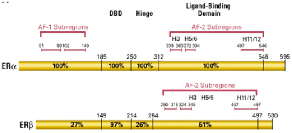
Figure 1. Schematic diagram of Estrogen Receptor alpha (ERα) and Estrogen Receptor Beta (ERβ) with the identified activated functions (AF) that bind coactivators, DNA binding domain (DBD) and ligand binding domain (LBD).
Both ER α and ER β like all the members of the nuclear receptor super family, are modular proteins sharing some common domains, named A/B, C,D and E/F. The N-terminal domain is involved in activation of gene transcription. The DNA binding domain (DBD/C) allows ER to dimerize and bind to the specific Estrogen response element sequence on DNA through two Zinc finger structures. The hinge domain is involved in receptor dimerization. The ligand binding domain, Estrogen binding domain together with N-terminal domain mediates the regulation of gene transcription. The ERα and the ERβ isoforms have the most contradictory functions, with
8
ERα having a growth promoting action and ERβ having a growth-inhibitory action in certain tissues (Weihua Z et al., 2003, Onate SA et al., 1995). Consequently, the tissue-selective ratio of ERα/ERβ can provide a tissue-selective function.
Genomic actions of Estrogens
The “genomic action” of steroid hormones occurs after a time lag of at least 2 hours after E2 stimulation and explains some of hormone functions in physiological and pathological situations. Upon binding of estrogen to its ER, it induces conformational change. Intracellular ligand-bound SHRs recognizes specific sites on chromatin, referred to as hormone response elements (HREs) located within the promoter or enhancers of target genes. This ligand bind SHR translocates as homo/heterodimer and binds promoter regions of the target genes thereby regulates gene transcription. (Simoncini.Tet al., 2003).
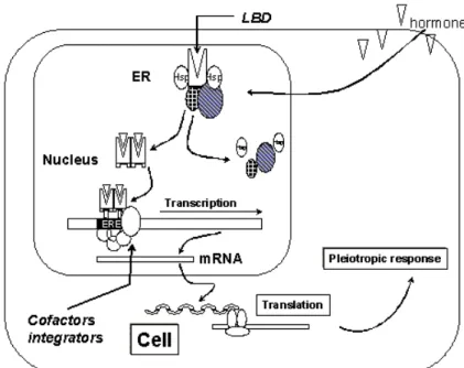
Figure 2. General Estrogen receptor mechanism.The mechanism behind estrogen action at the cellular level starts with an estrogen (ligand) entering the cell and the nucleus and binding to estrogen receptor isoforms (ER α or ER β) via their respective "Ligand Binding Domain" (LBD). Subsequently, the receptor dimerizes and binds via its "DNA Binding Domain" (DBD) to specific sequences on the DNA called "Estrogen Receptor Elements" (ERE). Parallel to the binding of the
9 ER to the ERE, a number of co-activators and/or co-repressors bind to the ligand bound ER, enhancing or repressing its transcriptional activity.
Rapid Non Genomic actions of steroid hormones.
Various signaling pathways are activated upon E2 binding to ERs. These rapid events may be
classified into four main signaling cascade: Phospholipse C (PLC)/protein Kinase C (PKCs), Ras/Raf/MAPK, phosphatidyl inositol 3 kinase (PI3K)/AKT and cAMP/protein kinase A (PKA) activation. The non-genomic effects of estrogen can regulate different cellular processes, such as proliferation, survival, apoptosis, and differentiated functions in diverse cell types, including breast cancer cells(Bjoinstorm L 2005, Driggers PH 2002, Hall JM 2001, Hayes MP 2000).Non genomic actions of steroid hormones are mediated by recruitment of signaling pathways associated with cell membrane receptors such as GPCR, ion channels or enzyme linked receptors (Watson CSet al., 1999). Through the recruitment of various membrane receptors, steroids rapidly mediate multiple cellular functions including the functions mediated by phosphorylation or dephosphorylation of several regulatory proteins thereby mediate non genomic cellular proliferation, migration and invasion (CastoriaG et al., 1999). Non-genomic signaling by steroid hormones are particularly important in tissue sites where there are relatively lower concentration of SRs (Ciocca DR &Vargas RoigLMet al., 1999).
Non genomic regulation in different cell settings.
The physiological significance of rapid membrane starting pathways has been clarified at least for some E2 targets. In the nervous system, E2 affects neural functions in part by inducing such rapid
responses. In the skeleton, ER alpha present in caveolae of bone forming osteoblasts, transmits survival signals through activation of the Src /Shc/ERK pathway and prolongs the life span of osteoblasts. At the same time, E2 delivers a pro-apoptotic signal to bone resorbing osteoclasts,
10 in vivo. In the liver, rapid E2-induced signals (i.e. PLC/PKC) are deeply linked to the expression of
the LDL receptor and to a decreased level of serum LDL-cholesterol. Finally, vascular protection by E2 in ischemia/reperfusion injury in vivo requires E2 induced activation of endothelial NOS, as
mediated by the PI3K/AKT pathway.
Estrogens and cardiovascular system
The cardiovascular actions of estrogens are prominent, but incompletely understood (Mendelsohn and Karas, 2005; Arnal et al., 2009; Luksha and Kublickiene, 2009; Simoncini, 2009). The presence of estrogen receptors (ERs) and of enzymes involved in estrogen synthesis in human blood vessels and endothelium is recognized (Diano et al., 1999). Between the many actions of estrogens on the vascular wall, facilitation of endothelial recovery after injury has been one of the first to be established (Mendelsohn and Karas, 2005; Arnalet al., 2009; Luksha and Kublickiene, 2009; Simoncini, 2009).
A number of studies show that estrogens enhance endothelial proliferation at sites of vascular injury, promoting endothelial recovery after structural damage (Mendelsohn and Karas, 2005; Arnal et al., 2009; Luksha and Kublickiene, 2009). This has been related to the activation of both long-term genomic actions and rapid signaling by estrogens through ERs (Mendelsohn and Karas, 2005; Arnal et al., 2009; Luksha and Kublickiene, 2009) or through the G-protein-coupled receptor 30 (GPR30) (Prossnitz and Maggiolini, 2009), but the mechanistic explanation of these processes has been scarcely investigated. The actin cytoskeleton forms the backbone of the cell, and its spatial organization is crucial for cell movement and migration. Modification of the form and positioning of actin fibers and their relationship with membrane-anchoring structures such as integrins and focal adhesion complexes allows cell movement in the extracellular environment (Pollard and Cooper, 2009). We have recently shown that estrogen controls actin remodeling in
11 human endothelial cells and the development of specialized membrane structures such as ruffles and pseudopodia, thus inducing endothelial cell migration (Simoncini et al., 2006). These phenomena depend on the activation of the actin-regulatory protein moesin (Simoncini et al., 2006).
Related to this set of actions of estrogens, another important factor of cell-membrane remodeling and cell movement is the turnover of focal adhesion complexes. These structures are dynamic in nature and their formation and breakdown are regulated by different stimuli, particularly by tyrosine kinases (McLean et al., 2005).
Estrogen exerts pleiotropic functions on the cardiovascular system through binding to estrogen receptors (ERs). Traditionally, ERs have been recognized as transcription factors regulating the expression of target genes. In the past decades, however, numerous studies have revealed rapid actions of estrogen in different systems, especially in non-reproductive tissues such as the cardiovascular system. At this level, estrogen triggers rapid vasodilatation, exerts anti-inflammatory effects, regulates vascular cell growth and migration, and confers protection to cardiomyocytes. These so-called 'extra nuclear actions do not require gene expression or protein synthesis and are independent of the nuclear localization of ERs. Indeed, some of these actions are elicited by ERs residing at or near the plasma membrane. Through complex interactions with membrane-associated signaling molecules lead to the activation of downstream cascades such as mitogen-activated protein kinase (MAPK) and phosphatidylinositol 3-OH kinase (PI3K). These cascades are responsible for important cardiovascular actions of estrogen, for instance, the activation of nitric oxide synthesis or the remodeling of the endothelial actin cytoskeleton. Moreover, these cascades play crucial roles in regulating the expression of target proteins implicated in cell proliferation, apoptosis, differentiation, movement and homeostasis. Recent advancements in the characterization of the molecular basis of the extra-nuclear signaling of
12 estrogen help us to understand the biological functions of estrogen and would be beneficial in elucidating current controversies on estrogen's clinical efficacy in the cardiovascular system.
Estrogens and breast cancer.
Breast cancer metastasis is a life threatening stage of cancer and is the leading cause of death in advanced breast cancer patients. Estrogen signaling and the estrogen receptor (ER) are implicated in breast cancer progression, and the majority of the human breast cancers start out as estrogen dependent and express estrogen receptor. Endocrine therapy has been shown to have a positive effect on the treatment of ER positive breast cancer. Despite these positive effects, initial or acquired resistance to endocrine therapies frequently occurs with tumors recurring as metastatic. Tumor metastasis includes a series of biological processes that migrates tumor cells from the primary neoplasm to a distant location and involves a multistep cascade of coordinated cell adhesion and contractility. Although substantial information is available on the process of metastasis, the molecular basis of breast cancer progression to metastasis and the role of estrogen signaling in this process remain poorly understood.
Estrogens and cell movement
Cell movement is required in relevant physiological processes such as embryonic development, tissue and organ differentiation, inflammation, immune response and wound healing, along with pathological phenomena, such as cancer metastatic spread. Cell motility is tightly controlled by a complex and often redundant array of intracellular signaling pathways largely devoted to the dynamic regulation of the actin cytoskeletal network and of its relationship with the cell membrane and the extracellular matrix. Sex steroids, particularly estrogen, are effective regulators of cell growth, proliferation, migration and tissue organization. Cells respond to the environment through the expression of trans-membrane receptors that sense extracellular stimuli and activate an
13 elaborate network of intracellular signaling molecules. Given the large number of different signaling molecules and the complex relationships between them, a major challenge is to understand how specific signals generate unique responses. Recent evidence indicates that this is in part obtained through the regulation of the cytoskeleton. Intriguingly, many of these regulatory actions related to cell movement are achieved through rapid, non-classical signaling of sex steroid receptors to kinase cascades, independently from nuclear alteration of gene expression or protein synthesis. The identification of the mechanistic basis for these rapid actions on cell cytoskeleton and cell movement has special relevance for the characterization of the effects of sex steroids in physiological conditions, such their role in the control of inflammation, brain or vascular cell remodeling, angiogenesis or wound healing, as well as in the context of pathological conditions such as steroid-sensitive cancer cell invasion and metastasis.
14 The ability of cancer cells to move and invade the surrounding environment is the basis of local and distant metastasis. Cancer cell movement requires dynamic remodeling of the cytoskeleton and cell membrane and is controlled by multi-protein complexes including focal adhesion kinase (FAK) or Moesin).
The architecture of signaling networks plays an important role in achieving signaling specificity The control of cell morphology and of the interaction of the cell with the extracellular environment requires a number of regulators orchestrating the different cytoskeletal components and their interactions with the cell membrane and with anchorage proteins. Recent findings indicate that extra-nuclear signaling of estrogen is a fundamental regulator of cell morphology in diverse cellular lines, including endothelial cells, breast cancer cells and neuronal cells and that many of these actions are played via rapid signaling to the actin cytoskeleton achieved via actin recruitment These morphological modifications are tightly dependent on the generation of dynamic structural changes of the actin cytoskeleton, via actin polymerization and de-polymerization that also represent the starting platform for other important cellular processes such as cytokinesis, adhesion and motility. When a cell remodels its shape, the actin cytoskeleton is reorganized, and protrusive membrane structures, such as lamellipodia and filopodia, are formed at the leading edge. These structures generate locomotive force in migrating cells and are critical in the generation of cell–cell interconnections.
Previous studies have shown the importance of PAI-1 on cellular migration in vascular smooth muscle cells. Peter Carmeliet et al have shown that, by affecting cellular migration, PAI-1 plays inhibitory role in vascular wound healing and arterial neo intima formation after injury and adenoviral transfer of PAI-1 gene reduces arterial neo intima formation in mice, however, in the issue of Arteriosclerosis, Thrombosis, and Vascular Biology, Tanaka and colleagues presented
15
data indicating that PAI-1 can paradoxically enhance, rather than impair, smooth muscle cell migration under selective circumstances.
In conclusion, estrogens controls different target cells through rapid and genomic signaling pathways. The cell context specific environment (e.g., differentiation, ER level, and ER co-expression) has an impact on the integration of rapid signaling by E2 from the membrane and on
subsequent nuclear transcription. This leads to different signal cascades, different gene expression in response to the same hormone, and different cell biological outcome. My Ph.D thesis comprises of two different projects which focuses on new ways of conventional and unconventional role of estrogens in the modulation of cell movement in different cell types.
16
PART-I
ESTROGEN REGULATES ENDOTHELIAL MIGRATION VIA
PLASMINOGEN ACTIVATOR INHIBITOR (PAI-1)
17 ABSTRACT
Aim: Endothelial plasminogen activator inhibitor (PAI-1) controls vascular remodeling, angiogenesis and fibrinolysis. PAI-1 blood levels in women are related to estrogen. The aim of this study is to characterize the signaling pathways through which estrogen regulates PAI-1 in endothelial cells. Furthermore, we aimed to investigate whether PAI-1 is implicated in the control of endothelial migration by estrogen.
Methods: Cultured human endothelial cells (HUVEC) and ovariectomized rats were used to test the effects of 17β-estradiol (E2) on PAI-1 expression and its role on endothelial migration.
Results: At physiological concentrations, E2 increases expression of PAI-1 in HUVEC within 6-12
hours through activation of a signaling cascade initiated by ERα and involving G proteins, phosphatidylinositol 3-OH kinase (PI3K) and Rho-activated kinase-II (ROCK-II). ROCK-II activation turns into an over-expression of c-Jun and c-Fos that is required for E2-induced
expression of PAI-1. Estrogen-induced PAI-1 expression is implicated in HUVEC horizontal migration. PAI-1 regulation is found also in vivo, in female rats, where ovariectomy is associated with reduced PAI-1 expression, while estrogen replacement counteracts this change.
Conclusions: Estradiol increases PAI-1 synthesis in human endothelial cells and in rodent aorta through a G protein-initiated signaling that targets early-immediate gene expression. This regulatory pathway is implicated in endothelial cell migration. These findings describe new mechanisms of action of estrogens in the vessels, which may be important for vascular remodeling and heamostasis.
18 INTRODUCTION
Plasminogen activator inhibitor-1 (PAI-1) is a serine protease inhibitor (SERPIN)that controls the fibrinolytic system by inhibiting tissue-type and urokinase plasminogen activators(Bergheim et al. , 2006, Dimova and Kietzmann, 2008, Xu et al. , 2010, Zorio et al. , 2008). By controlling the plasminogen cascade, PAI-1 is also implicated in physiological and pathological vascular processes such as thrombosis, inflammation, vascular remodeling and wound healing (Diebold et al. , 2008). A prominent source of PAI-1 in these conditions is represented by vascular endothelial cells, which synthesize the protease inhibitor in response to a variety of stimuli (Gramling and Church, 2010, Lyon and Hsueh, 2003, Marx et al. , 1999, Stiko-Rahm et al. , 1990, Ye et al. , 2002).
Beyond being a modulator of the fibrinolytic cascade, PAI-1 has been recently identified as a controller of cell movement and migration. Modulation of cell migration and angiogenesis by PAI-1 appears to be implicated both in vascular remodeling (Diebold, et al., 2008) as well as in promotion of tumor growth, invasion and metastasis (Kwaan and McMahon 2009). This newly identified set of activities is exerted through interactions with cell surface receptors affecting cell motility as well as through multifaceted interactions with extracellular matrix proteins (Czekay, etal., 2011). In addition, signaling events that turn into modifications of the shape of the cytoskeleton and of the cell membrane modulate the expression of PAI-1 (Samarakoon, et al., 2010). A link exists between estrogen concentrations and PAI-1 in women. Menopausal estrogen withdrawal is associated with increased blood levels of PAI-1, while a decrease is found with estrogen therapy (Gebara, et al., 1995)(Koh, et al., 1997)(Sowers, et al., 2005a). These changes have been interpreted as potential reasons for the beneficial vascular effects of endogenous estrogens in fertile women (Sowers, et al., 2005b). In addition, a link between PAI-1 and hormonal
19 status has been identified in the presence of coronary heart disease in women (Grancha, et al., 1999) and experimental animals (Chua, et al., 2011).
On the other side, PAI-1 is also a known negative prognostic factor in endocrine sensitive cancers, such as breast (Manders, et al., 2004)(Eljuga, et al.,2011), and endometrial cancer (Steiner, et al.,2008), with higher expression of the protease inhibitor being associated with poorer response to chemotherapy and prognosis. Based on preliminary evidence, estrogen seems to cross talk with growth factors in regulating PAI-1 synthesis in breast cancer cells (Burdette and Woodruff 2007), but little is known on the signaling mechanisms through which estrogen controls PAI-1 nor is this regulatory action extends to other cell types.
The purpose of the current study was to explore the regulatory actions of estrogen on PAI-1 in human endothelial cells and to characterize the signaling steps through which these actions are enacted.
20 MATERIALS AND METHODS
Cell cultures and Treatments
Human umbilical vein endothelial cells were extracted with collagenase. HUVEC were seeded in Dulbecco’s Modified Eagle’s Medium (DMEM) containing fetal bovine serum (FBS) for 24 hours. Before experiments investigating transcriptional effects, medium was replaced by DMEM containing 10% FBS stripped with charcoal for 4-6 h. Whenever an inhibitor was used, the compound was added 30 minutes before starting the active treatments. 17β Estradiol (E2), pertussis
toxin (PTX), ICI 182,780 (ICI), 4,49,40-(4-propyl-[1H]-pyrazole-1,3,5-triyl) trisphenol (PPT) and 2,3-bis(4-hydroxyphenil)-propionitrile (DPN), Phosphatidylinositol 3-OH kinase inhibitor (Wortmannin), Rho kinase inhibitor (Y-27632) were from Sigma-Aldrich (Saint-Louis, MO). 4-amino-5-(4-chlorophenyl)-7-(t-butyl) pyrazolo (3,4-d) pyrimidine, (PP2) and PD98059 was purchased from Calbiochem (Nottingham, UK).
Animal treatments
Fertile female Wistar rats, weighing 175–199 g (Harlan,S. Pietro al Natisone, Italy), were kept under 14 h of illumination per day (06.00–20.00 h) and had free access to standard rat chow and tap water. After 14 days the rats were either sham-operated or ovarectomized in the same oestrous cycle stage. Ovariectomized rats were treated with E2 at a concentration of 0.01mg/kg, 0.05 mg/kg,
and 0.1mg/kg).Estradiol was dissolved in pure ethanol and administered orally after dilution in water. Animals were euthanized by decapitation under pentobarbital anesthesia (30 mg/kg) and abdominal aorta was dissected. Aortas were snap-frozen in dry ice and stored at -80 C. Animals were maintained in accordance with the National Institutes of Health Guide for the Care and Use of Laboratory Animals (www.nap.edu/readingroom/books/labrats).
21 Immunoblottings
After treatments, cells were lysed with lysis buffer containing Sodium Dodecyl Sulfate (SDS), glycerol and Tris-EDTA. The concentration of total protein was quantified by Bichinconic acid (BCA) assay, and 30µg of proteins were loaded into each well and separated by denaturing SDS-PAGE. Antibodies against the following proteins were used: c-Fos (sc-448), c-Jun (sc-74543), Actin, all from Santa Cruz Biotechnology (Santa Cruz, CA), PAI-1 (ab82218), ERα (ab2746) and ERβ (ab288)were purchased from Abcam (Cambridge, UK). Primary and secondary antibodies were incubated with the membranes, followed by three 5-min washes with TBS-Tween 20. Immunodetection was accomplished using enhanced chemiluminescence and was recorded with a quantitative digital imaging system (Quantity One; BioRad, Hercules, CA)
Gene silencing with RNA interference
Gene silencing was performed using synthetic siRNAs targeting c-Fos, c-Jun or ERβ purchased from Santa Cruz. ERα siRNAs were from Dharmacon (Lafayette, CO). All siRNAs were used at the final concentration of 30nM according to the manufacturer’s instructions. HUVEC were transfected with Lipofectamine 2000 (Invitrogen, Carlsbad, CA) in opti-MEM without antibiotics or anti-mycotics. Efficacy of gene silencing was checked by western blotting. Viability of cells was checked throughout transfection experiments.
Transfection experiments
Dominant negative Gα13(Gα13Q226L/D294N) was obtained from the Guthrie DNA Resource
Center (www.cdna.org). The insert was cloned in pcDNA3.1+. The plasmid (10 µg) was transfected into HUVEC cells using Lipofectamine (Invitrogen). Cells were treated 48 h after transfection.
22 Cell migration assays
Cell migration was assayed with razor scrape assays as previously described (Xiao-dong fu et al 2008). Briefly, a razor blade was pressed through the confluent T47-D breast cancer cell monolayer into the plastic plate to mark the starting line. T47-D cells were swept away on one side of that line. Cells were washed, and 2.0 mL of DMEM containing steroid-deprived FBS and gelatin (1 mg/mL) were added. Cytosine b-D-arabinofuranoside hydrochloride (Sigma)(10 mM), a selective inhibitor of DNA synthesis which doesn’t inhibit RNA synthesis was used 1 h before the test substance was added. Migration was monitored for 24 hours. Every 12 h fresh medium and treatment were replaced. Cells were digitally imaged and migration distance was measured by using phase-contrast microscopy.
Statistical analysis
All values are expressed as Mean ± SD. Statistical differences between mean values were determined by ANOVA, followed by the Fisher's protected least significance difference (PLSD). All differences were considered significant at P<0.05.
23 RESULTS
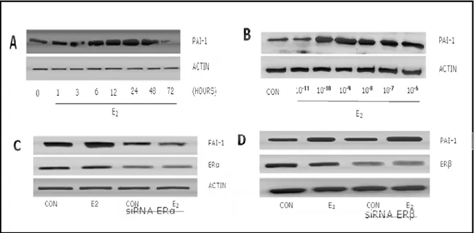
Figure 1. E2 induces PAI-1 expression in endothelial cells via estrogen receptor α.
Human umbilical vein endothelial cells (HUVEC) were treated with 17-β estradiol (E2) for
different times and protein lysates were obtained. Expression of PAI-1 gradually increased during exposure to estradiol and peaked at 24-48 hours (Fig 1A). Endothelial PAI-1 expression was increased in the presence of physiological concentrations (Fig 1B).
In order to check which estrogen receptor isoform mediates the induction of PAI-1 we silenced ERα or ERβ with small interfering RNAs. Silencing of ERα blocked PAI-1 induction by E2 (Fig 1C),
whereas silencing of ERβ was ineffective (Fig 1D).
Figure 1. A) HUVEC were treated with E2 for different times (0-72 hours). B) HUVEC was
treated with different concentrations of E2 (10-11 – 10-6M). C-D) HUVEC were transfected with
ERα or ERβ siRNAs for 48 hours before the addition of E2 1nM for an additional 24 hours protein
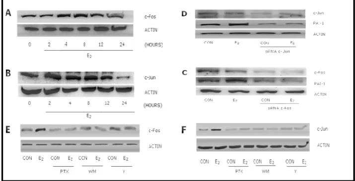
24 Figure 2. E2 induces PAI-1 through up-regulation of c-Fos and c-Jun.
PAI-1 gene promoter region is controlled by c-Jun and c-Fos through two dedicated consensus sequences. We checked whether E2 regulates PAI-1 through these transcription factors performing
time course experiments with 1 nM E2 and analyzing expression of c-Fos and c-Jun in HUVEC. E2
induced the expression of c-Fos and c-Jun as early as after 2 hours, with a peak at 8 hours (Fig 2 A-B). In parallel, silencing of c-Fos and c-Jun with siRNAs blocked PAI-1 induction by E2 (Fig 2
C-D).
Figure 2. A-B) HUVEC were treated with 1nM E2 for different times. C-D) HUVECs were transfected with siRNAs targeted against c-Fos and c-Jun (30 nM) for 30 hours before the addition of E2 for an additional 24 hours E-F) HUVEC were treated with 10 µM Y27632, 30 nM Wortmannin (WM) and 100 ng/ml pertussis toxin (PTX). Protein extracts were used to quantitate expression of c-Fos, c-Jun, Actin and PAI-1 with Western analysis.
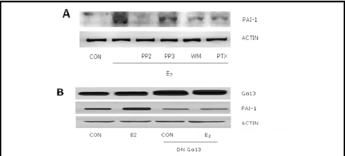
25 Figure 3. E2 regulates c-Jun, c-Fos and PAI-1 expression via signaling to G proteins,
Phosphatidylinositol-3-OH-kinase and Rho –associated kinase II.
To search for the signaling pathways involved in E2 mediated c-Fos and c-Jun induction, we
blocked different signaling intermediates that have an established role in regulating Jun and c-Fos. E2 failed to induce c-Fos expression in HUVEC when G proteins, PI3K or ROCK-II were
inhibited with pertussis toxin (PTX), wortmannin (WM) or Y27632, respectively (Fig 2E). Similar results were found with c-Jun (Fig 2 F).
In agreement, the increased expression of PAI-1 associated with E2 treatment was prevented by
PTX, wortmannin or PP2, which blocks the activity of the PI3K controller, c–Src (Fig. 3A). As a control, the PP2–related, inactive compound, PP3, was ineffective (Fig. 3A). In addition, transfection of HUVEC with a dominant negative Gα13 construct indicated that E2 uses Gα13 to
induce PAI-1 expression (Fig. 3B).
Figure 3.A) HUVEC were treated with the c-Src inhibitor, PP2, the inactive PP2-analogue PP3 (5µM), with the PI3K inhibitor, wortmannin (WM, 30nM) or with the G proteins inhibitor, pertussis toxin (PTX -100 ng/ml). B) HUVEC were transfected with a dominant negative construct (Gα13 D294N) for 48 hours using Lipofectamine before the addition of E2 for a additional 24 hours. Protein lysates were used to check for expression of PAI-1, Gα13 and Actin with Western analysis.
26
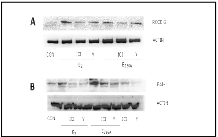
Figure 4. E2controls PAI-1 via ROCK-2
Administration of either normal E2 or of a membrane-impermeable form of E2 (E2 covalently cross-
linked to bovine serum albumin, E2-BSA) to HUVEC turned into increased expression of ROCK-2
and its expression was blocked by the ER antagonist (ICI 182,780). As a control, the ROCK–2 inhibitor Y27632 did not block the increase of expression of ROCK–2 associated with E2 (Fig. 4A).
Consistent with this result E2 as well as E2-BSA induced PAI-1 expression through ROCK–2, as
shown by the loss of effect in the presence of Y27632 (Fig. 4B).
Figure 4. A) HUVEC were treated with the estrogen receptor antagonist ICI 182,780 10 nM and with the ROCK-2 inhibitor Y27632 (10 µM) for 30 minutes before the addition of E2 (1nM) or of E2 conjugated with BSA (E2-BSA-1nM) for additional 24 hours. B) HUVEC were treated with ICI 182,780 (10 nM) or Y27632 (10 µM) for 30 minutes before the addition of E2 or E2-BSA for additional 24 hours. Protein extracts were obtained and used with western analysis to control expression of Rock-2 or PAI-I
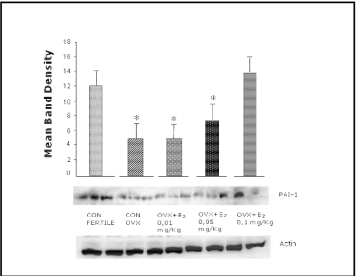
27 Figure 5. E2 regulates PAI-1 expression in vivo.
To check whether estrogen also regulates PAI-1 in an in vivo setting, we administered E2 at different
doses for 14 days to ovariectomized (OVX) rats, and compared the expression of PAI-1 in the abdominal aorta versus that of intact or of placebo-treated OVX rats. PAI-1 expression was present in the aorta of fertile rats and was decreased in OVX rats. OVX rats treated with oral E2 showed a
dose-related increase of PAI-1 expression in the abdominal aorta (Fig. 5).
Figure 5. Ovariectomized mice were fed with 17β-estradiol at different doses (0.01, 0.05 or 0.1 mg/kg) for 14 days. Abdominal aorta was obtained after sacrifice sham-operated fertile mice and placebo-treated ovariectomized mice served as controls.
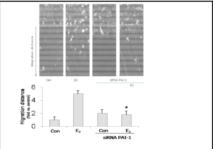
28 Figure 6. E2 induces endothelial cell migration through PAI-1.
HUVECs were culture dish and then treated with 1nM E2. The mean migration distance from the starting line taken from 5 random microscopic fields was assayed and expressed as mean ± SD. Average migration distance ± SD was expressed as the ratio between active treatments and control. Representative images are shown and the mean migration distance is shown in bar graphs.* = p<0.05 vs. E2(non-silenced cells).
Figure 6. HUVECs were transfected with siRNAs against PAI-1 for 48 hours. Endothelial cells were scraped from the culture dish and then treated with 1nM E2. The mean migration distance from the starting line taken from 5 random microscopic fields was assayed and expressed as mean ± SD. Average migration distance ± SD was expressed as the ratio between active treatments and control. Representative images are shown and the mean migration distance is shown in bar graphs.* = p< 0.05 vs. E2 (non-silenced cells).
29 DISCUSSION
This paper identifies a direct regulation of PAI-1 expression by estradiol in human endothelial cells, which is exerted through an extra-nuclear signaling to the early-immediate genes c-Jun and c-Fos.
Regulation of PAI-1 by estrogen has been suggested in a variety of clinical settings; however, it is unclear how this hormone directs PAI-1 expression in tissues. A recent paper indicates that in endothelial cells estrogen has a double-sided effect on PAI-1, triggering its expression through ERα while repressing it through ERβ (Smith, et al. 2004). We here extend this concept showing that estrogen drives ERα to enact rapid, extra-nuclear signaling involving G proteins and the Rho-A-associated kinase (ROCK-2). The cross talk between this cytoplasmic recruitment of ROCK-2 and the subsequent activation of c-Jun and c-Fos expression mediates PAI-1 increase in endothelial cells. This proposed regulatory pathway is consistent with the published hypothesis of a prominent role of ERα as positive regulator of PAI-1. Furthermore, it provides possible ways to selectively modulate PAI-1 in endothelial cells by either using preferential ERα or ERβ ligands, or by associating pharmacological tools that interfere with the rapid signalling of ERα to ROCK-2. Such strategies may be helpful in conditions where estrogen-related induction of PAI-1 may either be desirable or undesirable.
Endocrine-sensitive cancers, such as breast or endometrial cancer, represent a straightforward situation where PAI-1 induction linked to estrogen may be unwanted (Manders, et al. 2004)(Eljuga, et al. 2011)(Steiner, et al. 2008). Inactivation of ERα-mediated signaling could turn into reduced ability to remodel the extracellular matrix and decreased cancer cell motility, thus possibly leading to less efficient cancer progression. If this hypothesis holds, our findings may suggest a role of endothelial cell-derived PAI-1 as a possible target of estrogen to support cancer
30 progression. However, our unpublished data (Gopalet al, personal communication) as well as work by others (Burdette and Woodruff 2007) suggest that estrogen also increases PAI-1 expression directly in breast cancer cells. This fits with the mounting evidence that sex steroids are effective enhances of cancer cell motility and invasion through a multi-modal control of cytoskeletal remodeling, membrane shaping and, now, extracellular matrix turnover (Flamini, et al. 2011; Sanchez, et al. 2010).
It is less clear how our results fit with the large body of evidence that endogenous or exogenous estrogens decrease PAI-1 blood levels (Gebara, et al. 1995)(Koh, et al. 1997) Our findings suggest that, contrary to what previously thought, estrogen-related decrease in circulating PAI-1 is not explained by an endothelial effect, but may rather rely on other phenomena, such as modified metabolic clearance or reduced production in other tissues (may be because of local prevalent ERβ activity).
However, increased endothelial-derived PAI-1 may still represent a biologically important mean through which estrogens regulate vascular function. Indeed, this action may result into locally reduced fibrinolysis, which may contribute to the pro-thrombotic actions of estrogens in veins (Mendelsohn 2002). On the other side, PAI-1-mediated extracellular matrix remodeling may add to the previously identified stimulatory effects of estrogens on endothelial cell motility (Simoncini, et al. 2006), thus helping arterial re-endothelialization processes, and thus affect the incidence of arterial events in women.
In conclusion, this paper provides new molecular cues on how estrogens control PAI-1 in endothelial cells. These findings may help to develop new therapeutic strategies to reduce unwanted side effects of estrogens in the cardiovascular system or on endocrine-sensitive cancers.
31
PART-II
THE ROLE OF UNCONVENTIONAL ESTROGEN RECEPTORS
IN CELL MOVEMENT IN ER NEGATIVE BREAST CANCER
32 ABSTRACT
Aim: The aim of this study is to characterize the signaling pathways through which estrogen induces actin remodeling in ER negative MDA MB468 cells. Furthermore, we aimed to investigate whether the E2 induced phosphorylation of Moesin and FAK is implicated in the control of cell invasion and metastasis.
Methods: Cultured human MDA MB 467 cells were used to test the effects of 17β-estradiol (E2)
on FAK and Moesin phosphorylation and its role on actin remodeling.
Results: At physiological concentrations, E2 induces phosphorylation of Moesin and FAK in
MDA MB468 cells within 30 minutes through rapid non genomic activation of a signaling cascade initiated by GPER and involving G proteins, phosphatidylinositol 3-OH kinase (PI3K) and Rho-activated kinase-II (ROCK-II). ROCK-II activation turns into E2-induced phosphorylation of
Moesin and FAK, thereby remodels actin filaments implicated in cell migration.
Conclusions: Estrogen induces phosphorylation of Moesin and FAK without the presence of endogenous Estrogen receptors viz ERα and ERβ in MDAMB468 cells. Our preliminary work clearly confirms the previous results reported by Filardo et al about GPER as estrogen receptors. We characterized the signaling events by which estrogen 17β estradiol induces Moesin and FAK phosphorylation. These regulatory pathways is implicated in remodeling of actin cytoskeleton and cell movement and invasion in ER negative breast cancer cells. These findings describe new mechanisms of action of estrogens in actin remodeling, in ER negative breast cancer cells, which may enable us to provide therapeutic strategies for ER negative breast cancer cells.
33 INTRODUCTION
Estrogens are important regulators of breast cancer cell progression. Many of the effects are mediated by the classical estrogen receptors viz ERα and ERβ, where they mediate genomic effects. In addition, it is well known that estrogens can induce rapid effects, which do not mediate gene transcription, since responses occur within seconds or minutes (Szegoet al., 1967; Pietras et al., 1977). Depending on the cell type, the rapid responses to estrogen involve mobilization of various second messengers, e.g. cAMP, Ca2+, PI3kinase (Morley et al., 1992; Lieberherr and Grosse, 1994).interaction with membrane receptor, e.g. the epidermal growth factor (EGF) receptor, or activation of intracellular kinase pathways (Nelson et al., 1991; Migliaccioet al., 1996; Ho and Liao, 2002). Such observations suggested that a G protein-coupled receptor (GPR) mediated these fast effects. GPRs represent one of the largest and most diverse super-families of cell membrane receptors in mammals, and they respond to a great variety of ligands.
G-protein coupled receptor (GPR30) also known as G-protein coupled estrogen receptor 1. The G protein-coupled estrogen receptor 1 (GPER) was first cloned in 1996 and initially named GPR30 (Owmanet al., 1996). GPR30 is an integral membrane protein that displays high affinity towards estrogens. GPR30 is encoded by GPER gene. This protein is a member of the rhodopsin like family of G protein-coupled receptors which binds estrogen, resulting in intracellular calcium mobilization and synthesis of phosphatidyl inositol (3,4,5)-triphosphate in the nucleus. This protein therefore plays a role in the rapid non-genomic signaling events widely observed following stimulation of cells and tissues with estrogen. The distribution of GPR30 is well established in the rodent, with high expression observed in the hypothalamus, pituitary gland, adrenal medulla, kidney medulla and developing follicles of the ovary (Hazell GGJ et al., 2002).
34 GPR30 sub-cellular localization
Although many GPCRs are localized in the plasma membrane, some GPCR like nuclear ER, localized at intracellular sites (Gobeil et al., 2006). Using sub-cellular markers Eric Prosnitz reported that GPR30 is expressed in intracellular tubule-reticular network. In contrast, previous reported the expression of GPR30 in the plasma membrane (Thomas et al., 2005), so there is no clear evidence reporting the site of GPR30 expression however it is certainly possible that under some conditions, intracellular GPR30 could exist or translocate to the plasma membrane or vice versa.
GPR30 and its ligand
Since no ligands was known for GPER, it was termed initially as orphan receptor. It was not until 2000 that possible function for GPER was identified. The proposed role of GPER in response to estrogen was based on the correlation of receptor expression with estrogen mediated signaling (Ylikomiet al., 2004). Revankaret al., in 2005, synthesized fluorescent derivative of ethinyl estradiol to test the hypothesis that GPR30 binds estrogen by confocal fluorescence microscopy and quantitative flow cytometry . They observed that fluorescent derivative of estrogen displays subcellular colocalization with ER in the nucleus or GPR30 in endoplasmic reticulum (Revankaret al., 2005).
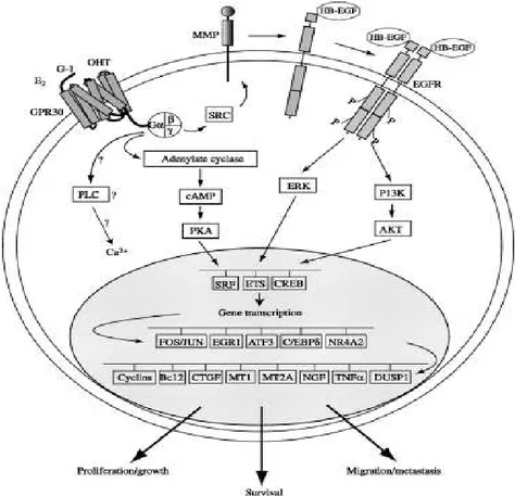
GPR30 signaling network
E2 induction leads to activation of SRC kinase, phosphorylation of adaptor protein SHC, and
through the activation of extracellular release of heparin bound EGF (fig 1). The release of HB-EGF allows the activation of MAPK pathway. Accumulating evidence suggest that GPR30 triggers short term and long term responses. One of the main early events mediated by E2 is the activation of PI3Kinase through GPR30 thereby activating anti apoptotic and anti proliferative AKT. The
35 specific GPR30 antagonist G15 inhibits the stimulation of PI3K by both E2 and GPR30 agonist G1 in SKBr3 breast cancer cells which endogenously express GPR30. The stimulation of PIP3 by E2 and G-1 and their inhibition by G15 could also be demonstrated in ER negative COS7 cells
Figure 1. GPR30 signaling pathway. Source: Maggiolini M, Picard DJ J Endocrinol 2010;204:105-114.
In ER-negative SKBr3 breast cancer cells, E2 induces c-fos expression through the
GPR30/EGFR/MAPK signaling cascade (Maggiolini et al., 2004).E2 and the partial ER antagonist
hydroxyl tamoxifen (OHT) both stimulate the expression of c-fos and elicit proliferative effects through GPR30 in thyroid and endometrial cancer cells (Vivacqua et al. 2006a,b). In ovarian cancer cells, both E2 and G-1 up-regulate numerous estrogen-responsive genes including c-fos,
36 pS2, and cyclins A, D1 and E, while other direct ERα target genes such as the progesterone receptor (PR) gene only respond to E2 (Albanito et al. 2007).
Actin cytoskeleton remodeling
Actin is a globular protein with an ATP binding site in the center of the molecule. Termed "G-actin" the monomer will dimerize or form a trimer. This serves as a site for nucleation and further growth of the actin proto filament. G-actin forms F-actin (the filament) in the presence of ATP, Mg and K. The concentration of actin is also critical. Above the critical concentration Cc of G-actin, the molecules will polymerize. ATP hydrolysis is not required for polymerization, but it is required to promote de-polymerization (if it is converted to ADP). In this regard, it behaves like microtubules and their need for GTP hydrolysis to de-polymerize. The plus end of G-actin is the end that is opposite the cleft that holds the ATP molecule. The minus end is the opposite end. Growth and polymerization is more rapid at the plus end. If you add a solution containing myosin to actin filaments, they will "decorate the filaments" and they will be pointing in the direction of the plus end.Two actin molecules bind weakly, but addition of a third stabilizes the complex. This trimer then adds additional molecules and forms a "nucleation site". This is the slow, or lag phase of the polymerization process. One could add fragments of actin filaments to speed this up, in vitro. Or, key actin binding proteins may help to speed this process. Addition of actin molecules to form a long helical polymer. After a period of growth, an equilibrium phase is reached in which de-polymerization controls the length as new monomers are added. Polymerization of actin filaments can occur via a network regulated by filamin. This protein works like a clip to connect the filaments at the cross-over points. Other proteins create bundles of actin filaments.
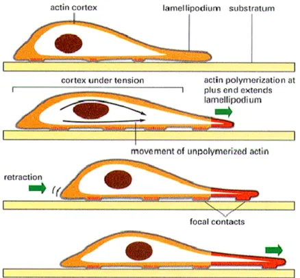
37 Figure.2. This is the most dense concentration of actin filaments. It lies just under the plasma membrane. It interacts with a number of different proteins associated with the membrane as well as proteins in signaling pathways. It may also be traversed by myosin as vesicles are brought to the periphery for secretion. As stated earlier, it may be organized in a network or bundles, depending on the area and the needs of the cell. At specific sites in the cell, the focal adhesions are formed, linking the cell to specific matrix proteins. It is this region that is primarily responsible for forward movement.
Cell migration is a key process in the development and maintenance of multi-cellular organisms. Tissue formation during embryonic development, wound healing and immune response all require the orchestrated movement of cells in particular directions to specific locations. Cell migration is required for cancer cell spread, invasion and metastasis and it is achieved through a dynamic remodeling of actin filaments and focal adhesion sites (Acconcia F et al., 2006). This process leads to rapid changes of cell membrane morphology, with the formation of specialized structures linked to cell movement such as pseudopodia and ruffles. Motile cells extend a leading edge by assembling a branched network of actin filaments that produces physical force as the polymers grow beneath the plasma membrane. Proteins that reconstitute actin cytoskeleton remodeling includes actin, Arp 2/3 complex and WASP proteins. WASP proteins regulate the activity of Arp2/3 complex to nucleate actin filaments from the side of pre-existing actin filaments. These
38 actin filaments push the membrane forward as they grow at their barbed ends (Pollard TD et al., 2003).
We previously characterized the estradiol mediated signaling events that induces actin cytoskeleton remodeling. We showed that in ER positive T47D breast cancer cells, estradiol induces rapid and dynamic actin cytoskeleton remodeling with the formation of specialized cell membrane structures like ruffles and pseudopodia (Giretti MS et al.,2005). They also showed that estradiol mediate actin cytoskeleton remodeling through ERα. This work strongly supports the fact that ER is required for estrogen mediated actin cytoskeleton remodeling.
Since many research work speculates that estrogens uses GPR30 receptor to mediate several estrogen mediated signaling events in ER negative breast cancer cells, we studied how estrogens induces rapid actin cytoskeleton remodeling in ER negative MDA MB 468 breast cancer cells.
39 MATERIALS AND METHODS
Cell cultures and Treatments
MDA MB468 were seeded in L15 medium containing fetal bovine serum (FBS) for 24 hours. Before experiments investigating non-transcriptional effects, medium was replaced byL15 with without FBS for4-6 h. Whenever an inhibitor was used, the compound was added 30 minutes
before starting the active treatments. 17β Estradiol (E2), pertussis toxin (PTX),
Phosphatidylinositol 3-OH kinase inhibitor (Wortmannin), Rho kinase inhibitor (Y-27632) were from Sigma-Aldrich (Saint-Louis, MO). 4-amino-5-(4-chlorophenyl)-7-(t-butyl) pyrazolo (3,4-d) pyrimidine, (PP2) and PD98059 was purchased from Calbiochem (Nottingham, UK).
Immunoblottings
Cell lysates were separated by SDS-PAGE. Antibodies used were: Moesin (clone 38, Transduction Laboratories, Lexington, KY), Thr558-P-moesin (sc-12895, Santa Cruz Biotechnology, Santa Cruz, CA),pFAK Tyr397(BD bioscience). House keeping protein was used as a loading control. All primary antibodies were used at a concentration of 1:1000 and secondary antibodies were used at a concentration of 1:4000. All HRP conjugated secondary antibodies were purchased from Santa Cruz Biotechnology, (Santa Cruz, CA). After transfer, membrane was blocked with 5% BSA in TBS Tween 20 for one hour and then incubated with primary and secondary antibodies. Immunodetection was accomplished with enhanced chemiluminescence.
Actin Immunofluorescence
MDA-MB468 cells were grown on cover slips and transfected with shRNA plasmid encoding GPR30. After transfection, cells were exposed to treatments. Cells were fixed and permeabilized with Triton X 100 for 10 min. Blocking was performed with 1% BSA for 30 minutes. Cells were
40 incubated with Texas Red-phalloidin (Sigma). The nuclei were counterstained with or 4′-6-diamidino-2-phenylindole (DAPI) (Sigma) and mounted with Vecta shield mounting medium (Vector Laboratories, Burlingame, CA). Immunofluorescence was visualized using an Olympus BX41 microscope and recorded with a high-resolution DP70 Olympus digital camera. After conversion to grey scale images, the cell membrane thickness and the gray levels of the extracellular area, cell membrane as well as cytoplasm were quantified using the Leica QWin image analysis and image processing software (Leica Microsystems, Wetzlar, Germany).
Transient Transfections
MDA-MB468 cells were transfected with plasmid (1 µg) using the Lipofectin reagent (Invitrogen, Carlsbad, CA) according to the manufacturer's instructions. The transfected plasmids were: shRNA plasmids encoding GPR30. The Plasmids were obtained from Origene. For GPR30, transfection efficiency was verified using GFP expression. 60 hours after transfection, cells were treated with G1 and E2 and cellular extracts were prepared according to the experiments to be performed.
Cell invasion assay
Cell invasion were assayed following the standard method by using the BD Bio CoatTM Growth Factor Reduced (GFR) MatrigelTM Invasion Chamber (BD Bioscience, USA). In brief, after rehydrating the GFR Matrigel inserts, the test substance was added to the wells. 0.5mL of MDA-MB468 cell suspension (2.5×10⁴ cells/mL) were added to the inside of the inserts. The chambers were incubated for 24 h at 37°C, 5% CO2 atmosphere. After incubation, the non-invading cells were removed from the upper surface of the membrane using cotton tipped swabs. Then the cells on the lower surface of the membrane were stained with Toluidine blue. The invading cells were observed and photographed under the microscope at 20X magnification. Cells were counted in the
41 central field of triplicate membranes. The invasion index was calculated as the % invasion test cell/ % invasion control cell.
Statistical analysis
All values are expressed as Mean ± SD. Statistical differences between mean values were determined by ANOVA, followed by the Fisher's protected least significance difference (PLSD). All differences were considered significant at P<0.05.
42 RESULTS
Figure 1. E2 activates Moesin and FAK phosphorylation in ER negative MDA MB 468 breast
cancer cells
In order to check at which concentration, 17 β E2 activates phosphorylation of Moesin and FAK,
we treated MDA MB468 cells with different concentrations of E2 for 30mins, we found that both phosphorylation of Moesin and FAK by E2 at a optimum concentration of 1µ. Fig 1. This clearly
shows that E2 activates Moesin and FAK phosphorylation even in the absence of endogenous
estrogen receptors.
Figure 1. Dose dependent experiment.MDA MB 468 cells were treated with E2 at different
43
Figure 2. E2 activates phosphorylation of Moesin and FAK through GPER
In order to check whether GPER is involved in E2 mediated phosphorylation of Moesin and FAK,
we silenced the GPER with plasmid encoding GPER shRNA. Figure 2 clearly shows that E2 and
G1 failed to induce phosphorylation of FAK and Moesin in the cell silenced with GPER shRNA.
Figure 2. MDA MB 468 cells were transfected with shRNA plasmid against GPER for 48 hours and then treated with 1uM E2 for 30mins.
44
Figure 3. E2 activates Moesin and FAK phosphorylation through GPER, PI3K, ROCKII and
c-SRC pathways
To search for the signaling pathways involved in E2 mediated activation of phosphorylation of
Moesin and FAK, we blocked different signaling intermediates that have an established role in the activation of phosphorylation of Moesin and FAK, G1 and E2 failed to induce Moesin and FAK
phosphorylation in MDA MB468 cells when GPER, PI3K, c-SRC and ROCK-II were inhibited with G15, WM, PP2 and Y27632, respectively. Where as PD inhibitor does not have any effect indicating that E2 mediated phosphorylation of FAK and Moesin is through
GPER/PI3K/c-Src/ROCK2. Fig (3A and 3B)
Figure. 3a.MDA MB468 cells were treated with G15 (GPER antagonist), PD (MAPK) inhibitor, PP2 (c-SRC) inhibitor, Wortmannin (PI3K) inhibitor, Pertussis toxin PTX (G protein) inhibitor, Y27632 (ROCK2) inhibitor for one hour before adding E2 for 30mins.
45 Figure. 3b. MDA MB468 cells were treated with G15 (GPER antagonist), PP2 (c-SRC) inhibitor, Wortmannin (PI3K) inhibitor, Pertussis toxin PTX (G protein) inhibitor, Y27632 (ROCK2) inhibitor for one hour before adding G1 for 30mins.
46 Figure 4. Rapid activation of GPER is linked to breast cancer cell cytoskeletal and cell membrane rearrangement
Actin stress fibers in ER negative MDA MB468 breast cancer at baseline were arranged longitudinally in the cytoplasm and the cell membrane was regular. Activation of GPER with G1
and E2 (1 µM) resulted in a rapid shift of the actin fibers toward the edge of the membrane,
whereas G1 and E2 failed to induce actin remodeling to the cell membrane in the cells transfected
with GPER shRNA plasmid. (Fig. 2A).
Figure. 4. Immunofluoresence. E2and G1 induces actin cytoskeleton remodeling. MDA MB 468
cells were transfected with shRNA GFP GPER for 48 hours in cover slips and then the cells were treated with G1 and E2 for 30mins at 1uM concentration. Cells were stained with Texas Red
conjugated with phalloidin and counterstained with DAPI.
Control and GPER control
G1shRNA GPER + G1control
47 Figure 5. E2 drives breast cancer cell invasion.
In the inserts with GFR MatrigelTM, recruitment of GPER with G
1 or E2 promoted invasion of the
matrix by cancer cells (Fig. 1B). The number of cells that invaded the matrix in the presence of E2
and G1 was higher than non treated cells.
Figure.5. Invasion assay. MDA MB 468 Cells were treated for 24 hours with G15 (GPER antagonist) and G1 and E2and then the cells were stained with toluidine blue.
48 DISCUSSION
My second part of the thesis identifies the activation of Moesin and FAK phosphorylation by 17β E2 and G1 in MDA MB468 breast cancer cell which is exerted through rapid non genomic
signaling to the various signaling intermediates.
Activation of Moesin and FAK phosphorylation and actin cytoskeleton remodeling by estrogens has been reported previously in our lab (Giretti MS and Xiao dong fuet al), normally estrogens activate rapid and non rapid signaling pathways through its endogenous estrogen receptors however it is not known how estrogens mediate this effects in the absence of estrogen receptors. Previous studies identified trans-membrane G protein-coupled estrogen receptor 1 (GPER, GPR30) has been implicated in rapid non-genomic effects of estrogens. Estrogen activation of GPER increased adenyl cyclase activity in ER negative, GPER positive breast cancer cells (Filardo, 2002; Filardo et al., 2000). This gave us an idea to test whether estrogen uses GPER in ER negative breast cancer cells.
We here show that estrogen induces actin remodeling and invasion of three-dimensional matrices of ER- breast cancer cells by phosphorylating the actin-binding proteins, Moesin and FAK. Moesin resides at the nexus of multiple pathways regulating cell attachment with the extracellular matrix and with nearby cells, cell motility and metastatic potential as well as cell survival. These functions are directed by Moesin and FAK though the modulation of actin cytoskeleton/plasma membrane interactions.
We here extend this concept showing that estrogen drives GPER to activate actin cytoskeleton remodeling and invasion in MDA MB 468 breast cancer cell through various signaling intermediates. Our previous work in ER positive T47D breast cancer cells shows that GPER/PI3K/c-Src/RhoA/ROCK/Moesin and FAK cascade is necessary for the cytoskeletal
49 remodeling and for the enhancement of breast cancer cell horizontal migration and invasion of three-dimensional matrices induced by estrogen (Giretti MS et al., 2007).We tested whether the same signaling pathways estrogens uses in MDA MB468 breast cancer cells. Interestingly the pathways used by E2 in ER positive T47D cells also used same pathway in ER- MDA breast cancer cells. In fact, the specific GPER antagonist G-15 inhibits estrogen activated Moesin and FAK phosphorylation and such stimulation was further augmented by the specific GPER agonist G-1.
In conclusion, the present results suggest that within the broader range of actions of estrogen receptors, the rapid extra-nuclear signaling to the actin cytoskeleton through the GPER/PI3K/c-SRC/RhoA/ROCK/Moesin/FAK cascade is relevant for the determination of estrogen receptor negative breast cancer cell movement and invasion that are related to cancer metastasis. This is a preliminary work to show the actions of Estrogens in ER- breast cancers. Further work needs to be done to support the notion the full mechanistic basis of cell movement and invasion in ER- and ER+ breast cancers. Interestingly, in ER+ GPER+ breast cancer cells, function of E2 is abolished
when ER is knockdown using siRNA against ER hence it would be very interesting to study why E2 does not activate several rapid non genomic intermediates in ER knock down GPER positive
breast cancer cells. The characterization of these actions increase our understanding of the effects of estrogens on breast cancer progression and might be useful to develop new tools to interfere with the ability to diffuse locally or at distant sites of ER− breast carcinomas and the discovery of the new membrane bound estrogen receptor GPER may allow understanding of the previously reported non-genomic membrane effects of estrogen.
50 CONCLUSIONS
In conclusion, this thesis shows that there are a number of emerging and yet poorly characterized actions of conventional and non-conventional estrogen receptors that control cell cytoskeleton architecture and cell motility. These actions apply to cell types such as endothelial and breast cancer cells. Through the activation of these mechanisms of action estrogens and other ligand for these receptors may contribute to physiological phenomena such as angiogenesis, vascular remodeling and cancer progression. The further understanding of this novel way of cell regulation by steroid receptors may open new approaches based on the engineering of selective agonists and antagonists of these receptors, which could be useful in cardiovascular disease and cancer.
51 REFERENCES
Acconcia F, Ascenzi P, Fabozzi G, Visca P, Marino M. S-palmitoylation modulates human estrogen receptor-alpha functions. BiochemBiophys Res Commun. 2004;316:878–883.
Acconcia F, Barnes CJ, Kumar R..Estrogen and tamoxifen induce cytoskeletal remodeling and migration in endometrial cancer cells. Endocrinology. 2006 Mar;147(3):1203-
Anthony W. Norman, Mathew T. Mizwicki& Derek P. G. NormanNature Reviews Drug Discovery3,27-41 (January 2004).Steroid-hormone rapid actions, membrane receptors and a conformational ensemble model.
Bjornstrom L, Sjoberg M. Mechanisms of estrogen receptor signaling: convergence of genomic and nongenomic actions on target genes. MolEndocrinol. 2005;19:833–842.
Bologa CG, Revankar CM, Young SM, Edwards BS, Arterburn JB, Kiselyov AS, Parker MA, Tkachenko SE, Savchuck NP, Sklar LA, Oprea TI, Prossnitz ER. Virtual and biomolecular screening converge on a selective agonist for GPR30. Nat Chem Biol. 2006;2:207– 212.
Burdette, J. E. Woodruff, T. K., 2007.Activin and estrogen crosstalk regulates transcription in human breast cancer cells. EndocrRelat Cancer. 14(3): 679-89.
Carmeci C, Thompson DA, Ring HZ, Francke U, Weigel RJ.Identification of a gene (GPR30) with homology to the G-protein-coupled receptor superfamily associated with estrogen receptor expression in breast cancer. Genomics. 1997;45:607–617
Castoria G, Barone MV, Di Domenico M, Bilancio A, Ametrano D, Migliaccio A. Non-transcriptionalaction of oestradiol and progestintriggersDNA synthesis. EMBO Journal 1999 18 2500–2510.
Chua, S., Wang, H.L., Lin, Y.C., Wu, C.H., Tsai, T.H., Chang, L.T., Kao, Y.H., Yen, C.H., Yip, H.K., Sun, C.K., 2011.Enhanced expression of plasminogen activator inhibitor may prevent cardiac rupture in female and castrated mice after myocardial infarction. Gend Med. 8 (4): 239-51