1
A critical retrospective and prospective review of designs and materials in in-line
solid-phase extraction capillary electrophoresis
L. Pont, R. Pero-Gascon, E. Gimenez, V. Sanz-Nebot and F. Benavente*
Department of Chemical Engineering and Analytical Chemistry, Institute for Research on Nutrition and Food Safety (INSA·UB), University of Barcelona, Martí i Franquès
1-11, 3rd floor, 08028, Barcelona, Spain
2
Abstract
Several strategies have been developed to decrease the concentration limits of detection (LODs) in capillary electrophoresis (CE). Nowadays, chromatographic-based preconcentration using a microcartridge integrated in the separation capillary for in-line solid-phase extraction capillary electrophoresis (SPE-CE) is one of the best alternatives for high throughput and reproducible sample clean-up and analyte preconcentration. This review covers different designs (geometrical configurations, with frits or fritless, capillary types, compatibility with commercial instrumentation, etc.) and materials (sorbents, supports, affinity ligands, etc.) applied for almost 30 years to prepare in-line SPE-CE microcartridges (i.e. analyte concentrators), with emphasis on the conventional unidirectional configuration in capillary format. Advantages, disadvantages and future perspectives are analyzed in detail to provide the reader a wide overview about the great potential of this technique to enhance sensitivity and address trace analysis.
Keywords: capillary electrophoresis / limit of detection / in-line solid-phase extraction /on-line
3
1. Introduction
Over the years, different strategies have been described to decrease the limits of detection in capillary electrophoresis (CE) [1–6]. High selective and sensitive detectors (e.g mass spectrometry (MS) and fluorescence detectors [7,8]) or the traditional off-line sample clean-up and preconcentration techniques have been widely applied [9–12]. At the same time have been developed the on-line electrophoretic [1–3,13–15] and chromatographic approaches to minimize sample handling and increase analysis throughput [4–6].
Electrophoretic preconcentration based on stacking, focusing or isotachophoresis has been widely used to improve CE sensitivity, with preconcentration factors higher than 10,000 times in some cases [1–3,13–15]. However, the dependence of these techniques on the analyte and sample matrix physicochemical properties, especially the electrical charge, hinders their performance in many applications. Chromatographic preconcentration techniques, which can be broadly categorized as on-line solid-phase extraction capillary electrophoresis (SPE-CE), show a more general applicability and can be also very efficient yielding high preconcentration factors (typically between 100 and 10,000 times) [4–6,16–21]. In on-line SPE-CE, a microcartridge (or analyte concentrator) is inserted near the inlet of the separation capillary. As the microcartridge is connected to or it is part of the separation capillary and the extraction happens immediately before the separation, the coupling has been referred as “on-line” by many authors since the late 80s, including N.A. Guzman, one of the pioneers in the field [6,22–32]. This nomenclature coexists in the literature with the term “in-line” that was introduced in the late 90s by U. A. Th. Brinkman and co-workers [16]. The term in-line
4
SPE-CE has been preferred in the last years by many authors because is more specific, as it allows differentiation from on-line devices using valves or more complex instrumental set-ups. In in-line SPE-CE the microcartridge is integrated in the separation capillary, the voltage is applied across to it and the system is operated without valves [4,5,17–21]. In either case, the microcartridge contains a sorbent to selectively retain the target analyte, hence enabling the hydrodynamic or electrokinetic introduction of a large volume of sample (∼50-100 μL). After sample loading, the capillary is rinsed to eliminate non-retained molecules and filled with background electrolyte (BGE). Then, the analyte is eluted in a small volume of an appropriate solution (~25-50 nL), resulting in sample clean-up and concentration enhancement before the electrophoretic separation and detection [4]. The higher the loaded volume while the analyte is retained without exceeding the breakthrough volume, the higher the preconcentration factors are obtained for a certain elution volume [21].
Nowadays, the main disadvantage for SPE-CE applicability is that there are no commercially available capillary columns modified with microcartridges, which must be fabricated by the interested users. However, the description of a wide variety of microcartridge designs and construction procedures, as well as the availability of appropriate commercial sorbents or preparation protocols and high selective detectors, especially MS detectors, have continuously broadened the applicability of SPE-CE [4,5,17–21]. Furthermore, it has been also demonstrated that SPE-CE can be combined with on-line electrophoretic preconcentration techniques, such as with transient isotachophoresis (t-ITP), to further enhance the sensitivity [33–39]. The possibility of this combination, together with the low cost, simplicity, valve-free operation and robustness (with an appropriate specific training to reproducibly construct the
5
microcartridges), makes SPE-CE highly advantageous versus similar approaches based on capillary or nano liquid chromatography-mass spectrometry and microfluidic separations.
The field of SPE-CE, using different detectors, has been comprehensively reviewed over the last 10 years, with a particular focus on the concept and applications [5,6,17– 21,23,24]. In addition, we have recently published a detailed experimental protocol focused on the construction of a double-frit packed microcartridge, as well as on method development for the analysis of peptides by C18-SPE-CE-MS [4]. Many of the practical tips and tricks given in this protocol are applicable for other configurations, designs and sorbents. As a unique complement to this information, the present review will cover in detail different fundamental aspects related with the designs and materials applied for almost 30 years to prepare the different microcartridge variants to perform SPE-CE, with emphasis on the conventional unidirectional configuration in capillary format. Advantages and disadvantages will be analyzed in detail to provide the reader a wide and critical overview, as well as a clear guidance, about the different alternatives for construction of the extraction microcartridges in SPE-CE. Finally, some general conclusions and future perspectives will be given.
2. SPE-CE designs and sorbents
This section analyzes in detail the most important designs and materials that have been employed in several laboratories to prepare microcartridges for SPE-CE.
6
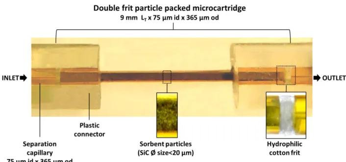
In the typical SPE-CE configuration, the microcartridge is mounted in series to the fused silica separation capillary, and sample loading is performed from the inlet to the outlet, in the same direction as later the separation (Figure 1). This could be detrimental when analyzing complex samples, because some of the matrix components could be irreversibly adsorbed in the inner wall of the separation capillary. Another inconvenient of this unidirectional or one-dimensional configuration is that the time to load a certain sample volume by pressure, which is the typical introduction mode because it is more general than electrokinetic introduction, depends on the applied pressure (e.g. typically 930 mbar in Agilent Technologies CE instruments) and the capillary dimensions (total length (LT) and internal diameter (i.d.)) [4]. In order to overcome these drawbacks, cruciform and staggered (i.e. zigzag or z-shaped) microcartridge configurations have been developed [6,23,24,29–31]. The operation in both cases is very similar, but in the staggered microcartridges the amount of sorbent, hence the extraction capacity, is intended to be higher [23]. The sample is introduced through a transport tube or passage tube (usually made of a polymeric material such as PEEK tubing), which is orthogonal to the separation direction, whereas elution and separation are performed in the other direction, applying the voltage across to the microcartridge between the inlet and the outlet extremes of the separation capillary. In such configurations sample loading time by pressure is not determined by the separation capillary dimensions. Furthermore, they potentially allow no carry-over of substances into the separation capillary, extended lifetime of the capillaries and the possibility of multidimensional separations with several microcartridges mounted in series for high-throughput analyses [6,23,24,29–31]. However, these orthogonal microextraction devices are more difficult to construct than the typical microcartridges, as they require controlling the fluid flow with microvalves and moving from capillaries to microfluidics. This type of microfluidic set-ups have
7
been described by different authors in articles [6,23,24,30,31] and patents [29,40,41], but, as other set-ups requiring specific instrumental multi-valve interfaces, they are out of the scope of this review, which will be focused on configurations for unidirectional SPE-CE with capillaries.
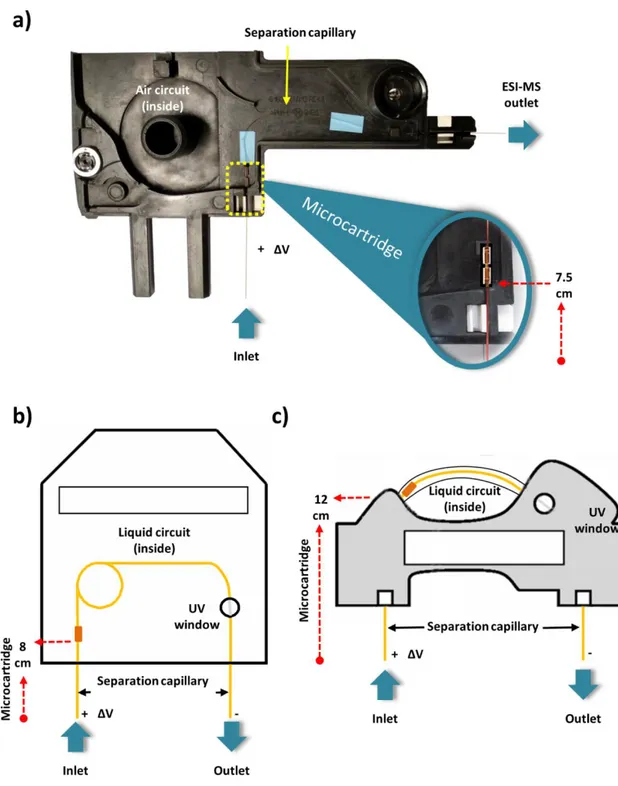
In addition to simplicity, another advantage of the conventional unidirectional configuration is that the capillaries modified with the microcartridges can be easily adapted to use the cartridge cassettes of the different commercial CE instruments (Figure 2). These cartridge cassettes allow thermostatizing the capillary to compensate Joule heating during separation while the capillary is folded or coiled to minimize the space requirements. As can be observed in Figure 2 a-c, the microcartridge is placed inside the cartridge cassette, as closest to the inlet as possible. Therefore, the microcartridge position from the capillary inlet differs between different instrument manufacturers. If the microcartridge is fabricated connecting with plastic tubing capillary fragments with different i.d. (Figure 1-a), it is not recommended to install the microcartridge outside the cassette, closer to the inlet, due to the electrode configuration (in general, parallel (and coaxial or not) to the capillary), the mechanical operation during the analyses (vial lift movement, vial cap puncher, etc.) and the particularities of the injection (the inlet capillary end is submerged in the solutions, pressure application during hydrodynamic injection, etc.). In the typical situation, as the microcartridge is placed a few centimeters beyond the separation capillary inlet, several authors, after injection of the eluent and before applying the separation voltage, prefer to push by pressure the injected plug from the inlet until it flows through the microcartridge to ensure an appropriate elution [4]. However, this little adjustment is not completely necessary to achieve good results. Thus, for example, the eluent using fused silica
8
capillaries in normal polarity (cathode in the outlet) migrates, after applying the voltage, towards the microcartridge with the velocity of the cathodic electroosmotic flow (EOF), which mainly depends on the BGE pH [4].
2.2. Microcartridge with frits and fritless
During the last almost 30 years, several microcartridge designs for SPE-CE have been described. In general, the microcartridges are mounted in 50-75 µm internal diameter (i.d.) x 365 µm outer diameter (o.d.) fused silica separation capillaries, using plastic tubing with an appropriate i.d. as zero death-volume connectors (e.g. Tygon® tube of 250 µm i.d.) (Figure 1) [4]. In many cases, the microcartridge body consists in a small piece (total length (LT)~2-10 mm) of fused silica capillary with a wider i.d. than the separation capillary (the most typical i.d. are 150 or 250 µm) that is connected with two plastic connectors to the separation capillary (Figure 1). Several authors have shown that extraction performance within this LT range is good, but for the shortest microcartridges (LT~2-4 mm) is better to introduce the microcartridge body completely inside a single plastic connector to reinforce the modified capillary [4,42]. With regard to the microcartridge i.d., we in general use microcartridges of 250 µm to ensure the maximum amount of sorbent inside [4]. Several authors have related the amount of different sorbents with an increase in sensitivity [43–45], but others have shown no effect [42] or worse results [46], because preconcentration factors depend on the compromise between the amount, porosity and packing of the sorbent for retention, and the volume necessary for an efficient elution and obtention of narrow electrophoretic peaks. Anyway, microcartridges of 250 µm i.d. guarantee with different sorbents a good compromise between extraction recoveries, effects on the EOF due to the chemical
9
features of the sorbent, separation efficiency and backpressure due to flow restriction. Excessive backpressure especially affects the electroosmotic flow, separation efficiency, migration time and reproducibility, and in some cases prevents current circulation after applying the separation voltage. As an alternative to these microcartridges made on fused silica, several authors have proposed to introduce the sorbent inside the plastic tube used as connector, between both pieces of the separation capillary [47]. However, these SPE-CE capillaries are prone to poor electrical performance, worse separation and limited reproducibility. With regard to the separation capillary material, the use of coated capillaries to prevent the adsorption in the inner capillary wall of the analytes or some of the matrix components that could be detrimental for the method performance has been described, but to a lesser extent [48–50]. J. Cai et al demonstrated for the first time the use of coated separation capillaries in SPE-CE-UV using capillaries coated with polyethylene glycol for the analysis of proteins with immobilized zinc affinity open-tubular microcartridges [48]. In this case, the permanently coated separation capillary is prepared before cutting and inserting the microcartridge to prevent sorbent degradation during the coating procedure, as we also demonstrated for the analysis of glycoproteins by SPE-CE-MS with immunoaffinity (IA) sorbents using capillaries coated with hexadimethrine bromide or an anionic derivative of polyacrylamide [49,50].
Once the microcartridge is assembled, if the plastic connectors provide a tight junction, no adhesive sealing is necessary and the microcartridge is completely replaceable (Figure 1 and 3-a). In other cases, especially if microcartridge position or capillary installation inside the commercial cartridge cassette compromise microcartridge mechanical integrity, it is recommended to completely cover the microcartridge and the
10
connectors with a thin layer of a two-component epoxy resin glue to reinforce the construction before installation. This is especially necessary when a liquid (Figure 2-b and 2-c), instead of air (Figure 2-a), circulates through the cartridge cassette in the capillary thermostatizing system, as in AB Sciex instruments (formerly Beckman Coulter instruments). We have recently shown that the internal thermostatizing system of the instrument or an external specific set-up designed for the microcartridge can be used to explore the influence of temperature in SPE-CE [51].
Figure 3 a-h show the most important microcartridge designs for SPE-CE that can be divided into two main groups: with frits and fritless.
2.2.1. Microcartridge with frits
Microcartridges with frits are packed with sorbent particles and frits are used to avoid particle leaking during the analyses (Figure 3 a-b). Microcartridges with frits can be tightly packed with smaller sorbent particles of a certain pore size, hence allowing a great active surface area for the extraction. However, extremely packed microcartridges, nonporous sorbents or too tighten frits promote flow restriction and excessive backpressure, hence EOF disturbance, bubble formation, current instability and power failures [4].
Frits form a porous wall or net structure (<100 µm thickness) that allow the passage of solution and analytes, without restricting the flow. For the same reasons as we suggested before for the location of the sorbent, we recommend placing the frits inside the microcartridge body (Figure 3-a and -b) [4]. However, with very small sorbent
11
particles (particle size<20 µm), it could be necessary to place the frits inside the plastic connector between the different capillary fragments (Figure 1-a) [52,53]. Frits can be prepared inside the microcartridge body sintering porous glass beads after heating in an oven or with a fiber-optic fusion splicer, nichrome wire or microelectric arc device. In the first microcartridge for SPE-CE ever described in the literature, N.A. Guzman et al used sintered porous borosilicate frits in a double-frit microcartridge (Figure 3-a) containing sorbent particles with an antibody against methamphetamine covalently attached to irregularly shaped glass beads [22]. However, the high temperature needed for the fabrication could degrade and inactivate the sorbent and several alternatives have been described to these sintered frits. Metal frits, as those used in chromatographic columns, have been scarcely applied because of the poor performance and electrical issues [54]. Frits made on glass fiber or wool [43,55], polymeric membranes [56,57], sol-gel polymers [55] and hydrophilic cotton have been demonstrated [53]. These materials allow construction of tighten frits, which are especially needed with the smallest sorbent particles (diameter size<20 µm), but have to be prepared with care to ensure an appropriate performance of the system. Alternatively, we have widely applied microfrits prepared after cutting into small pieces (diameter size~100 μm) the original polyethylene filters of the commercial cartridges for off-line SPE [4]. These polyethylene microfrits allow a good electrical and flow performance with double-frit microcartridges packed with the typical sorbent microparticles that can be found in the commercial cartridges for off-line SPE [37,58,59] or with the microparticles that are commercialized as solid supports to prepare IA sorbents [49,50,60–62] (diameter size>30 µm, but typically between 50-100 µm, and pore size>100 Å). In general, double-frit microcartridges are preferred because sorbent leaking is totally prevented. However, packed microcartridges with a single frit in the outlet perform well when no
12
sorbent leaking though the inlet or poor packing is observed. Single-frit microcartridges can be necessary with certain sorbents and frit materials to avoid an excessive backpressure with two frits (Figure 3-b) [53,63]. The feasibility of the single-frit design using a C4 sorbent (diameter size=13 µm) and a sintered frit was first demonstrated by He et al. for the preconcentration and chiral separation by SPE-CE-UV of the enantiomeric drug verapamil in human plasma using C4 sorbent particles and a cyclodextrin containing background electrolyte (BGE) [63]. We have recently used a single-frit microcartridge with a silicon carbide (SiC) sorbent (diameter size<20 µm) and a hydrophilic cotton frit in the outlet end of the microcartridge for the analysis of circulating microRNAs in serum samples by SPE-CE-MS (single-frit version of the double-frit microcartridge presented in Figure 1) [53].
Table 1-a shows a representative overview of some selected SPE-CE applications using microcartridges with frits that are of relevance to the field.
2.2.2 Fritless microcartridges
As an alternative to the microcartridge with frits, several authors have described the use of fritless microcartridges in SPE-CE. In some cases, the assembly of these fritless microcartridges can be much simpler, and certain designs can prevent some of the typical drawbacks of microcartridges with frits related with flow restriction. Fritless microcartridges have been fabricated using different designs (Figure 3 c-h): packed with sorbent particles with a slightly larger size than the inner diameter of the separation capillary (Figure 3-c) [42,46,47,64–71] or using sorbent membranes (Figure 3-d) [27,33–36,46,72–75] and fibers [76–78], as well as coatings [79–86] or monoliths
13
(Figure e and -f) [39,87–102] or retaining magnetic particles with a magnet (Figure 3-g and -h) [44,45,103–110].
The most straightforward way to prepare a fritless particle-packed microcartridge is to pack sorbent particles with a slightly larger size than the inner diameter of the separation capillary (Figure 3-c) [42,46,47,64–71]. If necessary, the sorbent particles can be sieved with an iron steel sieve of an appropriate size (sieve pore size>i.d. separation capillary). [42,69,70]. In general, 50 μm i.d. separation capillaries are preferred to avoid using extremely large sorbent particles that could generate loose packings. K. Jooß et al showed for the analysis of organic sulfonates and glycans by SPE-CE-MS that this can be prevented using fritless microcartridges with a bead string design (weak anionic-exchange sorbent particle size>90 μm, 4 mm LT packing x 100 µm i.d. connected to a 50 μm i.d. separation capillary) [70]. In 1994, A.T. Tomlinson et al described for the first time the fritless microcartridge design [47]. They used a fritless microcartridge packed with C18 particles inside a Teflon tube to analyze haloperidol and standard solutions of synthetic peptides. In the last years, the fritless particle-packed microcartridges made of fused silica have become very popular because of the widespread availability of sorbent microparticles, preparation simplicity and good performance, and different applications have been described using from conventional chromatographic sorbents (C18, hydrophilic-lipophilic balance (HLB), cationic exchange, etc.) [42,46,47,66–70], to molecular imprinted polymers (MIPs) [64,71] or IA [65] sorbents for the analysis of drugs, herbicides and biomarkers (e.g. Aminopirene trisulfonate (APTS)-labeled glycans, amino acid derivatives or peptides), in standard solutions, biological fluids and foodstuff with UV and MS detection (Table 1-b). Recently, we investigated the potential of sheathless SPE-CE-MS with this
14
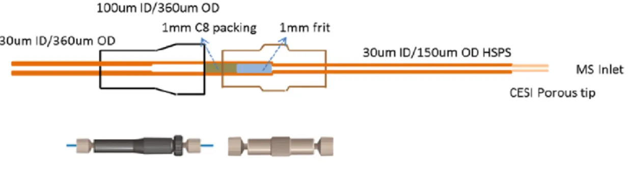
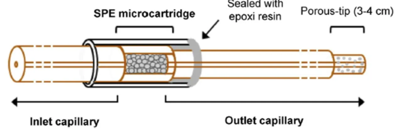
microcartridge design for the highly sensitive analysis of opioid peptides in aqueous solutions [111]. This high sensitivity porous sprayer (HSPS) sheathless CE-MS interface necessarily runs with a 30 µm i.d. x 150 µm o.d. capillary, ended with a porous-tip in the outlet, instead of the typical 50 or 75 µm i.d. x 365 µm o.d. capillaries. As an alternative to the double-frit C8 microcartridge (C8 particle size~5 µm, 1 mm LT packing x 100 µm i.d. x 365 µm o.d.) proposed by Y. Wang et al for these special capillaries, which needed special connectors (Figure 4) [38], we proposed a novel fritless C18 microcartridge (C18 particle size>50 μm, 4 mm LT packing x 150 µm i.d. x 365 µm o.d.), which was inserted in the separation capillary just employing the typical plastic tubing and an epoxy resin to seal the capillaries with different o.d. (Figure 5) [111]. In this case, LODs for the analyzed opioid peptides could be decreased down to the picomolar level, which represented an improvement of 5,000-fold with respect to the LODs achieved by sheathless CE-MS without preconcentration, which was five times more sensitive than sheathflow CE-MS [37]. Nowadays, this HSPS sheathless CE-MS approach provides the most promising performance in terms of sensitivity of SPE-CE with on-line MS detection [38,111]. Other authors have also shown remarkable results in SPE-CE-MS with different types of microcartridges and sheathless interfaces [73,112]. P. Viberg et al described a double-frit C18 microcartridge (C18 particle size~5 µm, 1-2 mm LT packing x 200 µm i.d. x 300 µm o.d.) to analyze heterocyclic aromatic amines until concentrations of 22 nM with a sheathless interface made with a nanospray emitter. The microcartridge was connected and sealed with epoxy glue near the separation capillary without plastic connectors, taking advantage of the diameter differences with the separation capillary (30 µm i.d. x 190 µm o.d), which was gold-coated and etched in the outlet end [112]. G. M. Janini et al tapered and etched 75 µm i.d. x 190 µm o.d fused silica capillaries in the outlet end to prepare 30 µm i.d spray tips
15
for sheathless SPE-CE-MS. They were using fritless microcartridges containing a sorbent membrane (3 mm, C18) (Figure 3-d), which will be discussed later in detail, to enhance sensitivity in the analysis of tryptic digests from diluted protein solutions (until 250 nM of digested protein) [73]. Despite all this progress, the increased reproducibility and robustness, as well as the lower cost of the instrumental set-up and materials required for sheathflow SPE-CE-MS, makes today very difficult a widespread use of sheathless SPE-CE-MS.
With regard to the performance of this particle-packed fritless microcartridge compared to microcartridges with frits, we have demonstrated that results with a certain sorbent sieved to select particles of certain diameters are not better than those that can be obtained with a double-frit microcartridge packed with the unsieved sorbent particles [42]. In that study we developed a C18-SPE-CE-MS method for the analysis of opioid peptides in cerebrospinal fluid [42]. A fritless microcartridge of 4 mm LT x 150 μm i.d. was packed with a C18 sorbent (particle size>50 μm) and was connected near the inlet of the separation capillary (50 μm i.d.). Under the optimized conditions, the LODs for the studied peptides were 0.5-1.0 ng/mL, while they were detected at 0.1 ng/mL by C18-SPE-CE-MS using a double-frit microcartridge (7 mm LT x 250 μm i.d. connected to a 75 μm i.d. separation capillary) that was packed without sieving the same C18 sorbent particles (20 μm<particle size<100 μm). This was mainly because the microcartridge with frits, which was longer and wider and was tightly packed with smaller unsieved sorbent particles, was connected to 75 μm i.d. separation capillaries that allowed to introduce in a shorter time a larger volume of sample.
16
Another way to prepare a fritless microcartridge is to insert a coated/impregnated sorbent membrane (1-3 mm) in the plastic tube connecting the two pieces of separation capillaries (Figure 3-d) [27,33–35]. This design was firstly used [33], and later widely demonstrated [27,34,35], by A.J. Tomlinson et al for the analysis of drugs and peptides by SPE-CE-MS using C18 or polymeric-impregnated membranes. Using these membranes, it was possible to minimize the sorbent bed volume at the inlet of the separation capillary and no flow restriction was reported. High retention capacity of the membranes permitted the analysis of the typical large sample volumes with double-frit or fritless packed microcartridges (>100 µL) and high preconcentration factors, without compromising either analyte resolution or separation efficiency. Furthermore, the authors demonstrated for the first time the combination of SPE-CE-MS with t-ITP to further enhance the preconcentration factors for the analysis of peptide mixtures [33]. The methanol-water (80:20 v/v) plug with the eluted peptides (~60 nL) was sandwiched between two small volumes of leading electrolyte (LE) (~60 nL, 1% ammonium hydroxide) and BGE (~90 nL 2 mM ammonium acetate-1% acetic acid (pH 2.9), which acted also as terminating electrolyte (TE). After voltage application, when the steady state was reached, peptides were preconcentrated as discrete narrow bands with the same velocity because the LE and TE ions moved faster and slower, respectively. Despite all the benefits of using sorbent membranes, only very few authors have tested this design [36,46,72–75], probably because of issues related with fabrication reproducibility and availability of impregnated membranes with appropriate pore size, permeability and active surface area. Other similar approaches based on fibers have been described but also with very limited success [76–78].
17
Open tubular microcartridges for SPE-CE (Figure 3-e) were first described by J. Cai et al [79], in parallel to the developments on the on-line coupling of open tubular enzymatic microreactors to CE [113,114]. They connected to the inlet of the separation capillary as a microcartridge a 50 µm i.d. x 20 cm LT piece of a fused silica capillary derivatized (i.e. coated) with C18 groups to analyze standard solutions of two herbicides (prometon and prometryne) by SPE-CE-UV [79]. After preparing a coated capillary of a certain length, the SPE-CE capillary column was very easy to assemble, as it was only necessary to cut a “plug-and-play” microcartridge to the desired length, connect and analyze. However, as the affinity surface area in these microcartridges is small, preconcentration factors are very limited (e.g. 10-35 times for prometon and prometryne [79]), and only a limited number of publications have been reported [80–87]. Most of the applications have been described with IA-SPE-CE immobilizing antibodies or antibody fragments in the open tubular microcartridge to analyze peptides in biological samples [80–84], especially with antibody fragments by T.M. Phillips and coworkers [81–84], before they moved to explore similar IA-SPE-CE approaches in microchips [115,116]. As an example of the limitations of open tubular microcartridges but with other sorbents, H. Wang et al presented a 100 cm LT x 50 µm i.d. open tubular capillary coated with gold nanoparticles for off-line SPE of monohydroxy-polycyclic aromatic hydrocarbons in spiked urine samples before CE-UV and enrichment factors were lower than 100 times [85]. In order to extend the active surface area with IA open tubular microcartridges, N.A. Guzman proposed to use as a microcartridge body multiple channels bored through a single glass rod or a bundle of multiple capillaries (25 µm (i.d.)) [25–27], but this extremely complex multi-bore design has demonstrated a very limited applicability. A simpler alternative to improve extraction capacity of single-bore capillaries is to increase the thickness and porosity of the sorbent layer in the inner wall
18
of the open tubular microcartridge. X. Zhang et al directly prepared in the separation capillary, using a novel light-emitting diode (LED) induced polymerization technology, a 3 mm LT x 75 µm i.d. open-tubular microcartridge coated with a MIP to preconcentrate until 200 times some testosterone derivatives in standards and spiked urine samples [86]. Similarly, we recently evaluated a 7 mm LT x 250 µm i.d. open-tubular C18-silica monolithic microcartridge for the analysis of neuropeptides in human plasma [87]. In this case the monolith was not prepared in situ, but ex situ as a column of a certain length that was later cut to obtain several “plug-and-play” microcartridges. The LODs by SPE-CE-MS with the C18-silica monolithic microcartridge were 100 times lower than without preconcentration, but 100 times higher than with a C18 double-frit particle-packed microcartridge [58,59]. In order to maximize the microcartridge capacity and extraction recovery, it is recommended that the monolith fills completely the lumen of the microcartridge body (Figure 3-f). The monolith length (typically from approximately 1 cm), structure and the number and type of active groups on the surface can be tailored to avoid flow restriction, as well as to maximize selectivity and extraction recoveries. Silica- and polymer-based monoliths have been described to prepare fully-filled microcartridges for SPE-CE [39,88–102]. However, polymer-based monoliths have been mainly preferred, because preparation by polymerization of monomers and cross-linkers is more simple and reproducible than the sol-gel procedure necessary to prepare silica-based monoliths [100]. It is well known that polymer-based monoliths can show a less uniform mesopore structure. However, polymer-based monoliths present improved stability, once selected appropriate solutions to prevent shrink or swell. Today, monoliths are probably the most promising way to prepare “plug-and-play” fritless microcartridges, but progress must be made improving extraction capacity and decreasing non-specific binding for the minute amount of
19
sorbent used in SPE-CE. In this sense, P.R. Haddad and coworkers have been exploring the use of nanoparticles (i.e. functionalized quaternary ammonium latex [98,99] or silica [90] nanoparticles) to expand the extraction capacity of polymer-based monolithic microcartridges while tuning their selectivity to analyze by ion-exchange SPE-CE-UV anions [98,99] or weak bases [90]. Thus, they reported a sensitivity enhancement between 1,500-1,900 with regard to conventional CE for a mixture of three neurotransmitters (dopamine, norepinephrine and metanephrine) with a microcartridge (8 cm LT x 75 µm i.d.) containing a polymeric monolith coated with silica nanoparticles (LODs were between 0.5-0.7 ng/mL) [90]. With regard to more selective monolithic microcartridges, recently A. Marechal et al described for the first time the use of an aptamer-based monolithic sorbent in SPE-CE [93]. They applied an aptamer silica-based monolithic microcartridge (1.5 cm LT x 75 µm i.d.) for the analysis by SPE-CE with laser-induced fluorescence (LIF) detection of ochratoxin A in standards, beer and wine at concentrations of a few ng/mL.
Nowadays, there are many commercially available magnetic particles (i.e. beads) with different surface chemistries ready for the immobilization of a wide variety of affinity ligands to develop bioanalytical applications [117,118]. The versatility and reasonable price of the commercial magnetic beads and the simplicity of operation are rapidly expanding their application in dispersive SPE [118]. As magnetic beads can be held in position by an external magnet or electromagnet, they have been used for preparation of fritless microcartridges (Figure 3-g and -h), in particular for IA-SPE-CE. Since the pioneering work of Rashkovetsky et al. [103], several authors have described the use of IA magnetic particles in IA-SPE-CE with fluorescence [44,103–105], ultraviolet (UV) [45,108,109] and MALDI-MS [106,107] detection. Recently, we reported for the first
20
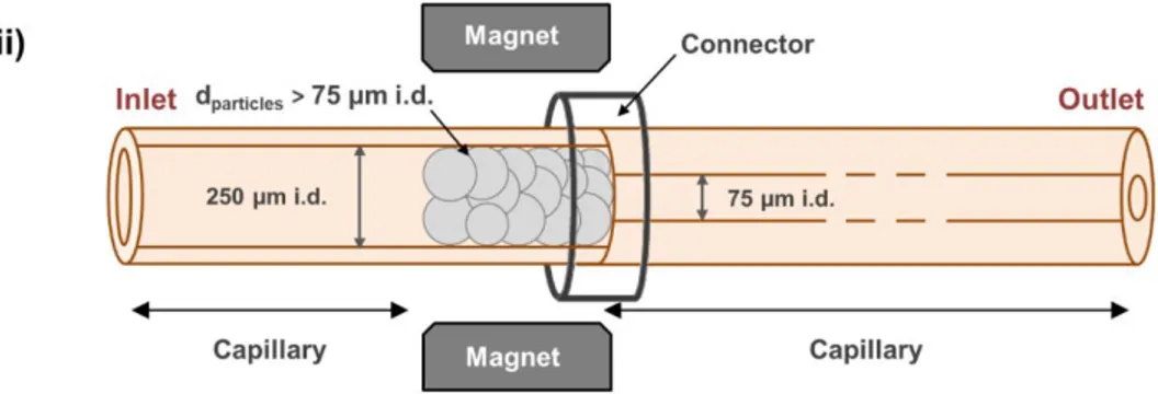
time a IA-SPE-CE-MS method, with on-line electrospray ionization mass spectrometry (ESI-MS), using magnetic agarose microparticles (45 μm<particle size<165 μm) with an intact antibody against human serum transthyretin (TTR), which performed better than magnetic silica microparticles of smaller size (particle size~2.8 μm) [110]. In this case, two fritless microcartridge designs (9 mm LT x 250 μm i.d.) were described. As agarose magnetic microparticles swell when suspended in aqueous solutions, one of the designs was taking advantage of their larger size than the inner diameter of the separation capillary (75 μm i.d., Figure 3-c). In the other case, a magnet was used to hold a 9 mm length of magnetic particles in the outlet end of a capillary fragment (8.5 cm LT x 250 μm i.d.) that was connected with a plastic tube to the separation capillary (75 μm i.d., Figure 3-h). In all cases, the performance of both fritless microcartridges was similar. However, the “magnetic” design (Figure 3-h) had several remarkable advantages, such as the presence of only one capillary connector and the simplicity to replace the sorbent through the inlet after certain number of analysis. Under the optimized conditions, LODs for TTR standard solutions were 25-fold lower than those obtained by CE-MS and the method could be applied for the analysis of serum samples [110]. With regard to non IA magnetic particles, Y. H. Tennico et al reported for the first time the use of C18 silica-coated Fe3O4 magnetic nanoparticles (particle size~500 nm) for the SPE-CE-UV analysis of parabens and drugs in standards [105]. However, these authors proposed to mix the magnetic nanoparticles with the sample in a vial before injecting a small plug of the reacted nanoparticles (length~1 mm in a 50 µm i.d. fused silica separation capillary) for SPE-CE-UV analysis and LODs were around 1 µg/mL for standards of parabens. T. Baciu et al proposed later a typical SPE-CE-UV procedure with C18 silica-coated Fe3O4 magnetic nanoparticles (particle size~75 nm) for the SPE-CE-UV analysis of drugs of abuse in urine samples at concentrations
21
ranging from 20 to 50 ng/mL. In this case, the magnetic nanoparticles were retained first inside the short section (length~1.5 mm) of the inlet of a 50 µm i.d. fused silica separation capillary (Figure 3-g) [45,109]. At the moment, the main limitations of using magnetic particles in SPE-CE is that many magnetic (micro or nano) particles commercialized for dispersive SPE are not porous, which could be a limitation for the active surface area when the amount of sorbent is small such as in SPE-CE. Another drawback affecting extraction capacity and reproducibility is particle aggregation [119]. In addition, the magnetic field holding the magnetic particles should be enough to avoid particle leaking during the washing and loading steps [44,45,104,108]. Magnetic particle leaking, especially with nanoparticles, could be a serious problem for the integrity of the mass spectrometer with on-line MS detection. Finally, little is known about the influence of the magnetic fields in immunoextraction in capillaries or microdevices [120,121].
Table 1-b shows a representative overview of some selected SPE-CE applications using fritless microcartridges reported to date in the literature.
2.3. SPE-CE sorbents
As noted above, many different sorbents have been described to analyze by SPE-CE a great variety of compounds, in a wide range of applications. Sorbents using particles as solid supports to immobilize the affinity ligands have been the most widely applied, from the low-selective conventional chromatographic sorbents (C8, C18, HLB, cationic or anionic exchange, etc.) until more selective sorbents with metals, molecular imprinted polymers, lectins, antibodies, antibody fragments, aptamers, etc. [4–6,17–
22
21,23,24]. In SPE, recovery is mainly governed by the compromise between the eluotropic strength and the volume of the eluent and the sorbent capacity, which depends on the affinity strength and selectivity of the target analyte to the sorbent and the number of active sites available for the extraction that in particle sorbents are related to the particle size and porosity [122]. From the method development perspective in in-line SPE-CE, the only requirement of a good sorbent, in addition to reproducibility, is the compatibility between the extraction (high recovery, rapid elution and low non-specific retention), separation (high resolution, short analysis time) and detection (high selectivity and sensitivity). However, in some cases, this is difficult to accomplish, for example, if the retained analytes are eluted while filling the capillary with BGE before the separation, or with on-line MS detection, which is negatively affected by the presence of salts and requires the use of volatile solutions and BGEs. In other cases, it is the sorbent that is prone to degradation due to the presence of certain components in the solutions and BGE, such as when IA sorbents are exposed for a long time to BGEs with extreme pH values or organic solvents. These are the reasons why, for example, IA-SPE-CE was widely demonstrated with ultraviolet and fluorescence detection [22,25–28,80–82,103] before the first IA-SPE-CE-MS application was described in 2000 [123]. In that study, N.A. Guzman reported the analysis of gonadotropin-releasing hormone (GnRH) in serum and urine using a double-frit microcartridge with sorbent particles containing antigen-binding antibody fragments (Fab) against the peptide hormone. He was using a volatile BGE of ammonium bicarbonate pH 8.0 with 1% acetonitrile combined with a 0.3 M glycine-HCl buffer (pH 2.5) for the elution, which was not very compatible with on-line MS detection. Since then, our group have been very active in IA-SPE-CE-MS, describing applications for small peptides and proteins with intact antibodies or Fab antibody fragments using different microcartridge designs
23
and neutral pH ammonium BGEs combined with eluents containing formic acid, acetic acid or ammonium hydroxide [50,60–62,65,110]. We recognize that making compatible and adapting the use of sorbents for in-line SPE-CE-MS is a challenge. In general, this challenge can be successfully faced with an appropriate method optimization. However first, as we mentioned before, the sorbent physical features (e. g. particle size, permeability and porosity) must allow operation without promoting flow restriction and backpressure, which may disturb the EOF and produce current instability or power failures.
In particle-packed microcartridges, the best compromise between active surface area, flow performance and durability is achieved using as a sorbent porous microparticles (e.g. pore size>100 Å and diameter size>50 μm), despite smaller microparticles [53,108], and even nanoparticles [45,109], have also been applied but to a lesser extent. Furthermore, if possible, our recommendation is to pack the microcartridge by vacuum using the dried sorbent particles instead of the slurry traditionally used to pack liquid chromatography columns [4]. With regard to continuous unitary sorbents, such as membranes and monoliths, there is very little information about the porosity of the membranes [27,33–35,46,74,75] or monoliths [39,87–102] used in microcartridges for SPE-CE, and probably this is one of the reasons of the limited amount of applications described. Our recommendation is preparing “completely full” monolithic microcartridges but avoiding extremely small pore sizes, as with particle sorbents, to ensure enough permeability and avoid excessive flow restriction. In either case, after preparing the SPE-CE capillaries with the microcartridge and before the analyses, it is highly recommended to flush water manually with a syringe through the system to check for abnormal flow restriction followed by a blank analysis to monitor current
24
stability [4]. Not all the SPE-CE methods described have been fully validated and applied to complex real samples, but many good examples can be found in the literature with different sorbents [4–6,17–21,23,24]. In general, a microcartridge with an appropriate sorbent after method optimization can be reused between 10 and 30 times with standards and until 10 times with samples, depending on the sample matrix complexity [4]. For the analysis of complex biological samples, it is recommended to apply before SPE-CE a minimum sample pretreatment to prevent microcartridge saturation with matrix compounds and extend the microcartridge lifetime [4,9,52,53,60– 62,110,124].
Nowadays, conventional chromatographic particle sorbents (C8, C18, HLB, cationic or anionic exchange, etc.) are the most commonly used for SPE-CE, as they fulfil most of the requirements for optimum performance. On the one hand, they provide a large active surface area, without interfering with the electrophoretic separation and detection. On the other hand, they are commercially available, have been optimized and widely used for the analysis of a great variety of compounds and the extraction methods are compatible with on-line MS detection (e.g. with C18 sorbents using formic and acetic acid BGEs and similar acidic hydroorganic mixtures with methanol for the elution). Especially reversed phase silica-based (e.g. C18) and polymeric sorbents (e.g. HLB) are widely recognized for their good recoveries, being the most common chromatographic sorbents used for SPE-CE-UV and -MS applications involving peptides (C18 [4,9,37,42,58,59,111,124]), antibiotics (C18 [125]), pharmaceuticals (C18 [126,127] and HLB [68]), drugs of abuse (HLB [69,128,129]), other bioactive compounds (e.g. ochratoxin A [130] or alkaloids [131] with C18 ), rare earth elements (C18 [132]) and
25
metabolites (C18 [133]) in different environmental and biological samples (see Table 1).
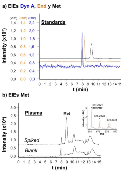
However, the major drawback of these chromatographic sorbents is their limited selectivity, which precludes the direct analysis of complex samples such as biological fluids. In such cases, a previous clean-up pretreatment, which can be more or less laborious, is required to purify and (optionally) enrich the target analytes in order to prevent microcartridge saturation. As an example, we have proposed a double-step sample pretreatment for the analysis of low molecular mass compounds in human plasma, which was optimized and validated for the analysis of neuropeptides by C18-SPE-CE-MS [4,9,59,124]. The method consisted of precipitation with acetonitrile followed by centrifugal filtration using 10,000 relative molecular mass cut-off filters. Using this double-step sample pretreatment, it was possible to obtain with preconcentration, LODs for these specific peptides in plasma samples between 100 and 1,000 times lower than the LODs obtained using CE-MS. Figure 6 shows detection at 1ng/mL of three neuropeptides in a standard mixture and a plasma sample by C18-SPE-CE-MS with a double-frit particle-packed microcartridge after this sample pretreatment (LODs by CE-MS and C18-SPE-CE-MS for standards were 50 ng/mL and 0.1 ng/mL, respectively [4,37]). As can be observed in Figure 6-b, different compounds from the plasma matrix still were detected after the sample pretreatment due to the limited selectivity of the C18 sorbent, and MS detection using an accurate mass and high resolution TOF mass spectrometer was necessary to reliably identify the different neuropeptides. This sample pretreatment has been also applied to analyze other low molecular mass compounds as plasma metabolites in untargeted metabolomics studies
26
by C18-SPE-CE-MS of samples from mice genetically modified to develop Huntington's disease (HD), an inherited neurodegenerative disorder [133].
In general, a certain type of sample pretreatment is even necessary when sorbents that present higher extraction selectivity are applied for the analysis of complex biological samples, such as when small molecules, peptides, proteins or microRNAs are analyzed in biological fluids using sorbents such as immobilized metals [52], SiC [53], antibodies [50,60,110] or antibody fragments [61,62]. Figure 7 shows the extracted ion electropherograms (EIEs) and the total ion electropherograms (TIEs) obtained by IA-SPE-CE-MS and C18-IA-SPE-CE-MS for two plasma samples containing two neuropeptides after the sample pretreatment explained above using a sorbent with a specific Fab antibody fragment and a double-frit particle-packed microcartridge. As can be observed in the EIEs of Figure 7-a both neuropeptides were selectively detected, and no interferences were observed in the TIEs of Figure 7-b ii. In contrast, many matrix compounds were still present, retained and detected by C18-SPE-CE-MS (Figure 7-b i). Improving sorbent selectivity allows targeting specifically a certain compound and obtaining interference-free electropherograms, which are extremely helpful even with the most selective detectors (e.g. MS). However, there is always a remaining analyte-sample matrix interaction and non-specific retention to the affinity ligand or the solid support that, with the small amount of sorbent in the microcartridges, precludes in most cases direct or dilute-and-shoot analyses of the most complex biological fluids (e.g. serum or plasma, and in less extent urine or cerebrospinal fluid), complex environmental samples, beverages and foodstuff. Figure 8 shows two examples of urine and beverage analysis with minimum sample pretreatment. As can be observed in the UV electropherograms of Figure 8-A, F. J. Lara et al were able to analyze by
SPE-CE-27
UV triazine herbicides at 10 µg/mL in spiked urine samples using a fritless microcartridge packed with MIP particles. In this case, it was necessary to acidify urine until pH 1 with HCl, instead of the optimized pH 7 with the standards (75 µM H3PO4 adjusted to pH 7 with 1M NaOH) [71]. In Figure 8-b are shown the LIF electropherograms obtained by A. Marechal et al who detected ochratoxin A at 0.5 ng/mL in white wine samples with an aptamer silica-based monolithic microcartridge, after diluting twice the sample with binding buffer (Tris 10 mM, NaCl 120 mM, CaCl2 20 mM, KCl 5 mM, pH 8.5) and readjusting pH to 9 [93]. As pointed out above for monoliths, improving extraction capacity and decreasing non-specific retention are again crucial to enhance the performance of these high selective sorbents in SPE-CE. Another difficulty of these sorbents, compared to the conventional chromatographic sorbents, and despite the advancements made in the last years, is the limited commercial availability of activated supports or specific affinity ligands to prepare these sorbents. Therefore, nowadays, the number of applications described using high selective sorbents in SPE-CE is markedly lower than with conventional chromatographic sorbents, and the number decreases dramatically considering SPE-CE-MS. In the last case, this is because SPE-CE-MS faces the additional difficulty of making compatible the requirements of the extraction, separation and detection. Thus, for example, as we introduced before, there is only a publication demonstrating the use of an aptamer-based sorbent [93] and another showing a lectin-based sorbent [27] in SPE-CE-LIF and -UV or very few with IA [50,60–62,110,123], metal affinity [52] or MIP [64] in SPE-CE-MS. The use of high selective sorbents in SPE-CE-MS is one the most exciting fields to investigate and expand the applicability and impact of SPE-CE in the coming years.
28
This review critically examines the different designs and materials applied to prepare in-line SPE-CE microcartridges since the pioneering studies of the early nineties. It is focused on unidimensional SPE-CE with capillaries because of simplicity, as no valves are required to control the fluid flow through the modified capillaries. This configuration is the best test bed at present for the future development of SPE-CE with microfluidic set-ups containing staggered microcartridges in series, which will allow routinely high-throughput multidimensional separations.
Nowadays, there are several well described microcartridge designs with frits and fritless for unidimensional SPE-CE with capillaries. Double-frit particle packed microcartridges have the potential to allow a greater active surface area for the extraction using tight packings of smaller porous sorbent particles. However, frit construction requires certain technical know-how to avoid flow restriction or sorbent degradation. The basic fritless microcartridge design which consists in packing sorbent particles with a slightly larger size than the inner diameter of the separation capillary smartly overcomes this issue and it is a straightforward way to construct the modified capillaries from commercially available sorbent particles. However, “plug-and-play” fritless microcartridges are probably the best allies to expand the applicability of SPE-CE. Specially (fully-filled) monolithic microcartridges are the best alternative to prepare such fritless microcartridges, but progress must be made improving extraction capacity and decreasing non-specific binding for the minute amount of monolithic sorbent used in SPE-CE. Keeping in mind this fact, the development of highly selective affinity sorbents, specially based on IA and aptamers, are another field to investigate in the coming years, with emphasis on using line MS detection and the combination of
on-29
line electrophoretic preconcentration to further enhance detection sensitivity. SPE-CE-MS with highly selective affinity sorbents combines the specificity of the extraction, the resolving power and speed of the separation and the selectivity and sensitivity of the detection. Therefore, provides a promising tool to reliably and accurately address the analysis of trace components of complex samples, preventing false positives of currently applied analytical techniques that lack separation and molecular mass confirmation (e.g. ELISA).
Acknowledgements
This study was supported by grants from the Spanish Ministry of Economy and Competitiveness (CTQ2014-56777-R and RTI2018-097411) and the Cathedra UB Rector Francisco Buscarons Ubeda (Forensic Chemistry and Chemical Engineering).
30
Table 1. Representative overview of some selected unidirectional SPE-CE applications using microcartridges a) with frits and b) without frits
(fritless).
a) With frits
Analyte Sample Microcartridge
design Sorbent Detection LODs Remarks [Ref.]
Methamphetamine Urine Double-frit
microcartridge mAb particles UV - The first report of SPE-CE [22,25] Glycopeptides Urine Double-frit
microcartridge Lectin (ConA) particles UV - The first report of lectin-SPE-CE [27]
Gonadotropin-releasing hormone (GnRH)
Serum and urine Double-frit microcartridge
Fab’ polyclonal antibody
(pAb) fragment particles MS 1 ng/mL The first report of IA-SPE-CE-MS [123]
Peptides Standards Double-frit
microcartridge C18 particles MS
Until 10 ng/mL
BGE was 20mM
N-[carbamoylmethyl]-2-aminoethanesulfonic acid (ACES) at pH 7.4 [32]
Opioid peptides Human plasma
Double-frit microcartridge C18 particles MS Until 0.01 ng/mL (with t-ITP)
Detailed experimental protocol [4]
Development and reoptimization of a double step plasma pretreatment (precipitation with acetonitrile followed by centrifugal filtration)
[9,58,59]
Evaluation of different sorbents: C2, C8, C18,
HLB, etc. [124]
31 Peptides Mixture of proteins and pyrococcus furiosus tryptic digests Double-frit microcartridge C8 particles MS -
Combination with t-ITP, separation capillary coated with poly-ethylenimine and HSPS
sheathless CE-MS interface
[38]
Erythropoietin Standards Double-frit
microcartridge pAb particles UV -
Separation capillary coated with
hexadimethrine bromide [49]
Transferrin Human plasma Double-frit
microcartridge pAb particles MS -
Separation capillary coated with an anionic
derivative of polyacrylamide [50]
β-carboline
alkaloids Standards
Double-frit
microcartridge C18 particles UV -
Use of the internal thermostatizing system of the instrument or an external specific set-up to
optimize T in SPE-CE
[51]
β-carboline
alkaloids Algae extracts
Double-frit
microcartridge C18 particles MS
Until 2 pg/mL
Undaria pinnatifida (wakame) algae were extracted with methanol followed by
centrifugal filtration
[131]
Amyloid peptide
fragments Human serum
Double-frit microcartridge
Ni (II) immobilized metal ion affinity (IMA)
particles
MS 2.5 µg/mL
For one of the tested sorbents frits were placed inside the plastic connector, not in the
microcartridge body
[52]
MicroRNAs Human serum Single-frit microcartridge
Silicon carbide (SiC)
particles MS 10 nM
A single hydrophilic cotton frit was placed
inside the plastic connector of the outlet [53]
Sulfonylurea drug Standards Double-frit
microcartridge C18 particles UV
Until 5 ng/mL
Sorbent in the plastic connector between two
32 Clorophenols River water Double-frit
microcartridge C18 particles UV
Until 17 pg/mL
Sol-gel frit in the outlet and glass wool frit in the inlet.
A hole is made after the microcartridge to speed-up sample loading and avoid sample
flowing through the separation capillary
[55]
Metallothioneins Sheep liver extracts Double-frit
microcartridge C18 particles UV - Nitrocellulose membrane frits [56]
Peptides
Yeast proteins separated by
two-dimensional gel electrophoresis (2D-PAGE) and digested
with trypsin Double-frit microcartridge C18 particles MS 33 amol/µL (MS) and 300 amol/µL (MS/MS)
Zytex teflon membrane frits [57]
Opioid peptides Human plasma Double-frit
microcartridge pAb particles MS
Until 100
ng/mL Polyethylene microfrits [60]
Opioid peptides Human plasma Double-frit microcartridge
Fab’ pAb fragment
particles MS
Until 1
ng/mL Polyethylene microfrits [61]
Transthyretin Human serum Double-frit microcartridgee
Fab’ pAb fragment
particles MS 0.5 µg/mL Polyethylene microfrits [62]
Verapamil
enantiomers Human plasma
Single-frit
microcartridge C4 particles UV 5 nM
The first single-frit microcartridge for SPE-CE.
A BGE with a cyclodextrin is applied to separate the enantiomers.
33 Heterocyclic
aromatic amines Human urine
Double-frit
microcartridge C18 particles MS
Until 22 nM
Glass-fiber frits.
Sheathless CE-MS interface [112]
Ceftiofur River water Double-frit
microcartridge C18 particles UV
until 10 ng/L
Polyethylene microfrits Comparison with Large volume sample
stacking
River water preconcentrated off-line by C18-SPE
[125]
Oxprenolol Human urine Double-frit
microcartridge C18 particles UV
Until 0.5 ng/mL
Sintered C18 particles frits. Urine was pretreated by liquid-liquid
extraction
[126]
Naproxen Tap water Double-frit
microcartridge C18 particles UV
Until 10 ng/L
Polyethylene microfrits.
Elution from the outlet without injection of elution plug.
Comparison with different stacking techniques.
Tap water preconcentrated off-line by C18-SPE
[127]
Ochratoxin A River water Double-frit
microcartridge C18 particles UV 1 ng/L
Polyethylene microfrits
Comparison between off-line SPE CE and in-line SPE-CE
[130]
Rare earth elements Tap water Double-frit microcartridge
C18 particles
UV Until 20 pg/L
Gadolinium and lanthanum form 2-(5-bromo-2-pyridylazo)-5-diethylaminophenol
complexes.
[132]
Metabolites Mouse plasma Double-frit
microcartridge C18 patricles MS -
Metabolomics of Huntington. Plasma pretreatment: precipitation with acetonitrile followed by centrifugal filtration.
34
b) Fritless
Analyte Sample Microcartridge
design Sorbent Detection LODs Remarks [Ref.]
Opioid peptides Human cerebrospinal fluid (CSF) Separation capillary i.d.<sorbent particle size C18 particles MS Until 0.5 ng/mL
Comparison between fritless and double-frit
microcartridges [42] Haloperidol and
synthetic analogs Standards
Separation capillary i.d.<sorbent particle
size
C18 particles MS - The first report of SPE-CE with this fritless
design [47] Peptides Erythropoietin (EPO) and novel erythropoiesis stimulating protein (NESP) tryptic digests
Separation capillary i.d.<sorbent particle
size
pAb particles MS Until 6 µg/mL
CNBr-Sepharose 4B solid support swells and
75µm i.d. separation capillary can be used [65]
3-nitrotyrosine Rat urine
Separation capillary i.d.<sorbent particle size Mixed-mode strong cation-exchange (MCX) particles UV Until 4.4 µM
Reversed phase and strong cationic exchange
mixed mode sorbent [66]
Quinolones Meat Separation capillary i.d.<sorbent particle size MCX particles MS Until 17 ng/mL (MS/MS)
Reversed phase and strong cationic exchange mixed mode sorbent.
Meat sample pretreatment: pressurized liquid extraction (PLE)
[67]
Pharmaceuticals River water
Separation capillary i.d.<sorbent particle
size
HLB particles UV Until 0.19 ng/mL
Reversed phase hydrophilic-lipophilic
polymeric sorbent [68]
Drugs Human urine
Separation capillary i.d.<sorbent particle
size
HLB particles MS 0.013 ng/mL
Reversed phase hydrophilic-lipophilic
35 Organic sulfonates and APTS-labeled glycans Standards Separation capillary i.d.<sorbent particle size Strata X/XL AW (weak anion-exchange mixed mode polymeric) particles
MS Low nM range
Reversed phase and weak anion exchange mixed mode sorbent.
Bead string microcartridge design. Separation capillary coated with polyvinyl
alcohol (PVA)
[70]
Triazine herbicides Human urine
Separation capillary i.d.<sorbent particle
size
MIP particles UV Until 0.2 µg/mL
Cetyltrimethylammonium bromide (CTAB) in the BGE to reverse the EOF. Comparison with HLB sorbent particles
(fritless microcartridges)
[71]
Quinolones Bovine milk
Separation capillary i.d<sorbent particle size MIP particles MS Until 1 µg/kg (MS/MS)
Milk sample pretreatment: protein
precipitation with acetic acid and defatting [64]
Sulfonamides Tap, bottled mineral and river waters
Separation capillary i.d<sorbent particle
size
HLB particles UV Until 0.3 µg/L
Comparison with Strata-X and MCX sorbents particles and SDB-XC membranes (fritless
microcartridges)
[46]
Opioid peptides Standards
Separation capillary i.d<sorbent particle
size
C18 particles MS Until 2
pg/mL HSPS sheathless CE-MS interface [111]
Cathinone
derivatives Human hair
Separation capillary i.d<sorbent particle
size
HLB particles UV Until 0.05 ng/mg
β-cyclodextrin in the BGE for chiral separation.
Hair sample pretreatment: pressurized liquid extraction (PLE)
[128]
Cocaine and
metabolites Human hair
Separation capillary i.d<sorbent particle
size
HLB particles UV Until 0.1 ng/mg
α-cyclodextrin in the BGE for chiral separation
Hair sample pretreatment: pressurized liquid extraction (PLE)
[129]
Peptides Standards Membrane Styrene Divinylbenzene
(SDB) membrane MS
Until 1 pg/mL
The first report of membrane-SPE-CE and of combination of SPE-CE with t-ITP Different variants are reviewed in [27,34,35]
[33]
36 Peptides and lipopolysaccharides Standards and pathogenic strains of Haemophilus influenza Membrane SDB-XC membrane MS Until 0.23 nM (peptides)
Comparison with C18 sorbent particles (fritless microcartridges). Complex sample pretreatment of the cells
[72]
Peptides Protein tryptic digests. Membrane C18 membrane MS - Sheathless CE-MS interface [73]
Peptides
Standards and bovine serum albumin (BSA)
tryptic digests
Membrane SDB-XC membrane MS - Comparison with LC-nanoESI-MS [74]
BSA Standard Membrane Cellulose acetate
membrane UV -
Membrane in the inlet and sample
electrokinetic loading from the outlet [75]
Proteins Standards Fiber Hollow fiber UV 100 ng/mL [76]
Phenols Standards Fiber Poly(acrylate)
(PA)-coated silica fiber UV -
The fiber is connected after the off-line
loading step [77]
Proteins Phycoerythrins in
cultured cyanobacteria Fiber Derivatized hollow fiber LIF
Until 3.5 pM
Capillary isoelectric focusing (CIEF) with laser-induced fluorescence (LIF) whole
column imaging detection (WCID)
[78]
Triazine herbicides Water Open-tubular
(single tube) C18 coating UV 0.1 µg/mL
The first single-tube open-tubular
microcartridge for SPE-CE [79] Methamphetamine Urine Open tubular
(capillary bundle)
Monoclonal antibody
(mAb) coating UV - The first report of SPE-CE [22,25]
IgE Human serum
Open tubular (capillary bundle or solid rod of glass with
multiple through holes)
mAb coating UV - - [25,26]
Proteins Standards Open-tubular
(single tube) Zn(II) coating UV 1 µg/mL
Separation capillary coated with polyethylene
glycol [48]
Atrazine Standards Open-tubular
(single tube) mAb coating LIF 1 nM
Separation capillary coated with C8 and BSA.
Combination with sample stacking [80] Cyclosporin A Human tears Open-tubular
(single tube)
Fab’ mAb fragment
37 Cytokines Plasma, serum, urine,
and saliva samples
Open-tubular (single tube)
Fab’ mAb fragment
coating LIF
Until 5
ng/µL - [82]
Cytokines Single-cell cultures Open-tubular (single tube)
Fab’ mAb fragment
coating LIF 0.1 fg/mL
Special incubation chamber and microdialysis sampling of the lymphocyte cells to induce
cytokine secretion by neuropeptides
[83]
Neurothrophins Human serum Open-tubular (single tube)
Fab’ mAb fragment
coating LIF 0.1 fg/mL - [84]
Testosterone
derivatives Human urine
Open-tubular
(single tube) MIP coating UV <50 ng /mL
Novel light-emitting diode-induced technology for in-capillary polymerization.
Silylated separation capillary.
[86]
Peptides Plasma samples
Open-tubular (Monolithic. Single tube) C18-silica monolithic coating MS Until 0.01 µg/mL - [87]
Peptides BSA and E. Coli tryptic digests Monolithic. (completely filled) Sulfonate-silica hybrid strong cation-exchange (SCX) monolith MS Low pg/mL range
Combination with dynamic pH junction. Electrokinetically pumped nanospray CE-MS
interface
[39]
S-propranolol Standards Monolithic.
(completely filled) Methacrylate monolith UV
Low nM range
Reverse elution (from the outlet) with the BGE
using the EOF as a pump (reversed polarity) [95]
Immunoglobulin G Human serum Monolithic. (completely filled)
Protein G glycidyl methacrylate (GMA)
monolith
UV 1 nM
Separation capillary coated with dextran and hexadimethrine bromide.
Serum samples were diluted 1:10 and heated at 95ºC [96] Histidine-containing peptides Standards and synthetic peptide mixture Monolithic. (completely filled)
Cu(II) iminodiacetic acid (IDA) di(ethylene glycol)
dimethacrylate (DEGDMA) and glycidyl
methacrylate (GMA) monolith
38 Inorganic anions Open ocean water Monolithic.
(completely filled)
Sulfonated methacrylate monolith coated with anion exchange latex
nanoparticles
UV Until 75 pM
Separation capillary coated with poly(diallyldimethylammonium) chloride
(PDDAC) or quaternary ammonium functionalized latex nanoparticles.
Combination with t-ITP
[98]
Organic anions Standards Monolithic. (completely filled)
Sulfonated methacrylate monolith coated with anion exchange latex
nanoparticles
UV Until 1.5 nM
Separation capillary coated with PDDAC.
Combination with t-ITP [99]
Carbamate
pesticides Tap and river water
Monolithic.
(completely filled) Divinylbenzene monolith UV
Until 0.01 ng/mL
The BGE contained high percentage of ACN
and SDS [101]
Methionine-enkephalin Human CSF
Monolithic.
(completely filled) Sol-gel silica monolith MS <1 ng/mL CSF sample was deproteinized [102]
Neurotransmitters Human urine Monolithic. (completely filled)
Methacrylate weak
cation-exchange monolith UV
Until 2.9 ng/mL
Combination with focusing by a pH step
gradient [88]
Cationic
compounds Standards
Monolithic. (completely filled)
Methacrylate weak
cation-exchange monolith UV
Until 8 ng/mL
Combination with focusing by a pH step
gradient [89]
Neurotransmitters Standards Monolithic. (completely filled)
Methacrylate weak cation exchange monolith templated with silica
nanoparticles
UV Until 0.5 ng/mL
Combination with focusing by a pH step gradient.
Caffeine in human urine, cola drink and wastewater were also analyzed
[90]
DNA fragments E. Coli cell lysates Monolithic.
(completely filled) Amino silica monolith LIF
Until 65 pg/mL
Linear poly(N-isopropylacrylamide) was used
as a sieving media in the BGE [91]
Phenols Tap, snow and river water Monolithic. (completely filled) Poly(4-vinylpridine-co-ethylene glycol dimethacrylate) (4-VP-coEGDMA) monolith UV Until 1.3