Review
Ultrasound-guided embryo transfer: summary of the
evidence and new perspectives. A systematic review
and meta-analysis
Mauro Cozzolino
a,*
, Amerigo Vitagliano
b, Maria Valentina Di Giovanni
b,
Antonio Simone Laganà
c, Salvatore Giovanni Vitale
c, Mija Blaganje
d,
Kristina Drusany Staric
ˇ
d, Kobal Borut
d, Tito Silvio Patrelli
e,
Marco Noventa
ba Instituto Valenciano de Infertilidad, IVI-RMA global, Avenida del Talgo 68-70, 28023, Madrid, Spain b Department of Woman and Child Health, University of Padua, Padua, Italy
c Department of Human Pathology in Adulthood and Childhood ‘‘G. Barresi” University of Messina, Messina, Italy d Division of Gynaecology and Obstetrics, University Medical Centre Ljubljana, Ljubljana, Slovenia
e Vicenza General Hospital, Vicenza, Italy
Mauro Cozzolino has recently completed a Fellowship in Reproductive Medicine in Spain at Instituto Valenciano de Infertilidad (IVI–Madrid), where he is currently working. He was previously awarded an MD degree from Uni-versity of Naples Federico II, Italy. Consecutively, he started a residency programme in Obstetrics and Gynaecology at University of Florence.
KEY MESSAGE
The quality of evidence supporting the effectiveness of ultrasound guidance during embryo transfer is moderate.
A B S T R A C T
Despite the purported advantages of ultrasound guidance during embryo transfer, and the large number of clinical trials published on this topic, rec-ommendations for the use of this technique in daily clinical practice are still under debate. We designed a meta-analysis based exclusively on evidence from published randomized controlled trials, with the aim of analysing the effect of trans-abdominal ultrasound guidance during embryo transfer versus clinical touch and of transvaginal ultrasound guidance (TV-US) versus the trans-abdominal approach on IVF outcomes. On the basis of 14 randomized trials, we found a moderate quality of evidence supporting the beneficial effects of transabdominal guidance during embryo transfer compared with conventional clinical touch in clinical pregnancy and ongoing or live birth rates. No significant differences were found in miscarriage and ectopic preg-nancy rate, with low or very low quality of evidence, respectively. On the basis of three randomized trials, we found the quality of evidence supporting
* Corresponding author.
E-mail address:[email protected](M Cozzolino). https://doi.org/10.1016/j.rbmo.2018.01.015
the equivalence of transvaginal versus transabdominal approach in clinical pregnancy and ongoing or live birth rates to be low. Finally, larger ran-domized controlled trials are necessary to explore the possible benefits of TV-US, three-dimensional ultrasound imaging modality, and uterine length measurement before transfer.
© 2018 Reproductive Healthcare Ltd. Published by Elsevier Ltd. All rights reserved.
Introduction
In recent years, despite great improvements in embryo manipula-tion techniques (Coates et al., 2017), ovarian stimulation protocols (Alviggi et al., 2016; Gizzo et al., 2014, 2015a, 2016), reproductive surgery (Coccia et al., 2015; Gizzo et al., 2015b) and infertility diag-nostic work-ups (Foresta et al., 2015; Noventa et al., 2016; Vitagliano et al., 2017), IVF success rates are only slowly increasing, with an overall delivery rate estimated at about 20% (Dyer et al., 2016). On the basis of different studies, such suboptimal results are mainly as-cribable to embryo aneuploidy, especially in women of late reproductive age (Coates et al., 2017), to defects in endometrial receptivity (Vitagliano et al., 2017) and possibly to imperfect embryo transfer techniques (Abou-Setta et al., 2007).
Embryo transfer has hardly changed since its first descriptions (al-Shawaf et al., 1993; Teixeira et al., 2015). Some variables, such as the avoidance of blood during the procedure (Plowden et al., 2016), the type of catheter used (Van Weering et al., 2002), the timing of cath-eter removal (Devranog˘lu et al., 2017; Knutzen et al., 1992) and the period of patient rest (Gaikwad et al., 2013) have been reconsidered and modified several times to improve IVF outcomes, encountering only frustrating or contrasting results.
The utility of ultrasound guidance during embryo transfer to improve pregnancy rates has been debated; the most studied tech-nique is transabdominal ultrasound (TA-US) (Abou-Setta et al., 2007; Brown et al., 2016; Teixeira et al., 2015). The possible advantages of ultrasound-guided embryo transfer may be related to the low costs of the technology, the vision and confirmation of the catheter inser-tion, the possibility of placing embryos at the desired level within the endometrial cavity and the potential for patients to watch the whole procedure, likely reducing levels of anxiety (Karavani et al., 2017; Lin et al., 2016). The disadvantages of ultrasound guidance may include the need for a second operator, a longer procedure time and the in-convenience of filling the patient’s bladder (Teixeira et al., 2015).
Despite the purported advantages of ultrasound-guided embryo transfer, and the large number of clinical trials published on this topic, recommendations for the use of this technique are still under debate (Drakeley, 2015; Sallam, 2015).
When analysing the quality of evidence derived from recent sys-tematic reviews and meta-analyses, it is possible to identify bias that may have influenced the overall quality of the results, such as the inclusion of meeting abstracts and observational or retrospective studies in data analysis (Abou-Setta et al., 2007; Brown et al., 2016; Teixeira et al., 2015). Moreover, the optimal ultrasonic approach between trans-abdominal and transvaginal for embryo transfer guid-ance (TV-US), as well as the potential advantages of the use of three-dimensional ultrasound imaging modality (3D-US), and the promising new technique, uterine length measurement before transfer (ULMbET), need to be further assessed in light of recently published high-quality clinical trials (Bodri et al., 2011; Karavani et al., 2017; Porat et al., 2010; Revelli et al., 2016; Saravelos et al., 2016).
The main objective of our systematic review and meta-analysis was to analyse the effect of TA-US versus clinical touch and TV-US versus TA-US on IVF outcomes based exclusively on evidence from published randomized controlled trials. Moreover, we provide an ex-tensive systematic literature review of all published studies that compared different ultrasound approaches with embryo transfer, such as TA-US versus clinical touch, TA-US versus TV-US, two-dimensional ultrasound (2D-US) versus 3D-US, and TA-US versus ULMbET, to elu-cidate the current state of this topic and promote knowledge for further research to optimize embryo transfer techniques.
Materials and methods
Study design
This is an updated systematic review and meta-analysis to assess a body of evidence concerning the use of ultrasound guidance for embryo transfer and to provide an appraisal of future perspectives in ultrasound-guided embryo transfer techniques.
The present study consists of three major sections according to different outcomes: in the first section, we conducted a meta-analysis of all published randomized controlled trials comparing the effects of TA-US-guided embryo transfer and clinical touch embryo transfer on IVF outcome. In the second section, we meta-analysed data from randomized controlled trials comparing the results of TV-US-guided embryo transfer versus TA-TV-US-guided embryo transfer. In the third section, we conducted an extensive qualitative analysis of all available trials (independently from study design) evaluating com-parisons between different ultrasound-guided embryo transfer techniques (TA-US versus clinical touch, TA-US versus TV-US, and 2D-US versus 3D-US).
The present systematic review and meta-analysis was reported according to the Preferred Reporting Items for Systematic Reviews and Meta-Analyses guidelines (Moher et al., 2009).
Ethical approval
As this study was a systematic review and meta-analysis of aggre-gated published data, formal ethical approval was not required.
Search strategy
A systematic English-language literature search was conducted in the electronic databasePubMed,MEDLINE,Embase,ScienceDirect, Cochrane Central Register of Controlled Trials (CENTRAL), ClinicalTrials.gov(http://clinicaltrials.gov/), theEU Clinical Trials Reg-ister(https://www.clinicaltrialsregister.eu/), and the World Health Organization International Clinical Trials Registry Platform (WHO ICTRP;http://www.who.int/ictrp/en/) to July 1, 2017 (without a date restriction).
525
The key search terms included ‘abdominal ultrasound/ ultrasonography/sonography’ (MeSH) and ‘embryo or blastocyst transfer or replacement.’ A second search was conducted using MeSH ‘trans-vaginal ultrasound/ultrasonography/sonography’ with the same subheadings mentioned above. Finally, the last search was con-ducted to identify papers using three-dimensional sonography for embryo transfer.
A manual search of the reference lists of the included studies and review articles was successively carried out. All references of the re-trieved studies were also reviewed to avoid missing relevant publications.
Inclusion criteria
For the meta-analysis (the first and second sections of the study), we considered eligible exclusively full-text, published, randomized con-trolled trials comparing the results of TA-US-guided embryo transfer versus clinical touch embryo transfer (first section) or TV-US-guided embryo transfer versus TA-US-TV-US-guided embryo transfer (second section). We included studies in which embryo transfer was achieved using the trans cervical technique, without restrictions on the number of clinicians involved in the procedure, type of catheter, embryonic stage, frozen or fresh embryos transferred, and homologous or het-erologous IVF. All review articles, case reports or series, and meeting abstracts were excluded.
For the systematic review (the third section), all longitudinal, ret-rospective, or randomized studies that compared any ultrasound-guided embryo transfer technique, e.g. TA-US, TV-US, 2D-US, and 3D-US) with clinical touch or other ultrasound-guided embryo transfer techniques were included. Full-text articles and meeting abstracts were both considered adequate for inclusion.
Study selection and data extraction
Two authors (MC and GT) independently screened the titles and ab-stracts of the studies obtained by the search strategy. The text of each potentially relevant study was obtained and assessed for inclusion in each section of the review independently by two authors (MC and GT). They also independently extracted data from the included studies. Another author (MN) independently reviewed the selection and data-extraction process. The results were compared, and any disagreements were discussed and resolved by consensus.
End-points of meta-analysis
Clinical pregnancy rate (per woman)
The clinical pregnancy rate per woman was defined as the pres-ence of a gestational sac on trans-vaginal ultrasound or other definitive clinical signs.
Ongoing pregnancy and live birth rate
The ongoing pregnancy and live birth rate per woman was defined as the presence of a living intrauterine fetus on transvaginal ultra-sound at week 12 of gestation or as the delivery of a vital fetus after 20 weeks of gestation.
Miscarriage rate
The miscarriage rate per woman was defined as fetal loss before the 12th week of gestation.
Ectopic pregnancy rate
The ectopic pregnancy rate (per woman) was defined as a preg-nancy that implanted outside of the uterus.
Risk of bias (quality of the included studies)
Two reviewers (MC and GT) independently judged the methodologi-cal quality of the studies included in the meta-analysis (the first and second sections) using the Cochrane Collaboration’s tool for bias risk assessment (Higgins and Green, 2011). The recommended ap-proach for assessing the risk of bias in studies included in the Cochrane Review is a two-part tool addressing seven specific domains, namely sequence generation and allocation concealment (selection bias), blind-ing of participants and providers (performance bias), blindblind-ing of outcome assessor (detection bias), incomplete outcome data (attri-tion bias), selective outcome reporting (reporting bias), and other sources of bias. Because none of the included studies was blinded, and such a factor is unlikely to generate bias (owing to the impos-sibility of blind ultrasound examination), we excluded performance bias and detection bias from the criteria for the methodological judge-ment of the studies. For the estimation of reporting bias, we evaluated study protocols when available. If not available, we compared the end-points of each study with the results provided by the authors to identify possible inconsistencies. Any discrepancies concerning the authors’ judgements were referred to a third reviewer (MN) and resolved by consensus.
Statistical analysis
Review Manager Version 5.2 (The Cochrane Collaboration, Software Update, Oxford, London, UK) was used for statistical analysis. The cri-teria for inclusion in the quantitative data analysis were the presence of at least three different studies investigating the specific out-comes analysed.
Dichotomous variables were analysed using the odds ratio, to-gether with a 95% confidence interval. The significance level was set atP< 0.05. To assess heterogeneity, the I2statistics was used (the value of I2describes the percentage of variability in point estimates, which is due to heterogeneity rather than to sampling error). We con-sidered the degree of heterogeneity as low when I2was less than 30%, moderate if between 30–50%, and high if I2was greater than 50%. When the heterogeneity was moderate or high, we reported both the random and the fixed outcomes to emphasize the role of heteroge-neity among the studies. When the heterogeheteroge-neity was low, the fixed and the random models gave similar values, and the results were re-ported only in a fixed-effects model using the more conservative value. Moreover, the influence of individual studies on the overall preva-lence estimate was explored by serially excluding each study in a sensitivity analysis.
Publication bias related to the size of the trials was assessed using a funnel plot (a plot of the effect estimate from each study against the standard error) when at least 10 studies were included in the meta-analysis. The results of tests for funnel plot asymmetry were interpreted by visual inspection as recommended by Cochrane’s Col-laboration (Cochrane Handbook. 10.4.3.1, ‘Recommendations on Testing for Funnel Plot Asymmetry’).
Grading of evidence
Two authors (MC and MN) independently assessed the body of evi-dence for each end-point of the meta-analysis using the GRADE
526
R E P R O D U C T I V E B I O M E D I C I N E O N L I N E 3 6 ( 2 0 1 8 ) 5 2 4 – 5 4 2(Grading of Recommendations Assessment Development and Evalu-ation working group, 2004;Schünemann et al., 2006; Guyatt et al., 2011) methodology. The GRADE criteria allow the evaluation of the cer-tainty of evidence in terms of study design, risk of bias, indirectness, imprecision and publication bias. Outlier studies were excluded from the final assessment of quality of evidence exclusively if they signifi-cantly reduced the heterogeneity of the analysed populations without weakening the final sample size and altering conclusive results. Dis-agreements between reviewers were resolved by discussion and the adjudication of a third reviewer (AV).
Results
Search results
The literature search of all the databases yielded 1522 publications, resulting in 868 papers after the removal of duplicates. The titles of these manuscripts were screened, resulting in 109 studies consid-ered potentially eligible for inclusion. Of the 109 relevant manuscripts identified, 59 studies were excluded after examination of the ab-stracts and 50 studies were further evaluated by retrieving the full text. Finally, after the exclusion of 12 studies, we identified 38 manu-scripts (published between 1989 and 2017) eligible for qualitative analysis (Wisanto et al., 1989; Al-Shawaf et al., 1993; Lindheim et al., 1999; Kan et al., 1999; Coroleu et al., 2000; Abdelmassih et al., 2001; Garcıa-Velasco et al., 2001; Kojima et al., 2001; Prapas et al., 2001; Tang et al., 2001; Coroleu et al., 2002; García-Velasco et al., 2002; Matorras et al., 2002; Sallam et al., 2002; Bar Hava et al., 2003; Cruickshank et al., 2003; Marconi et al., 2003; Mirkin et al., 2003; Weissman et al., 2003; de Camargo Martins et al., 2004; Moraga-Sanchez et al., 2004; Li et al., 2005; Maldonado et al., 2005; Flisser et al., 2006; Lambers et al., 2006; Chen et al., 2007; Davar et al., 2007; Kosmas et al., 2007; Drakeley et al., 2008; Eskandar et al., 2008; Zakova et al., 2008; Porat et al., 2010; Bodri et al., 2011;Ammar et al., 2013; Revelli et al., 2016; Saravelos et al., 2016; Karavani et al., 2017, Larue et al., 2017). Qualitative synthesis of the included studies is shown inTables 1 and 2.
According to abovementioned inclusion criteria, a total of 17 studies were included in the quantitative analysis: 14 were included in the meta-analysis comparing TA-US-guided embryo transfer versus clini-cal touch embryo transfer (Ammar et al., 2013; Chen et al., 2007; Coroleu et al., 2000, 2002; de Camargo Martins et al., 2004; Davar et al., 2007; Drakeley et al., 2008; Eskandar et al., 2008; García-Velasco et al., 2002; Kosmas et al., 2007; Li et al., 2005; Matorras et al., 2002; Tang et al., 2001; Wisanto et al., 1989). Three other studies were in-cluded in the meta-analysis comparing TV-US-guided embryo transfer versus TA-US-guided embryo transfer (Bodri et al., 2011; Karavani et al., 2017; Porat et al., 2010).
The study selection is shown inFigure 1. The specific reasons for study exclusion from the quantitative synthesis are reported in the meta-analysis sections.
Assessment of study quality and publication bias
Overall, the included studies were of good quality when assessed for random sequence generation, with only three out of 17 trials con-sidered at high risk (Eskandar et al., 2008) or unclear risk (Coroleu et al., 2000; Li et al., 2005) of bias. Conversely, most studies were
con-sidered at unclear risk (Chen et al., 2007; Coroleu et al., 2000, 2002; Eskandar et al., 2008; Karavani et al., 2017; Kosmas et al., 2007; Li et al., 2005; Matorras et al., 2002; Porat et al., 2010; Tang et al., 2001; Wisanto et al., 1989) or high risk (de Camargo Martins et al., 2004) of bias concerning allocation concealment, with only five studies judged at low risk of bias in this case (Ammar et al., 2013; Bodri et al., 2011; Davar et al., 2007; Drakeley et al., 2008; García-Velasco et al., 2002). Similarly, risk of attrition bias (incomplete outcome data) was judged low only for the study byBodri et al. (2011), unclear for eight trials (Coroleu et al., 2002; de Camargo Martins et al., 2004; Drakeley et al., 2008; Eskandar et al., 2008; Karavani et al., 2017; Kosmas et al., 2007; Matorras et al., 2002; Porat et al., 2010), and high for eight studies (Ammar et al., 2013; Chen et al., 2007; Coroleu et al., 2000; Davar et al., 2007; García-Velasco et al., 2002; Li et al., 2005; Tang et al., 2001; Wisanto et al., 1989).
Considering reporting bias, we identified two studies at low risk (de Camargo Martins et al., 2004; Porat et al., 2010), 10 studies at unclear risk (Ammar et al., 2013; Bodri et al., 2011; Chen et al., 2007; Coroleu et al., 2002; Davar et al., 2007; Drakeley et al., 2008; Eskandar et al., 2008; García-Velasco et al., 2002; Karavani et al., 2017; Matorras et al., 2002), and five studies at high risk of bias (Coroleu et al., 2000; Kosmas et al., 2007; Li et al., 2005; Tang et al., 2001; Wisanto et al., 1989) (Supplementary Figures S1a and S1b).
Publication bias was assessed for the outcome clinical preg-nancy rate in the meta-analysis comparing TA-US-guided embryo transfer with clinical touch embryo transfer by the construction of a funnel plot. According to ‘Recommendations for examining and in-terpreting funnel plot asymmetry in meta-analyses of randomised controlled trials’ (Whiting et al., 2011), the visual inspection of tri-angle (centred on a fixed-effect summary estimate and extending 1.96 standard errors either side) did not geometrically include 95% or more of included studies, showing the presence of publication bias. Mini-mizing the heterogeneity of studies by removingDrakeley et al. (2008) from funnel plot computation, the fixed-effect assumption was reached (Supplementary Figures S2a and S2b).
Meta-analysis of randomized controlled trials comparing TA-US-guided embryo transfer versus clinical touch
Study selection
A total of 31 manuscripts compared TA-US versus clinical touch embryo transfer (Wisanto et al., 1989; al-Shawaf et al., 1993; Lindheim et al., 1999; Kan et al., 1999; Coroleu et al., 2000; Abdelmassih et al., 2001; Garcıa-Velasco et al., 2001; Prapas et al., 2001; Tang et al., 2001; Coroleu et al., 2002; García-Velasco et al., 2002; Matorras et al., 2002; Sallam et al., 2002; Bar Hava et al., 2003; Cruickshank et al., 2003; Marconi et al., 2003; Mirkin et al., 2003; Weissman et al., 2003; Moraga-Sanchez et al., 2004; de Camargo Martins et al., 2004; Li et al., 2005; Maldonado et al., 2005; Flisser et al., 2006; Lambers et al., 2006; Chen et al., 2007; Davar et al., 2007; Kosmas et al., 2007; Drakeley et al., 2008; Eskandar et al., 2008; Zakova et al., 2008; Ammar et al., 2013). Of these, seven studies were excluded owing to non-randomized study design (Flisser et al., 2006; Kan et al., 1999; Lambers et al., 2006; Lindheim et al., 1999; Mirkin et al., 2003; Prapas et al., 2001; Sallam et al., 2002). Nine additional manuscripts were excluded because they were published exclusively in abstract form (Abdelmassih et al., 2001; Bar Hava et al., 2003; Cruickshank et al., 2003; Garcıa-Velasco et al., 2001; Maldonado et al., 2005; Marconi et al., 2003; Moraga-Sanchez et al., 2004; Weissman et al., 2003; Zakova et al., 2008). Finally, 14 randomized controlled trials (Ammar et al., 2013; Chen et al., 2007;
527
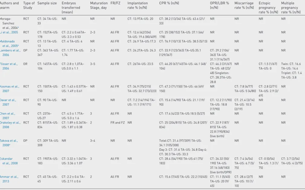
Table 1 – General features and clinical outcome of studies evaluating computed tomography versus transabdominal ultrasound embryo–transfer (focusing on implantation rate, pregnancy rate, ongoing pregnancy rate, multiple pregnancy rate, miscarriage rate and ectopic pregnancy rate).
Authors and yearm
Type of Study
Sample size Embryos transferred (mean) Maturation Stage, day FR/FZ Implantation rate % (n/N) CPR % (n/N) OPR/LBR % (n/N) Miscarriage rate % (n/N) Ectopic pregnancy rate % (n/N) Multiple pregnancy rate % (n/N) Wisanto et al., 1989 RCT CT: 98 TA–US: 98 NR NR NR NR CT: 9.2 (9/98) TA–US: 19.4 (19/98) NR NR NR NR Al–Shawaf et al., 1993 OP CT: 89 TA–US: 152 CT: 2.6 TA–US: 2.5 NR FR: 178 FZ: 63 NR CT: 30.3 (27/89) TA–US:28.9 (44/152) NR NR NR NR
Kan et al., 1999 OP CT:97TA–US: 98 CT: 2.6± 0.7TA– US: 2.7± 0.6 NR All FR CT: 16.2 (41/253) TA–US: 20.4 (53/260) Total: CT: 28.9 (28/97) TA–US: 37.8 (37/98) Single embryo transfer: CT: 16.7 (2/12) TA–US:20 (1/5) Double embryo Transfer: CT: 28.6 (4/14) TA–US: 20.8 (5/24) Triple embryo transfer: CT: 31.0 (22/71) TA–US: 44.9 (31/69) NR NR NR NR Lindheim et al., 1999 OR CT: 43TA–US: 95 CT: 3.7± 0.2TA– US: 4.05± 0.1 3–5 All FR CT: 17.5 (29/166) TA–US: 27.2 (106/390) CT: 34.9(15/43) TA–US: 61.1 (58/95) NR NR CT: 0 (0/15) TA–US 1.7 (1/58) CT: 46.7 (7/15) TA–US: 46.6 (27/58) Coroleu et al., 2000 RCT CT: 180TA–US: 182 CT: 2.9TA–US: 2.8 2–6 All FR CT: 18.1 (92/509) TA–US:25.3 (134/529) Total: CT: 33.9 (61/180) TA– US:50.0 (91/182) Single embryo transfer: CT: 26.7 (4/15) TA–US: 23.1 (3/13) Double embryo transfer: CT: 11.1 (3/27) TA–US: 46.4 (13/28) Triple embryo transfer: CT: 40.2 (45/112) TA–US: 54.8 (57/104) Four or more embryo transfers: CT: 34.6 (9/26) TA–US: 48.6 (18/37) CT: 85.2 (52/61) TA–US: 93.4 (85/91) NR NR NR Tang et al., 2001 RCT CT:400TA–US: 400 Total: CT: 2.3± 0.6 TA–US: 2.2± 0.6 FR cycle: CT: 2.1± 0.4 TA–US: 2.1± 0.4 FZ cycle: CT: 2.4± 0.6 TA–US: 2.5± 0.6 NR FR: 441FZ: 359 Total: CT: 12 (108/ 903) TA–US: 15.3 (136/891) FR cycle: CT: 13.7 (62/451) TA–US. 17.9 (83/464) FZ cycle: CT: 10.2 (46/452) TA–US: 12.4 (53/427) FR vs FZ: Total CT: 22.5 (90/400) TA–US: 26 (104/400) FR cycle: CT: 24.1 (52/216) TA–US: 28.9 (65/225) FZ cycle: CT: 20.7 (38/184) TA–US: 22.3 (39/175) For embryo transfer Single embryo transfer CT: 0 (0/24) TA–US: 13 (3/23) Double embryo transfer: CT: 25.8 (65/252) TA–US: 26.9 (70/260) Triple embryo transfer: CT: 20.2 (25/124) TA–US S: 26.5 (31/117) Total: CT: 19 (76/400) TA– US: 23.5 (94/ 400) FR cycle: CT: 20.8 (45/ 216) TA–US: 26.2 (59/225) FZ cycle: CT: 16.8 (31/184) TA–US: 20 (35/ 175) Total: CT: 11.1 (10/90) TA–US: 7.7 (8/104) FR cycle: CT: 7.7 (4/52) TA–US: 7.7 (5/65) FZ cycle: CT: 15.8 (6/38) TA–US: 7.7 (3/39) Total: CT: 4.4 (4/90) TA–US: 1.9 (2/104) FR cycle: CT: 5.8 (3/52) TA–US: 1.5 (1/65) FZ cycle: CT: 2.6 (1/38) TA–US: 2.6 (1/39) Total: CT: 22.2 (20/90) TA–US: 29.8 (31/104) FR cycle: CT: 25.0% (13/52) TA–US: 29.2% (19/65) FZ cycle: CT: 18.4% (7/38) TA–US: 30.8% (12/39) Prapas et al., 2001 OP CT: 636TA–US: 433 Day 3: CT: 2.94± 1.28 TA–US: 2.78± 1.01 Day 4: CT: 3.23± 1.46 TA–US: 3.47± 1.23 Day 5: CT: 3.55± 1.27 TA–US: 3.46± 1.09
3–5 All FR Day 3: CT: 15.8 TA– US: 23.3 Day 4: CT: 15.7 TA–US: 21.6 Day 5: CT: 23.6 TA– US. 26.7 Total: CT: 36 (229/636) TA–US: 47 (204/433) Day 3: CT 31.7 (100/315) TA–US 46 (120/261) Day 4: CT: 28 (27/98)–US: 43.5 (37/85) Day 5: CT: 45.7 (102/223) TA–US: 56.3 (49/87) NR NR NR NR
(continued on next page)
528
REPRODUCTIVE BIOMEDICINE ONLINE 3 6 ( 2018) 524–542Table 1 – (continued) Authors and yearm
Type of Study
Sample size Embryos transferred (mean) Maturation Stage, day FR/FZ Implantation rate % (n/N) CPR % (n/N) OPR/LBR % (n/N) Miscarriage rate % (n/N) Ectopic pregnancy rate % (n/N) Multiple pregnancy rate % (n/N) Abdelmassih et al., 2001a RCT CT: 40TA–US: 69
NR NR All FR CT: 7TA–US: 25 CT: 30 (12/40) TA–US: 52.2 (36/69) NR NR NR NR
Garcıa-Velasco et al., 2001a RCT CT: 106TA–US: 109 CT: 3.2± 0.04TA– US: 3.2± 0.04
2–6 All FR CT: 23.1TA–US: 28.3 CT: 52.8 (56/106) TA–US: 56.9 (62/109) NR CT: 17.9 (10/56) TA–US: 19.4 (12/62) CT: 3.6 (2/56) TA–US: 0 (0/62) CT: 19.6 (11/56) TA–US: 22.6 (14/62) García-Velasco et al., 2002 RCT CT: 187TA–US: 187 CT: 2.7± 0.02TA– US: 2.8± 0.02 Day 2: CT 63.1 (118/187) TA– US: 60.4 (113/ 187) Day 3: CT 31.6 (59/187)– US 33.2 (62/ 187) Day 6: CT 5.3 (10/187) TA–US 6.4 (12/187)
All FR CT: 26.3TA–US: 30.6 CT: 55.1 (103/187) TA–US: 59.9 (112/187) NR CT: 8.7 (9/103) TA–US: 10.7 (12/112) CT:2.7 (5/187) TA–US: 0 (0/187) CT: 22.5TA–US: 21.4 Matorras et al., 2002 RCT CT: 260TA–US: 255 CT: 3.40± 1.20TA– US: 3.54± 1.07 2–6 All FR CT: 7.5 (66/884) TA– US: 11.1 (100/903) CT: 18.1 (47/260) TA–US: 26.3 (67/255) CT: 14.2 (37/ 260) TA–US: 22.4 (57/255) CT: 21.3 (10/47) TA–US: 14,9 (10/67) CT: 0.4 (1/260) TA–US: 0.8 (2/255) CT: 29.8 (14/47) TA–US: 32.8 (22/67) Sallam et al., 2002 OP CT: 320USS: 320 CT: 2.8± 1.4TA– US: 2.9± 1.4 NR All FR CT: 9.8 (88/896) TA– US: 13.8 (128/928) CT: 18.4 (59/320) TA–US: 26.3 (84/320) NR NR CT: 0.9 (3/320) TA–US: 0.3 (1/320) NR Coroleu et al., 2002 RCT CT: 91TA–US: 93 CT: 2.2± 0.9 TA– US: 2.1± 0.8
NR All FZ CT: 11.7TA–US: 19.1 Total CPR: CT: 19.8 (18/91) TA–US: 34.4 (32/93) Singleton CPR: CT: 13.1 (12/91) TA–US: 28 (26/93) Twin CPR: CT: 6.6 (6/91) TA–US: 6.5 (6/93) NR CT: 22.2 (4/18) TA–US: 21.9 (7/32) CT: 0 (0/91) TA–US: 0 (0/93) NR Marconi et al., 2003a RCT CT: 42TA–US: 41 CT: 3.65± 0.69TA– US: 3.48± 0.67
3 All FR CT: 8.4TA–US: 25.6 CT: 34.7 (15/42) TA–US: 61 (25/41) CT: 25.6 (12/42) TA–US: 53.7 (22/41) NR NR NR Cruickshank et al., 2003a OP CT: 124 TA–US: 257 NR NR FR and FZ NR CT: 27.4 (34/124) TA–US: 37.7 (97/257) CT: 16.1 (20/ 124) TA–US: 27.6 (71/257) NR NR NR
Bar Hava et al., 2003a RCT CT: 66 TA–US: 65 CT: 2.0± 1.2 TA–US: 2.2± 1.0 NR All FR CT: 11.8TA–US: 13.6 CT: 22.7 (15/66) TA–US: 33.8 (22/65) NR NR NR NR Mirkin et al., 2003 OR CT: 456 TA–US: 367 CT: 3.1 TA–US: 3.04 3 All FR CT: 20 (284/1420) TA–US:22 (245/1115) CT: 44.1 (201/456) TA–US:48 (176/367) NR CT: 13.9 (28/ 201) TA–US: 15.9 (28/176) CT: 0.9 (four cases) TA–US: 0.5 (two cases) CT: 28.9 (58/ 201) TA–US: 27.8 (49/176) Weissman et al., 2003a RCT CT: 67 TA–US: 88 CT: 2.7± 0.8 TA– US: 2.7± 0.9 2–3 All FR CT: 17.8 (42/236) TA–US: 24.9 (44/177) CT: 40.3 (27/67) TA–US: 38.6 (34/88) NR CT: 11.1 (3/27) TA–US: 20.6 (7/34) NR NR de Camargo Martins et al., 2004 RCT CT: 50 TA–US: 50 CT: 2.3± 0.6 TA– US: 2.6± 0.9
NR All FR CT: 16.3 TA–US:19.6 CT: 30 (15/50) TA–US: 42 (21/50) NR CT: 13.3 (2/15) TA–US: 4.8 (1/21)
NR NR
(continued on next page)
529
REPRODUCTIVE BIOMEDICINE ONLINE 3 6 (2018) 5 24–542Table 1 – (continued) Authors and yearm
Type of Study
Sample size Embryos transferred (mean) Maturation Stage, day FR/FZ Implantation rate % (n/N) CPR % (n/N) OPR/LBR % (n/N) Miscarriage rate % (n/N) Ectopic pregnancy rate % (n/N) Multiple pregnancy rate % (n/N) Moraga-Sanchez et al., 2004a RCT CT: 34 TA–US: 33 NR NR NR CT: 13.9TA–US: 20 CT: 38.2 (13/34) TA–US: 63.6 (21/ 33) NR NR NR NR Li et al., 2005 RCT CT: 152TA–US: 178 CT: 2.2± 0.46TA– US: 2.3± 0.53 2–3 All FR CT: 12.6 (42/334) TA–US: 19.6 (80/409) CT: 25 (38/152) TA–US: 37.1 (66/ 178) NR NR NR NR Maldonado et al., 2005a RCT CT: 13 TA–US: 13
CT: 4 TA–US: 4 NR All FR CT: 26.9 TA–US:17.3 CT: 76.9 (10/13) TA–US: 38.5 (5/13) NR NR NR NR
Lambers et al., 2006 OP CT: 363 TA–US: 367 CT: 1.77 TA–US: 1.74
2–3 All FR CT: 24.2TA–US: 24.3 CT: 33.9 (123/363) TA–US:35.1 (129/367) CT: 29.2 (106/ 363) TA–US: 31.1 (114/367) NR NR NR Flisser et al., 2006 OR CT: 145TA–US: 104 CT: 2.8± 1,0TA– US:3.0± 1.1
3–5 All FR CT: 26TA–US: 23.5 CT: 46.20 (67/145TA–US: 46.1 (48/ 104) CT: 46.3 (31/67) TA–US: 48 (23/ 48) Singleton: CT: 28.3TA–US: 28.8 NR CT: 1.5 (1/67) TA–US: 0 Twin: CT: 16.6 TA–US: 14.4 Triplet: CT: 1.4 TA–US: 3.8 Kosmas et al., 2007 RCT CT: 150TA–US: 150 CT: 1.43± 0.57TA– US: 1.49± 0.61 NR All FR CT: 34.9 (75/215) TA–US: 32.7 (73/223) CT: 47.3 (71/150) TA–US: 46 (69/ 150) NR CT: 7.8 (6/77) TA–US: 5 (4/80) CT: 2.8 (2/71) TA–US: 2.9 (2/ 69) NR Davar et al., 2007 RCT CT: 90 TA–US: 90 NR NR NR CT: 7.2 (14/194) TA– US: 11.1 (19/171) CT: 15.6 (14/90) TA–US: 21.1 (19/ 90) CT: 12.2 (11/90) TA–US: 18.8 (17/90) CT: 21.4 (3/14) TA–US: 10.5 (2/19) NR NR Chen et al., 2007 RCT CT: 23TA– US:27 CT: 4.5± 1.7TA– US: 5.0± 1.6 3 All FR NR CT: 17.4 (4/23) TA–US:18.5 (5/27) NR NR NR NR Drakeley et al., 2008 RCT CT: 815TA–US: 834 CT: 1.89± 0.36TA– US: 1.87± 0.38 2 FR and FZ NR CT: 25 (204/815) TA–US: 24.8 (207/ 834) CT: 22.9 (187/ 815) TA–US: 22.8 (190/834) (live birth) NR NR NR Zakova et al., 2008a OP CT: 309 TA–US: 308 NR 3–4 NR NR Total:CT: 31.4 (97/309) TA–US: 34.1 (105/308)
Day 3: CT: 31.6 TA–US: 34.8 Day 4: CT: 30.3 TA–US: 33.3 NR NR NR NR Eskandar et al., 2008 RCT CT: 190TA–US: 183 CT: 3.32± 1.04TA– US: 3.36± 1.07 3 All FR NR CT: 28.4 (54/190) TA–US:41 (75/ 183) CT: 26.32 (50/ 190) TA–US: 37.16 (68/183) (live birth/OPR) CT: 7.4 (4/54) TA–US: 6.7 (5/ 75) CT: 0 (0/54) TA–US: 1.3 (1/ 75) CT: 3.7 (2/54) TA–US: 4 (3/75) Ammar et al., 2013 RCT CT: 45 TA–US: 45 CT: 2.2± 0.6 TA– US: 2.11± 0.6 2 All FR NR CT: 15.6 (7/45) TA–US: 22.2 (10/45) CT: 11.1 (5/45) TA–US: 20 (9/ 45) CT: 28.6 (2/7) TA–US: 10 (1/ 10) NR NR
aPapers published only in abstract conference form.
CT, clinical touch; CPR, clinical pregnancy rate; FR, fresh embryo transfer; FZ, frozen embryo transfer; LPR: live birth rate; NR: not reported; OP: observational prospective study; OR: observational retrospective study; OPR: ongoing pregnancy rate; RCT: randomized contolled trial; TA-US: transabdominal ultrasound guided; TV: transvaginal ultrasound guided.
530
REPRODUCTIVE BIOMEDICINE ONLINE 3 6 ( 2018) 524–542Table 2 – General features and clinical outcome of studies evaluating transabdominal ultrasound (TA–US) versus transvaginal ultrasound (TV–US) or clinical touch versus transvaginal or two-dimensional versus three-dimensional or uterine length measurement before transfer TV–US (ULMbET) versus TA–US embryo transfer (focusing on implantation rate, pregnancy rate, ongoing pregnancy rate, multiple pregnancy rate, miscarriage rate, ectopic pregnancy rate).
Authors and year
Type of study Sample size Embryos transferred (mean) Maturation stage, day FR/FZ Implantation rate % (n/N) CPR % (n/N) OPR/LPR % (n/N) Miscarriage rate % (n/N) Ectopic pregnancy rate % (n/N) Multiple pregnancy rate % (n/N) Porat et al., 2010 RCT TV–US: 93 TA– US: 93 Day 3: TV–US: 3.1± 1.1 TA–US: 3.5± 1.1 Day 6: TV–US: 2.2± 0.6 TA–US: 2.2± 0.7 3–6 FR and FZ TV–US: 31.1± 3.9 TA–US: 33.1± 4.1 TV–US: 45.2 (42/93) TA–US: 48.4 (45/93) TV–US: 38.7 (36/93) TA–US: 30.1 (28/93) NR TV–US: 0 TA– US: 0 NR Bodri et al., 2011 RCT TV–US: 165 TA–US: 164 NR Day 2: TV–US: 47.3 (78/165) TA–US: 47.6 (78/164) Day 3: TV–US: 52.7 (87/165) TA– US: 53 (87/164) All FR TV–US: 34.5 (114/330) TA– US: 31.4 (103/ 328) TV–US: 50.9 (84/165) TA–US: 49.4 (81/164) TV–US: 43 (71/165)TA– US: 42.7 (70/164) TV–US: 15.5 (13/84)TA–US: 13.6 (11/81) TV–US: 0 (0/ 165)TA–US: 0.6 (1/164) TV–US: 33.3 (28/84) TA–US: 25.9 (21/81) Revelli et al., 2016 RCT TV–US (ULMbET): 828 TA–US: 820 TV–US: 1.9 (1.5–2.2) TA– US: 1.9 (1.5–2.2) 2–3 All FR TV–US: 24.8 (390/1573) TA– US: 25.2 (393/ 1558) TV–US: 38.2 (316/828) TA–US: 38.9 (319/820) TV–US: 33.1 (274/828) TA–US: 34.8 (285/820) TV–US:13.3 (42/316) TA– US: 10.7 (34/ 319) TV–US: 0 (0/ 828) TA–US: 0.4 (3/820) TV–US: 23.4 (74/316) TA– US: 23.2 (74/ 319) Karavani et al., 2017 RCT TV–US:60 TA– US:60 TV–US: 1.7± 0.67 TA–US: 1.66± 0.71 2–3 All FR TV–US: 33(31/ 94)TA–US: 23.4 (22/94) TV–US:36.7 (22/60) TA–US: 30 (18/60) TV–US: 31.7 (19/60)TA– US: 25.0 (15/60) TV–US: 5.0 (3/ 600) TA–US: 5.0 (3/60) NR NR Larue et al., 2017 OR TV–US: 800 TA–US: 3910 NR NR NR NR TV–US: 38% TA–US: 30% NR NR NR NR Kojima et al., 2001 OR CT: 444 TV–US: 402 CT: 2.33± 0.7 TV–US: 2.4 ± 0.7 2–5 All FR CT: 7 (71/1015) TV–US: 15.2 (145/953) CT: 13.1 (58/444) TV– US: 28.9 (116/402) CT: 10.6 (47/444) TV–US: 22.4 (90/402) NR CT: 0.7 (3/444) TV–US: 2.7 (11/ 402) NR Saravelos et al., 2016 RCT 2D TA–US: 237 3D TA–US: 237 Single embryo: 2D: 161 (67.9%) 3D: 158 (66.7%) Double embryo: 2D: 76 (32.1%) 3D: 79 (33.3%) Day 3: 2D: 86 (36.3%) 3D: 93 (39.2%) Day 5: 2D: 151 (63.7%) 3D: 144 (60.8%) FR and FZ 2D: 38.0%± 3.0 3D: 37.1%± 3.0 2D: 44.3 (105/237) 3D: 43.5 (103/237) 2D: 37.1 (88/237) 3D: 35.4 (84/237) 2D: 13.7 (14/ 102) D: 16 (16/ 100) 2D: 2.9 (3/105) 3D:2.9 (3/103) Twins 2D: 6.9 (7/102) 3D: 5 (5/100) Triplets 2D:1 (1/102) 3D: 1 (1/100) CT, clinical touch; CPR: clinical pregnancy rate; FR, fresh embryo transfer; FZ, frozen embryo transfer; LPR, live birth rate NR, not reported; OR, observational retrospective study; OPR, ongoing pregnancy rate; RCT, randomized controlled trial; TA–US: transabdominal ultrasound guided; TV–US: transvaginal ultrasound guided; ULMbET: uterine length measurement before transfer by TV–US; 2D; two-dimensional; 3D, three-dimensional.
531
REPRODUCTIVE BIOMEDICINE ONLINE 3 6 (2018) 5 24–542Coroleu et al., 2000, 2002; de Camargo Martins et al., 2004; Davar et al., 2007; Drakeley et al., 2008; Eskandar et al., 2008; García-Velasco et al., 2002; Kosmas et al., 2007; Li et al., 2005; Matorras et al., 2002; Tang et al., 2001; Wisanto et al., 1989) were included in the meta-analysis.
Characteristics of included studies
The 14 articles included in the meta-analysis involved a total of 5503 participants, with sample sizes ranging from 50 (Chen et al., 2007) to 1649 (Drakeley et al., 2008). A total of 2731 patients underwent clini-cal touch embryo transfer and 2772 women underwent TA-US embryo transfer.
All 14 studies reported clinical pregnancy rates (Ammar et al., 2013; Chen et al., 2007; Coroleu et al., 2000, 2002; de Camargo Martins et al., 2004; Davar et al., 2007; Drakeley et al., 2008; Eskandar et al., 2008; García-Velasco et al., 2002; Kosmas et al., 2007; Li et al., 2005; Matorras et al., 2002; Tang et al., 2001; Wisanto et al., 1989), seven studies evaluated ongoing/live birth rates (Ammar et al., 2013; Coroleu et al., 2000; Davar et al., 2007; Drakeley et al., 2008; Eskandar et al., 2008; Matorras et al., 2002; Tang et al., 2001), nine manuscripts de-scribed miscarriage rates (Ammar et al., 2013; Coroleu et al., 2002; de Camargo Martins et al., 2004; Davar et al., 2007; Eskandar et al., 2008; García-Velasco et al., 2002; Kosmas et al., 2007; Matorras et al., 2002; Tang et al., 2001), and five studies evaluated ectopic preg-nancy rates (Eskandar et al., 2008; García-Velasco et al., 2002; Kosmas et al., 2007; Matorras et al., 2002; Tang et al., 2001).
Synthesis of results
Clinical pregnancy rate. Analysis involved 5503 patients (n= 2772 in the TA-US embryo transfer group andn= 2731 in the clinical touch embryo transfer group) with 1632 events (n= 897 in the TA-US embryo transfer group andn= 735 in the clinical touch embryo transfer group). The overall pregnancy rate was 29.66% (32.36% in the TA-US embryo transfer patients versus 26.91% in the CT embryo transfer pa-tients). In random-effects modelling, the clinical pregnancy rate was significantly higher in the TA-US embryo transfer group, with an OR of 1.41 (95% CI 1.19 to 1.67P< 0.0001); a moderate degree of het-erogeneity was found among the studies (I2= 37%). Excluding the study byDrakeley et al. (2008)in a sensitivity analysis, heterogeneity was reduced to a minimum (I2= 0%), and the clinical pregnancy rate showed a further increase among TA-US embryo transfer patients (35.60% versus 27.71%) using fixed-effects model, with an odds ratio of 1.48 (95% CI 1.29 to 1.71;P< 0.00001) (forest plots are shown inFigures 2a and 2b). The systematic exclusion of other studies from the analy-sis did not show significant modifications of results.
Ongoing and live birth rate. A total of 3969 patients were included (n= 1989 in the TA-US embryo transfer group andn= 1980 in the clinical touch embryo transfer group) in the data analysis, with 938 events (n= 520 in the TA-US embryo transfer group andn= 418 in the clinical touch embryo transfer group). The overall ongoing and live birth rate was 23.63% (26.14% in the TA-US embryo transfer Figure 1 – Preferred Reporting Items for Systematic Reviews and Meta-Analyses flow diagram of study selection.
patients versus 21.11% in the clinical touch embryo transfer pa-tients). Using a random-effects model, the estimated outcome was significantly greater in the TA-Us embryo transfer patients (OR 1.49; 95% CI 1.15 to 1.93;P= 0.003) even if a high degree of heterogene-ity among studies was found (I2= 57%). The sensitivity analysis showed that, as a result of excluding the study byDrakeley et al. (2008), het-erogeneity was reduced to zero (I2= 0%) and the advantages of TA-US embryo transfer became significantly higher (28.57% versus 19.82% in terms of the ongoing and live birth rate), with an odds ratio of 1.64 (95% CI 1.35 to 1.99;P< 0.00001) using a fixed-effects model (Figures 3a and 3b). The systematic exclusion of other studies from the meta-analysis was not correlated with modifications of the results.
Ectopic pregnancy rate. A total of 792 pregnancies were analysed (n= 427 in the TA-US embryo transfer group andn= 365 in the
clini-cal touch embryo transfer group), with only 19 events (n= 7 in the TA-US embryo transfer patients andn= 12 in the clinical touch embryo transfer patients). The overall ectopic pregnancy rate was 2.40% (1.64% in the TA-US embryo transfer patients versus 3.29% in the clinical touch embryo transfer patients). A data comparison did not show sig-nificant differences between the two techniques for the present outcome (OR 0.52; 95% CI 0.21 to 1.28; I2= 0%). The serial exclusion of each study in the sensitivity analysis did not provide significant modi-fications in the estimated outcome. Forest plots are shown inFigure 4. Miscarriage rate. A total of 945 pregnancies (n= 520 in the TA-US embryo transfer group andn= 425 in the clinical touch embryo trans-fer group) with a total of 100 events (n= 50 in the TA-US embryo transfer patients andn= 50 in the clinical touch embryo transfer pa-tients) were analysed. The overall miscarriage rate was 10.58% (9.61% Figure 2 – (a) Transabdominal ultrasound (TA-US) versus clinical touch embryo transfer. Meta-analysis of clinical pregnancy rate, random effects model; (b) TA-US versus clinical touch embryo transfer. Meta-analysis of clinical pregnancy rate, sensitivity analysis, fixed effects model.
533
in the TA-US embryo transfer patients versus 11.76% in the clinical touch embryo transfer patients). A data comparison did not show sig-nificant differences between the two techniques for the present outcome (OR 0.76; 95% CI 0.50 to 1.15; I2= 0%). The serial exclusion of each study in the sensitivity analysis did not provide significant modi-fications in the estimated outcome. Forest plots are shown inFigure 5. Meta-analysis of randomized controlled trials comparing TV-US versus TA-TV-US-guided embryo transfer
Study selection
A total of four studies compared TV-US versus TA-US-guided embryo transfer (Bodri et al., 2011; Karavani et al., 2017; Larue et al., 2017;
Porat et al., 2010). One study was excluded (Larue et al., 2017) owing to its study design (observational), and three randomized controlled trials were included in the quantitative analysis (Bodri et al., 2011; Karavani et al., 2017; Porat et al., 2010).
Characteristics of included studies
The studies included a total of 635 patients, with a range between 120 (Karavani et al., 2017) and 329 (Bodri et al., 2011). Overall, 318 pa-tients underwent TV-US-guided embryo transfer and 317 underwent TA-US-guided embryo transfer.
Data on clinical pregnancy rates and ongoing pregnancy rates and live birth rates were reported in all three studies (Bodri et al., 2011; Karavani et al., 2017; Porat et al., 2010), whereas two studies reported Figure 3 – (a) Transabdominal ultrasound (TA-US) versus clinical touch embryo transfer. Meta-analysis of ongoing and live birth rate, random effects model; (3b) TA-US versus clinical touch embryo transfer. Meta-analysis of ongoing and live birth rate, sensitivity analysis, fixed effects model.
Figure 4 – Transabdominal ultrasound versus clinical touch embryo transfer. Meta-analysis of ectopic pregnancy rate, fixed effects model.
534
R E P R O D U C T I V E B I O M E D I C I N E O N L I N E 3 6 ( 2 0 1 8 ) 5 2 4 – 5 4 2miscarriage rates (Bodri et al., 2011; Karavani et al., 2017), and two provided data about ectopic pregnancy rates (Bodri et al., 2011; Porat et al., 2010).
Synthesis of results
Clinical pregnancy rate. Analysis involved a total of 635 patients (n = 318 in the TV-US-guided embryo transfer group andn= 317 in the TA-US-guided embryo transfer group) with 292 events (n= 148 in the TV-US embryo transfer group andn= 144 in the TA-US embryo trans-fer group). The overall pregnancy rate was 45.98% (46.54% in the TV-Us embryo transfer patients versus 45.43% in the TA-US embryo transfer patients). A data comparison did not show significant dif-ferences between the two techniques for the present outcome (OR 1.05; 95% CI 0.76 to 1.43; I2= 0%). Forest plots are shown inFigure 6.
Ongoing pregnancy and live birth rate. The analysis included the same patients evaluated for the preceding outcome. The number of
events was 239 (n= 126 in the TV-US embryo transfer group andn= 113 in the TA-US embryo transfer group). The overall ongoing preg-nancy and live birth rate was 37.64% (39.62% in the TV-US embryo transfer patients versus 35.65% in the TA-US embryo transfer pa-tients). A comparison of the two techniques did not show statistical differences (OR 1.19; 95% CI 0.86 to 1.64; I2= 0%). Forest plots are shown inFigure 7.
Ectopic pregnancy rate. The present end-point was evaluated ex-clusively by two studies (Bodri et al., 2011; Porat et al., 2010) and a meta-analysis was not conducted.Porat et al. (2010)did not report any event.Bodri et al. (2011)reported no significant differences among the groups.
Miscarriage rate. Only two studies (Bodri et al., 2011; Karavani et al., 2017) reported data on miscarriages; accordingly, we did not proceed with a quantitative analysis.Bodri et al. (2011)reported no signifi-cant differences (a miscarriage rate of 15.48% versus 13.58%, Figure 5 – Transabdominal ultrasound versus clinical touch embryo transfer. Meta-analysis of miscarriage rate, fixed effects model.
Figure 6 – Transvaginal ultrasound versus transabdominal ultrasound embryo transfer. Meta-analysis of clinical pregnancy rate, fixed effects model.
Figure 7 – Transvaginal ultrasound versus transabdominal ultrasound embryo transfer. Meta-analysis of ongoing and live birth rate, fixed effects model.
535
respectively, in the TV-US-guided embryo transfer group and the TA-US-guided embryo transfer group), as didKaravani et al. (2017)(a miscarriage rate of 5% in both groups).
Overall quality of evidence
Comparison between TA-US-guided embryo transfer versus clinical touch embryo transfer
We found a moderate amount of evidence supporting the improve-ment of the clinical pregnancy rate and the ongoing pregnancy and live birth rate using TA-US-guided embryo transfer. Conversely, con-cerning other outcomes (the ectopic pregnancy rate and the miscarriage rate), the resulting evidence was very low/low (Table 3).
Comparison of TA-US-guided embryo transfer and TV-US embryo transfer
We judged the quality of evidence on the comparable results of TA-US-guided embryo transfer and TV-TA-US-guided embryo transfer in terms of the clinical pregnancy rate and the ongoing pregnancy/live birth rate to be low (Table 4).
Systematic literature review
TA-US-guided embryo transfer versus clinical touch embryo transfer
General and descriptive data on all 31 manuscripts that compared TA-US-guided embryo transfer versus clinical touch embryo trans-fer are detailed inTable 1. The study design of 21 articles were randomized controlled trial (Abdelmassih et al., 2001; Ammar et al., 2013; Bar Hava et al., 2003; Chen et al., 2007; Coroleu et al., 2000, 2002; de Camargo Martins et al., 2004; Davar et al., 2007; Drakeley et al., 2008; Eskandar et al., 2008; Garcıa-Velasco et al., 2001; García-Velasco et al., 2002; Kosmas et al., 2007; Li et al., 2005; Maldonado et al., 2005; Marconi et al., 2003; Matorras et al., 2002; Moraga-Sanchez et al., 2004; Tang et al., 2001; Weissman et al., 2003; Wisanto et al., 1989), but seven were published only in abstract form (Abdelmassih et al., 2001; Bar Hava et al., 2003; Garcıa-Velasco et al., 2001; Maldonado et al., 2005; Marconi et al., 2003; Moraga-Sanchez et al., 2004; Weissman et al., 2003). The design of seven papers was observational prospective (al-Shawaf et al., 1993; Kan et al., 1999; Prapas et al., 2001; Sallam et al., 2002; Cruickshank et al., 2003;
Table 3 – Findings and grading of the evidence for the main outcomes (transabdominal ultrasound versus clinical touch embryo transfer) in embryo transfers.
Outcomes Anticipated absolute effectsa(95% CI) Relative effect
(95% CI)
Number of participants (studies)
Quality of the evidence (GRADE)b
Risk with clinical touch
Risk with transabdominal ultrasound guidance
Clinical pregnancy rate 277 per 1.000 362 per 1.000 (331 to 396) OR 1.48 (1.29 to 1.71) 3854 (13 RCTs) ⨁⨁⨁·MODERATEc
Ongoing/live birth rate 198 per 1.000 289 per 1.000 (246 to 335) OR 1.64 (1.32 to 2.04) 2320 (Seven RCTs) ⨁⨁⨁·MODERATEc
Ectopic pregnancy rate 33 per 1.000 17 per 1.000 (7 to 42) OR 0.52 (0.21 to 1.28) 792 (Five RCTs) ⨁···VERY LOWc,d
Miscarriage rate 118 per 1.000 92 per 1.000 (63 to 133) OR 0.76 (0.50 to 1.15) 945 (Nine RCTs) ⨁⨁··LOWc,d
aThe risk in the intervention group (and its 95% confidence interval) is based on the assumed risk in the comparison group and the relative effect of the
intervention (and its 95% CI).
bhttps://gradepro.org.
cMost RCTs showed low risk of bias only concerning random sequence generation; for most of the other items, the risk of bias was considered unclear or
high.
dHigh imprecision owing to the very low number of events; RCTs not powered enough to detect this outcome.
RCT, randomized controlled trial.
Table 4 – Findings and grading of the evidence for the main outcomes (transabdominal versus transvaginal embryo transfer) in infertile women undergoing embryo transfer.
Outcomes Anticipated absolute effectsa(95% CI) Relative effect
(95% CI) Number of participants (studies) Quality of the evidence (GRADE)b,c
Risk with transabdominal ultrasound guidance
Risk with transvaginal ultrasound guidance
Clincal pregnancy rate 454 per 1.000 466 per 1.000 (387 to 543) OR 1.05 (0.76 to 1.43) 635 (Three RCTs) ⨁⨁··LOWd,e
Ongoing/live birth rate 356 per 1.000 397 per 1.000 (323 to 491) OR 1.19 (0.86 to 1.74) 635 (Three RCTs) ⨁⨁··LOWd,e
aThe risk in the intervention group (and its 95% confidence interval) is based on the assumed risk in the comparison group and the relative effect of the
intervention (and its 95% CI).
bhttps://gradepro.org.
cGRADE Working Group grades of evidence: high quality: we are very confident that the true effect lies close to that of the estimate of the effect; moderate
quality: we are moderately confident in the effect estimate. The true effect is likely to be close to the estimate of the effect, but there is a possibility that it is substantially different; low quality: our confidence in the effect estimate is limited. The true effect may be substantially different from the estimate of the effect; very low quality: we have very little confidence in the effect estimate. The true effect is likely to be substantially different from the estimate of effect.
d
All three RCTs showed low risk of bias only concerning random sequence generation; for most other items, the risk of bias was considered unclear.
e
Low number of events; RCTs not sufficiently powered to detect differences between the two techniques. RCT, randomized controlled trial.
Lambers et al., 2006; Zakova et al., 2008), two of which were pub-lished only in an abstract form (Cruickshank et al., 2003; Zakova et al., 2008). Finally, three papers were retrospective series (Flisser et al., 2006; Lindheim et al., 1999; Mirkin et al., 2003).
The number of included patients ranged from 26 (Maldonado et al., 2005) to 1649 individuals (Drakeley et al., 2008) per study. Consid-ering the observational prospective studies (al-Shawaf et al., 1993; Kan et al., 1999; Prapas et al., 2001; Sallam et al., 2002; Cruickshank et al., 2003; Lambers et al., 2006; Zakova et al., 2008), the first results were described byal-Shawaf et al. (1993)and did not find a differ-ence in clinical pregnancy rates between TA-US and clinical touch both in fresh and frozen embryo transfer (al-Shawaf et al., 1993). Simi-larly,Kan et al. (1999)found no difference in clinical pregnancy rates, overall or when stratifying the data for the number of embryos trans-ferred and patient age. Subsequently,Prapas et al. (2001)in a large sample size study (636 clinical touch versus 433 TA-US) demon-strated that TA-US yielded a higher overall pregnancy rate versus clinical touch (47% versus 36%,P< 0.001). This difference, however, was significant for day 3/4 embryos but not for day 5; the study found similar results for implantation rates (Prapas et al., 2001). Data from the remaining observational studies conflict, with one demonstrat-ing the possible benefits of TA-US in ongodemonstrat-ing pregnancy rates (Cruickshank et al., 2003); the other two found no benefit from this procedure (Lambers et al., 2006; Zakova et al., 2008).
In a retrospective study,Lindheim et al. (1999)showed that TA-US guidance significantly improved implantation and pregnancy rates (27.2% versus 17.5% and 61.1% versus 34.9%, respectively,P< 0.05) compared with clinical touch. Conversely,Mirkin et al. (2003), in a large sample size study, found no significant differences in implantation, clinical pregnancy and ectopic pregnancy rates. These data were con-firmed byFlisser et al. (2006)in a retrospective study, which found no differences between the TA-US and clinical touch groups in im-plantation and pregnancy rate, also stratifying data for day of embryo culture.
Coroleu et al. (2000)published the first randomized controlled trial aiming specifically to identify the possible superiority of TA-US versus clinical touch (apart from the preliminary data byWisanto et al., 1989) The study demonstrated a significantly higher pregnancy rate (50% versus 33.9%) and implantation rate (25.3% versus 18.1%) among the TA-US group compared with the clinical touch group (Coroleu et al., 2000). Interestingly, the study also confirmed the same benefit in the case of frozen embryos (Coroleu et al., 2002). A slight benefit con-cerning the implantation rate was also reported byTang et al. (2001) (15.3% in the TA-US group versus 12% in the clinical touch group), but the study did not report significant differences in the clinical preg-nancy rate, ongoing pregpreg-nancy rate, multiple pregpreg-nancy rate, ectopic pregnancy rate and miscarriage rate. Similar results were found by Garcia-Velasco et al. (2002);Kosmas et al. (2007); andde Camargo Martins et al. (2004). In addition, the largest randomized controlled trial published on this topic (815 clinical touch versus 834 TA-US embryo transfer) failed to find differences in clinical pregnancy or live birth rates between the TA-US and clinical touch groups (Drakeley et al., 2008).
Conversely,Matorras et al. (2002)(260 clinical touch versus 255 US patients) and Sallam et al. (320 clinical touch versus 320 TA-US patients) in randomized trials both confirmed a significant increase in the implantation rate and clinical pregnancy rate in the TA-US group. Matorras et al. (2002)confirmed an increase in the ongoing preg-nancy rate in the TA-US group. The study found no benefits in miscarriage and ectopic pregnancy reduction (Matorras et al., 2002).
Li et al. (2005)found that the rate of implantation and clinical preg-nancy in the TA-US group (19.6% and 37.1%, respectively) was significantly higher than in the clinical touch group (12.6% and 25%, respectively;P< 0.05). These results were subsequently confirmed in a study byEskandar et al. (2008)that demonstrated a signifi-cantly higher live birth and ongoing pregnancy rate, and clinical pregnancy rate in TA-US (37.16% and 40.98%, respectively) versus clinical touch (26.32 and 28.42%, respectively). The study did not find differences in ctopic pregnancies and miscarriages (Eskandar et al., 2008).
According toSallam (2015), the results provided byDrakeley et al. (2008)(no difference found between TA-US-guided embryo transfer and clinical touch embryo transfer in clinical and live birth rates) are potentially affected by the different embryo transfer catheters used in the two study arms, potentially violating the rules of randomiza-tion (Cook Soft-Pass EchoTip catheters in the TA-US-guided embryo transfer group and Wallace or Rocket catheters in the clinical touch embryo transfer group). For this reason, we opted to exclude it from the grading of evidence. The strength of this study, however, is the number of patients and events, and we, therefore, included it in the meta-analysis, emphasising its high heterogeneity compared with other studies.
TV-US versus TA-US-guided embryo transfer
Three randomized controlled trials compared TV-US versus TA-US (Bodri et al., 2011; Karavani et al., 2017; Porat et al., 2010).Porat et al. (2010), comparing 93 TV-US and 93 TA-US, found no differences in implantation rates, clinical pregnancy rates and live birth rates between the two approaches. These results were subsequently confirmed by Bodri et al. (2011)in 165 TV-US versus 164 TA-US and byKaravani et al. (2017). In addition,Porat et al. (2010)andBodri et al. (2011)found no significance differences in ectopic pregnancy rates.
Only one retrospective trial was available on this topic.Larue et al. (2017), in a large retrospective study, found a significant increase in pregnancy rates from a comparison of TV-US (pregnancy rate 38%) with TA-US-guided embryo transfer (pregnancy rate 30%) (P< 0.001) (Table 2).
Evaluation of patient discomfort comparing TV-US with TA-US-guided embryo transfer
Porat et al. (2010)found no differences between groups concerning the degree of uterine cramping (1.2± 0.5 versus 1.2 ± 0.4) and pain (1.4± 0.7 versus 1.3 ± 0.5).Bodri et al. (2011)found that the uterine cramping rate was comparable between the groups (27.2% versus 18.3%), and reported 41% of light, 16% of moderate and 6% of severe discomfort related to bladder distension in the TA-US group.
These results have recently been confirmed and expanded by Karavani et al. (2017), in a randomized controlled trial specifically de-signed to evaluate patient discomfort. The study reported that TV-US was significantly associated with a better visualization of the uterus and embryo transfer location (9.57 versus 8.42 and 9.58 versus 8.82, respectively) and a significant reduction in pain, discomfort and anxiety (measured through a questionnaire based on a visual analogue scale) especially during procedure preparation and performance.
TV-US versus clinical touch embryo transfer
Only one observational retrospective study compared TV-US versus clinical touch embryo transfer. The study compared 444 patients un-dergoing clinical touch with 402 patients unun-dergoing TV-US embryo transfer, and found significantly higher pregnancy and implantation
537
rates in the TV-US group (28.9% and 15.2%, respectively) compared with the clinical touch group (13.1% and 7.0%, respectively). The study did not show differences in ectopic pregnancy rates (Kojima et al., 2001).
Two-dimensional versus three-dimensional TA-US-guided embryo transfer
Only one study compared 3D-US-guided embryo transfer with 2D-US-guided embryo transfer. This randomized controlled trial found no significant differences in the ongoing pregnancy rate (35.4% versus 37.1% in three-dimensional versus two-dimensional, respectively), im-plantation rate, clinical pregnancy rate, miscarriage rate and ectopic pregnancy rate (Saravelos et al., 2016).
TV ULMbET versus TA-US-guided embryo transfer
Only one large randomized controlled trial evaluated this new and promising technique.Revelli et al. (2016)in 1648 patients, com-pared classical TA-US embryo transfer (820 women) with ULMb ET (828 women). With ULMbET, transvaginal scanning is used to measure the length of the cervix and the distance between the internal uterine orifice and the fundal endometrial surface. Then, using clinical touch, the investigators discharge the embryos at a point obtained by sub-tracting 1.5 cm from the total length of the cavity. The study found comparable clinical pregnancy rates (38.2% versus 38.9%), implan-tation rates (24.8% versus 25.2%), and ongoing pregnancy rates (33.1% versus 34.8%) between groups. The study also found a significant re-duction in the discomfort intensity score (2.6 versus 1.5 visual analogue scale points;P= 0.045) and in the proportion of patients with moderate-to-severe discomfort during embryo transfer through the application of ULMbET (19.8% versus 1.2%,P= 0.003).
Discussion
Over the past decade, despite great improvement in IVF settings and treatments, embryo implantation and related mechanisms are not well understood (Abou-Setta et al., 2007). Several factors can affect embryo implantation, but the actual trend is to focus specifically on the embryo (Huang et al., 2017), leaving aside the fundamental role of endome-trial receptivity (Edwards, 1995; Hoozemans et al., 2004; Irani et al., 2017) and the possible improvement derived by different embryo trans-fer techniques (Tang et al., 2001). Indeed, the transfer of a good-quality euploid embryo does not ensure a successful implantation or an ongoing pregnancy (Teh et al., 2016).
As reported previously, despite some attempts to improve embryo transfer methodology (Matorras et al., 2002), the results are often con-flicting and poorly reproducible. In addition, ultrasound-guided embryo transfer remains a topic of debate. Although it seems like an easy and feasible technique, most published studies have produced con-trasting results that cause more confusion than certainty.
The reasons for such uncertainties are likely attributable to the low or moderate quality of both clinical trials and review articles. Trials are limited by low sample sizes, probably too low to detect signifi-cant improvement derived by ultrasound. This limitation represents a fundamental issue, especially when attention is focused on lower frequency events such as live birth rates and even more miscar-riage rates and ectopic pregnancy rates.
Another limitation is the design of the published studies. In ad-dition to 17 published randomized controlled trials (Ammar et al., 2013;
Bodri et al., 2011; Chen et al., 2007; Coroleu et al., 2000, 2002; de Camargo Martins et al., 2004; Davar et al., 2007; Drakeley et al., 2008; Eskandar et al., 2008; García-Velasco et al., 2002; Karavani et al., 2017; Kosmas et al., 2007; Li et al., 2005; Matorras et al., 2002; Porat et al., 2010; Tang et al., 2001; Wisanto et al., 1989), several observational and retrospective studies (also with good sample sizes) reported con-trasting findings. (al-Shawaf et al., 1993; Kan et al., 1999; Lindheim et al., 1999; Prapas et al., 2001; Sallam et al., 2002; Mirkin et al., 2003; Lambers et al., 2006; Flisser et al., 2006). The main limitation of the published meta-analyses is the small number of included studies (Abou-Setta et al., 2007; Buckett, 2003; Sallam et al., 2002), the design of some included studies, i.e., observational (Buckett, 2003) and the inclusion of non-full text studies, e.g. meeting abstracts (Brown et al., 2016; Teixeira et al., 2015).
In the analysis of studies (14 randomized controlled trials) com-paring TA-US versus clinical touch-guided embryo transfer, we found that the application of TA-US guidance significantly increases the chances of achieving clinical pregnancy, with an odds ratio of 1.41. The results, however, were affected by moderate heterogeneity (I2= 37%) that was eliminated (I2= 0%) by the exclusion of a single study (Drakeley et al., 2008) through sensitivity analysis. It did not produce substantial modification in pooled odds ratio (OR = 1.48) and did not affect the precision of comparison (sample size of 3854 patients ex-cludingDrakeley et al., 2008). Similarly, the ongoing pregnancy rate and live birth rate was consistently higher in TA-US-guided embryo transfers patients (OR 1.49; I = 57%). In this case, heterogeneity was lowered to 0% by the exclusion of the same study (Drakeley et al., 2008) without affecting the validity of the comparison (OR 1.64, 2320 patients). Concerning the ectopic pregnancy rate, the number of events (19 ectopic pregnancies) reported in all randomized controlled trials (Eskandar et al., 2008; García-Velasco et al., 2002; Kosmas et al., 2007; Matorras et al., 2002; Tang et al., 2001) was too small to detect any differences; the calculated odds ratio suggests a possible protec-tive effect related to ultrasound guidance application. A randomized controlled trial specifically designed to understand this event is nec-essary. The same considerations can be applied to the parameter miscarriage rate.
Therefore, our results suggest a moderate but significant im-provement in the clinical pregnancy and ongoing and live birth rates related to TA-US guidance application. Disadvantages of this tool may be related to the need for a second operator and his or her experi-ence with the procedure, a longer embryo transfer execution time, and the inconvenience to the patients of needing a full bladder (Teixeira et al., 2015). Moreover, some investigators have suggested that, to display the catheter optimally, it is necessary to move it several times, leading to a higher possibility of damaging the endometrial surface (García-Velasco et al., 2002). Finally, the use of echogenic catheters may be preferred by some clinicians for guided embryo transfer, rep-resenting an additional cost to the transfer cycle (Teixeira et al., 2015). Apart from greater visibility, however, the effectiveness of echogenic catheters in terms of easier transfer and increased pregnancy rates has not been proven.
When analysing data comparing TV-US versus TA-US-guided embryo transfer from three randomized controlled trials, we calcu-lated an equal efficacy in clinical pregnancy (OR 1.05) and ongoing and live birth rates (1.19) (Bodri et al., 2011; Karavani et al., 2017; Porat et al., 2010).
This comparison, however, was affected by the small number of participants included (n= 635), as well as by some limitations in the study methodology. The final results byPorat et al. (2010)were affected
538
R E P R O D U C T I V E B I O M E D I C I N E O N L I N E 3 6 ( 2 0 1 8 ) 5 2 4 – 5 4 2by the small number of patients (186 in the interim analysis). The study byBodri et al. (2011)was not designed to detect the superiority of either technique, but only to demonstrate their equivalence. Finally, Karavani et al. (2017)carried out a power analysis aimed only at in-vestigating the reduction of patient discomfort in the TV-US group. Considering these limitations and a recently pubished large retro-spective study (Larue et al., 2017) describing opposite results (with an advantage for the TV-US technique), we cannot reach a definitive conclusion. It is interesting to note, however, that all the studies (in particular,Karavani et al., 2017) suggested the benefits of TV-US as feasibility (no second operator), better visualization of the uterus and embryo transfer location, and reduction of patient pain, anxiety and discomfort levels (Bodri et al., 2011; Porat et al., 2010). Therefore, it is mandatory to better understand all clinical applications of this new approach; it seems promising, if not in terms of IVF outcome, at least in terms of patient compliance and satisfaction.
Concerning the outcomes, the clinical pregnancy rate and ongoing and live birth rate evidence quality was judged as moderate. Most of the randomized controlled trials showed a low risk of bias concern-ing random sequence generation; in contrast, in most other studies, the risk of bias was considered unclear or high. Evidence on ectopic pregnancy rates and miscarriage rates was found to be very low or low because of serious concerns about imprecision related to the small number of events. Indeed, the available randomized controlled trials did not detect this outcome (Table 3).
Evidence relating to clinical pregnancy rates and ongoing and live birth rates was judged to be low owing to the unclear risk of bias in most studies and the small number of patients and events included. Ectopic pregnancy rates and miscarriage rates were not evaluated owing to lack of data (Table 4).
A possible biological explanation for the better results obtained in the TA-US group could be related to a less traumatic approach derived by ultrasound guidance. It is widely accepted that non-traumatic embryo transfer is correlated with better reproductive outcomes (Matorras et al., 2002), possibly because reducing the trauma related to the passage of the catheter decreases the occurrence of myometrium contractions (Ijland et al., 1996; Lesny et al., 1999). It has been noted that high-frequency uterine contractions visualized by ultrasound are associated with lower ongoing and clinical preg-nancy rates (Chung et al., 2017; Fanchin et al., 1998); many clinicians suggest that during embryo transfer, touching the fundus is abso-lutely contraindicated to prevent the onset of myometrial contractile activity (Coroleu et al., 2002; Kovacs, 1999). The reduction in implan-tation rates and clinical pregnancy rates associated with clinical touch may be explained by the possibility of myometrial contractions that can be related to an implantation failure because it is impossible to be sure of the correct embryo deposition inside the uterine cavity.
One advantage of ultrasound, which can explain our results, is the confirmation of the correct tip position inside the uterine cavity and embryo deposition, especially in women with acute uterine–cervical angulations, cervical stenosis or anatomical distortion of the cervi-cal canal and uterus (Waterstone et al., 1991; Woolcott and Stanger, 1997). This is likely related to an increase in the frequency of ‘easy’ embryo transfers (Matorras et al., 2002; Sallam et al., 2002). Indeed, Coroleu et al. (2002)suggested that implantation rates were signifi-cantly higher when embryo transfer was carried out about 15– 20 mm from the fundus compared with 10 mm, and no implantation was observed when transfer occurred over 20 mm from the fundus; this procedure requires ultrasound guidance to be carried out (Coroleu et al., 2002). Another speculative, but possible and realistic,
advan-tage may be related to psychological factors. The periods of egg retrieval, embryo transfer, and pregnancy tests after IVF are all recognised as vulnerable times linked to high levels of patient stress. Infertile women are under pressure throughout all phases of IVF, and embryo transfer is the final step, causing much anxiety (Gourounti et al., 2011). Finally, no study reported any direct adverse effects of ultrasound-guided embryo transfer.
Two recent review articles (Brown et al., 2016; Teixeira et al., 2015) investigated the advantages of TA-US guidance compared with tra-ditional clinical touch embryo transfer. Although the results of the comparison were quite similar, our study and methods were de-signed to avoid the potential bias of previous reviews.
Concerning the first meta-analysis, the authors presented sepa-rate outcomes for different types of catheter. Interestingly, for the clinical pregnancy rate, they reported a high (RR 1.32) and moder-ate (RR 1.04) quality of evidence if the same or different type of catheter was used. Similarly, for the live birth rate, and based only on two papers, the authors reported moderate evidence in favour of TA-US (RR 1.48) using the same catheter and, based on only one study, mod-erate evidence of no benefit using a different type of catheter. Moreover, the researchers reported no evidence of the effect on the miscarriage rate. In contrast toTeixeira et al. (2015), we conducted a meta-analytic calculation only if three or more randomized con-trolled trials were available. The performance of a meta-analytic calculation including only two studies, even those of good quality, is certainly questionable. Moreover, in addition to the collection and analysis of full-text studies, the researchers also included con-gress abstracts. In our opinion, this may represent a considerable source of bias owing to the lack of clear and evaluable methods and outcomes. Finally, in contrast to our study, the researchers did not evaluate ectopic pregnancy rates (Teixeira et al., 2015).
The study byBrown et al. (2016), as withTeixeira et al. (2015), also included data from the meeting abstract. In our opinion, including data from abstracts in a meta-analysis may potentially affect the overall quality of the evidence provided. Moreover, if included, it would be appropriate to analyse and discuss the results separately, elucidat-ing criteria adopted for methodological quality assessment.
Finally, the comparison between TA-US and TV-US and other novel ultrasound techniques, e.g. three-dimensional guidance, were not evaluated by both review articles. Originality, rigorous methodol-ogy, and a large amount of evidence are the main strengths of our study. To the best of our knowledge, this is the first systematic review including all available randomized controlled trials comparing TV-US versus TA-TV-US. Moreover, the strict inclusion criteria for meta-analysis (including only full-text articles) ensured the minimization of additional sources of bias.
Finally, we included all studies with different designs from ran-domized controlled trials in a separate systematic review section to provide a comprehensive synthesis of all available data on ultrasound-guided embryo transfer techniques.
The main limitation of our analysis is represented by the low meth-odological quality of the included studies. Because of the small number of events described, we provided only weak evidence about the ectopic pregnancy and miscarriage rates, potentially underestimating the pos-sible beneficial effects of US application.
Another possible limitation may also be the publication date of the randomized controlled trials, which ranged from 1989 to 2013. Most of the literature focused on day 2–3 embryo transfer, so the appli-cability of our results to programmes using more extended cultures and elective blastocyst transfer may raise some doubts. Some